Kinesiology Exam 1
1/119
There's no tags or description
Looks like no tags are added yet.
Name | Mastery | Learn | Test | Matching | Spaced | Call with Kai |
|---|
No analytics yet
Send a link to your students to track their progress
120 Terms
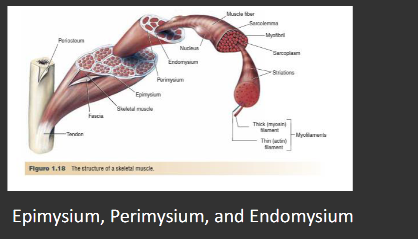
The connective tissues that wraps around the whole
muscle, muscle fiber bundles, and individual muscle
fibers, respectively are?
The body contains ______ bones.
206
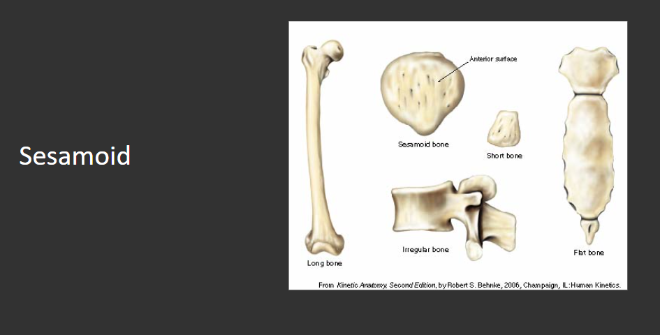
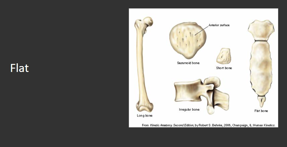
Bones are classified by their shapes. What are the 5 groups of bones?
Long, Short, Flat, Irregular, and Sesamoid
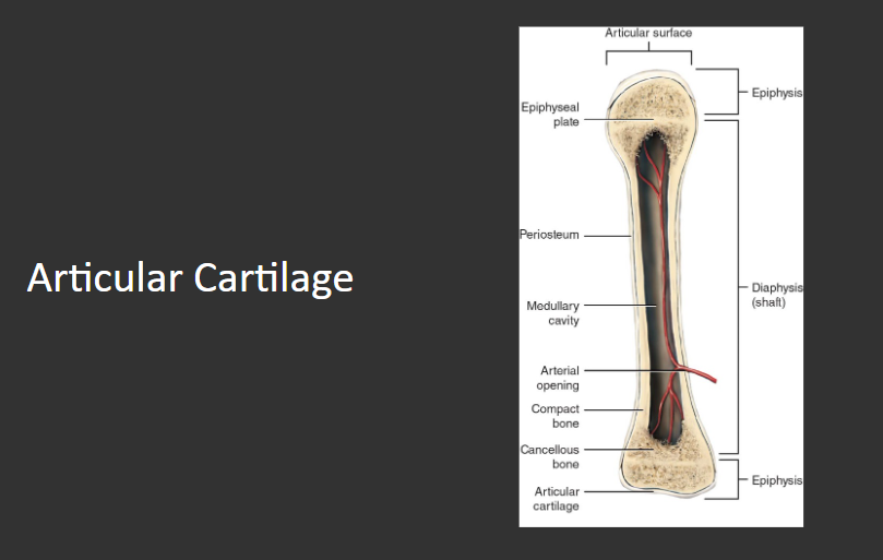
The very ends of long bone’s epiphyses are covered with ____________
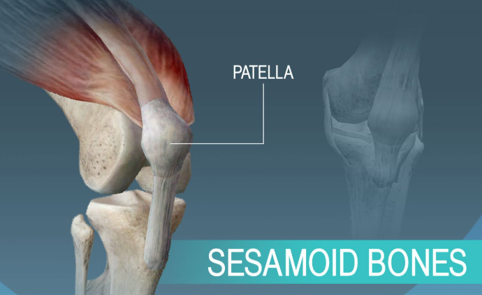
What is the name of the small, nobular bones usually found within tendons of muscles?
The ribs are an example of which type of bone?
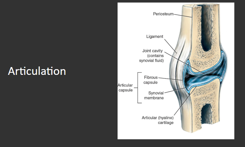
The place where two or more bones join together anatomically is referred to as an?
What are the two major forms of joints when classified by structure?
Diarthrodial & Synarthrodial
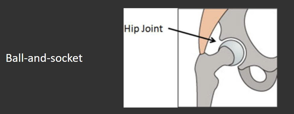
The hip joint is an example of which diarthrodial subdivision?
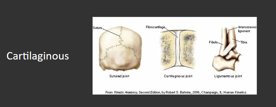
This type of synarthrodial joint contains fibrocartilage
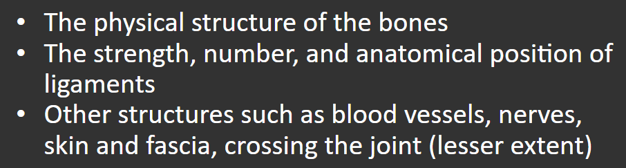
Joint strength is determined by what three factors. Name 2 of them.
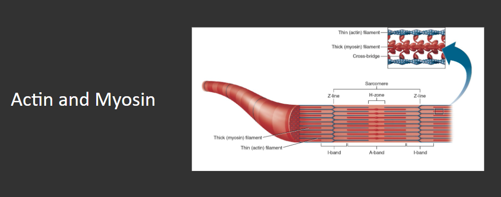
Name the two contractile protein filaments
Muscle tissue is categorized into what three types?
Smooth, Cardiac, and Skeletal
The muscle doing the most work/generating the most tension is called _______.
Prime mover/agonist
The two primary types of skeletal muscle fibers are?
Fast-twitch and Slow-twitch
________ are considered the functional units of “skeletal muscle”.
Sarcomeres
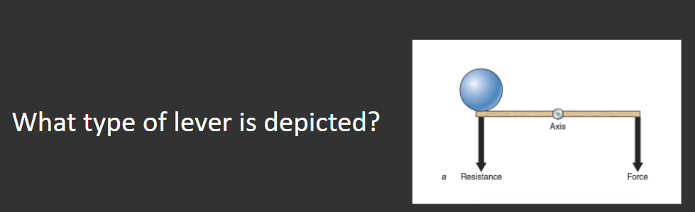
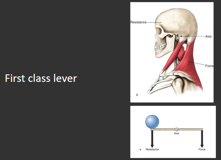
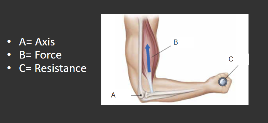
All three type of levers are found in the body, but most levers are _______ class.
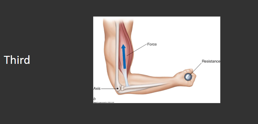
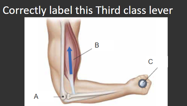
The bicep curl exercise is an example of which lever system?
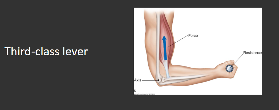
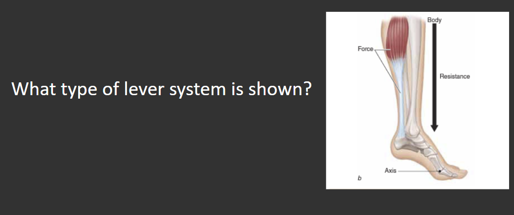
Second class lever
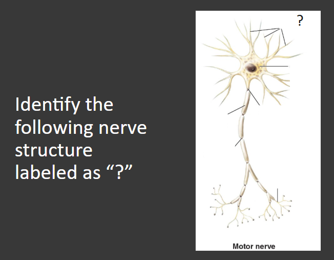
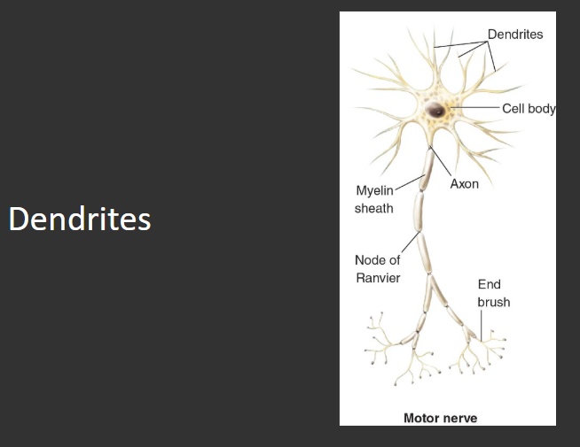
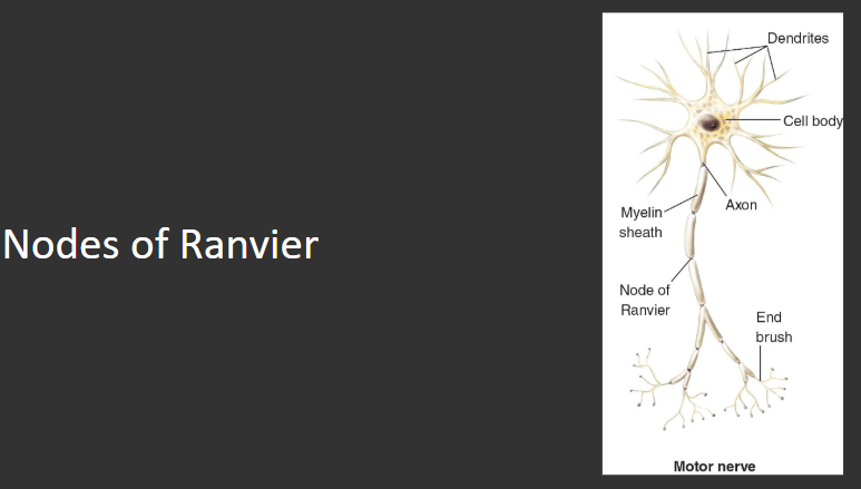
Dendrites
_______ receive information from surrounding tissue and conduct the nerve impulse to the nerve’s cell body.
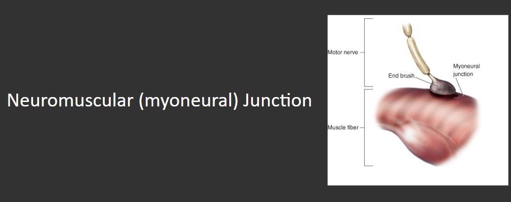
The nerve innervating a muscle is referred to as a ______.
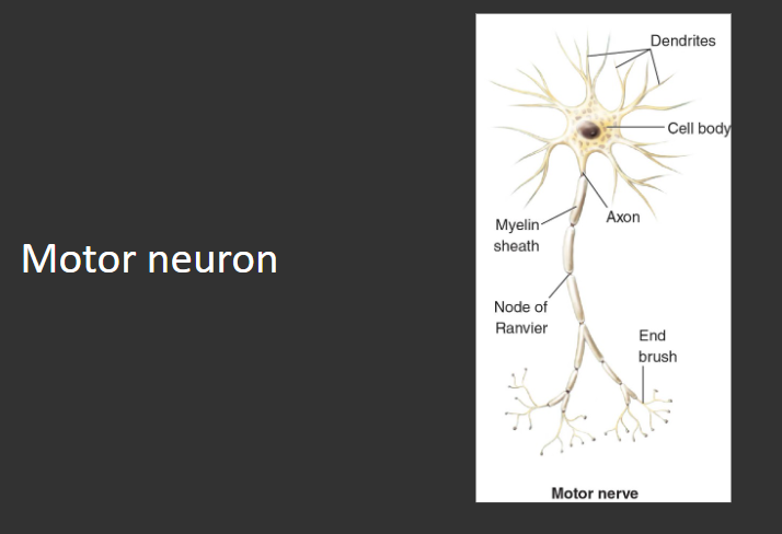
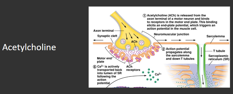
What is the name of the connection between the nerve fibers and the muscle fibers?
Nerve impulses “leap” along the myelin sheath (across the _________), allowing the impulses to travel at higher speeds than they would across an unmyelinated axon.
These blood vessels carry blood to the tissues.
Arteries
Name the components of the vascular tree.
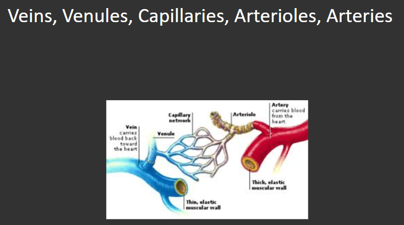
What is the purpose of the blood vessels?
Bring nutrients to the muscle tissue and carry away the waste products produced as the muscle tissues expend energy.
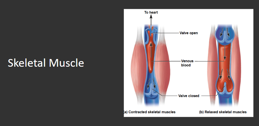
The veins contain small valves that permit blood to flow in only one direction. ___________ act as venous pumps that squeeze blood upward past each valve.
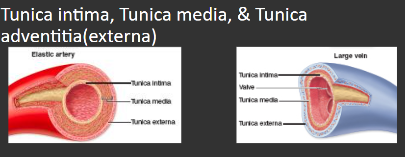
What are the three tissue layers of the walls of arteries, veins, and capillaries?
The _______ the ratio of muscle fibers to motor neurons, the greater the precision of movement.
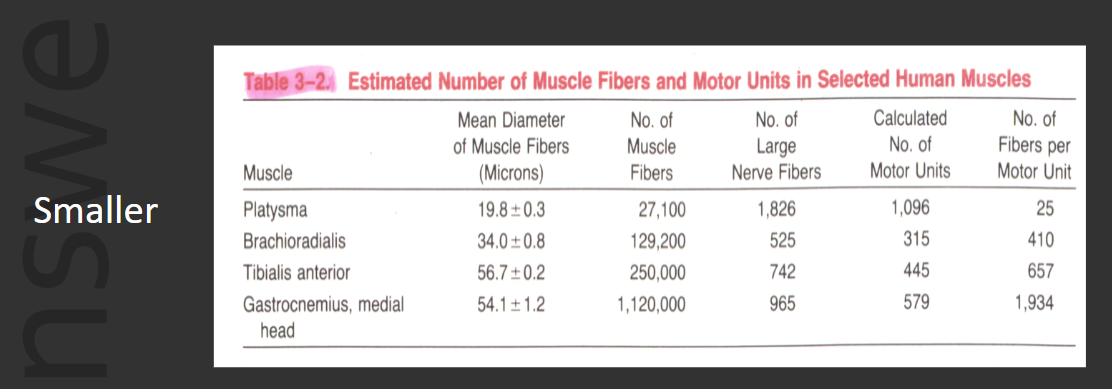
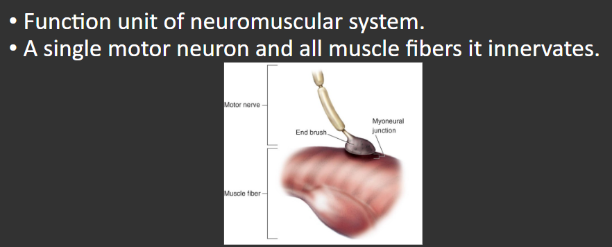
Define a Motor Unit.
The movement of what two ions is primarily responsible for an action potential
Sodium (Na) and Potassium (K)
Gradations in strength of muscular contractions affected by two factors:
Number of motor units
Frequency of stimulation
The release of _________ and its binding to receptors in the motor end plate, triggers an action potential in the muscle cell
Term used to refer to something that is above or higher than another structure
Superior
If a structure is closer to the midline of your body than another structure, the structure is ________.
Medial
In anatomical position, your hand is ______ to your elbow.
Distal
Pronation refers to turning the ________ surface of the hand face down
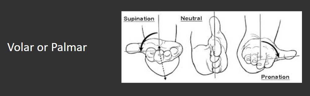
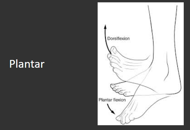
In the foot, the volar aspect of the foot is also referred to as ________.
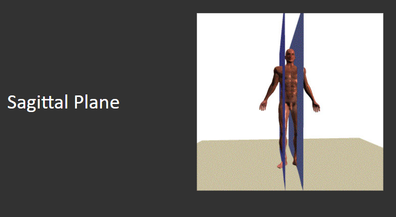
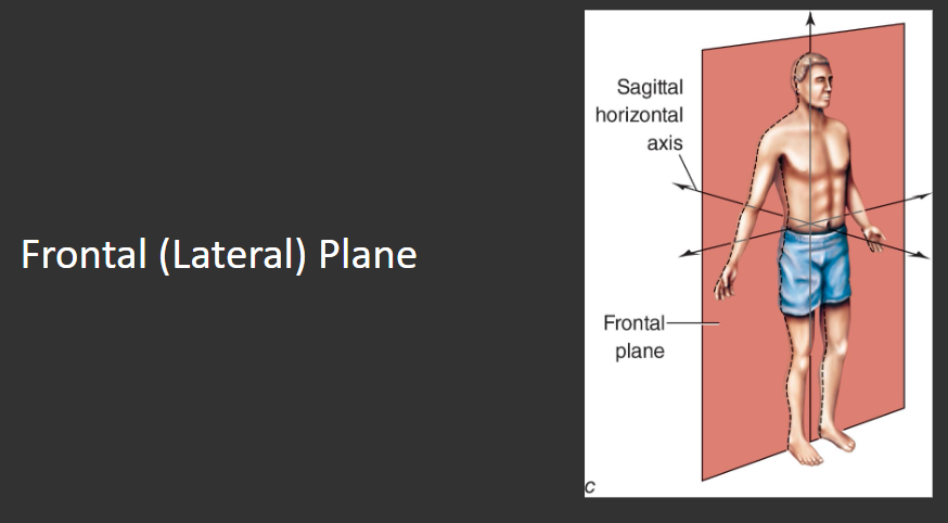
The term _______ refers to one plane that divides the body into exactly one half of the body
Cardinal
This plane passes from the front through the back of the body, creating a left and a right side of the body.
The point at the intersection of all three cardinal planes is the body’s ___________.
Center of Gravity
Which plane rotates about a vertical axis?
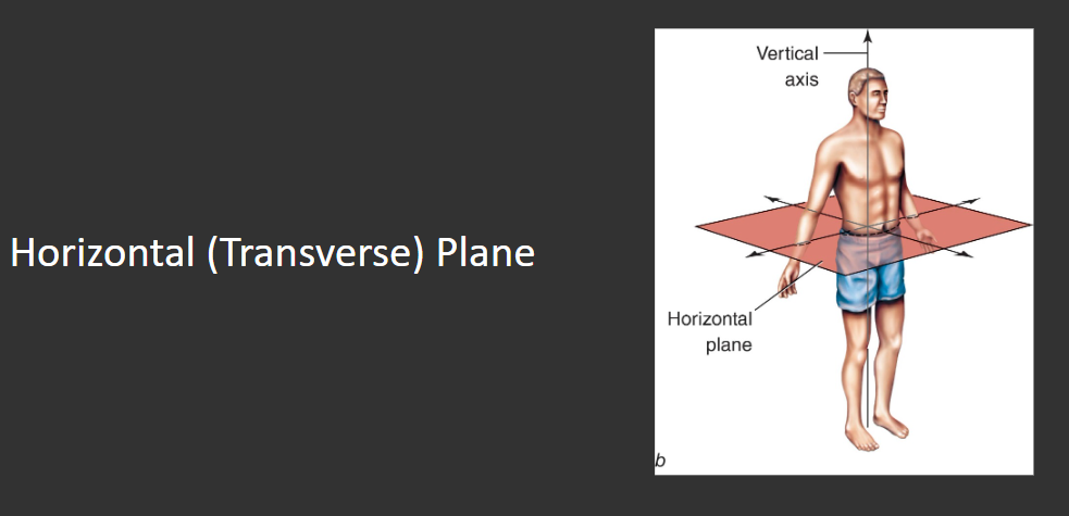
A lateral raise exercise (Shoulder Abduction) is performed in which plane?
A bicep curl is an example of elbow _____.
Flexion
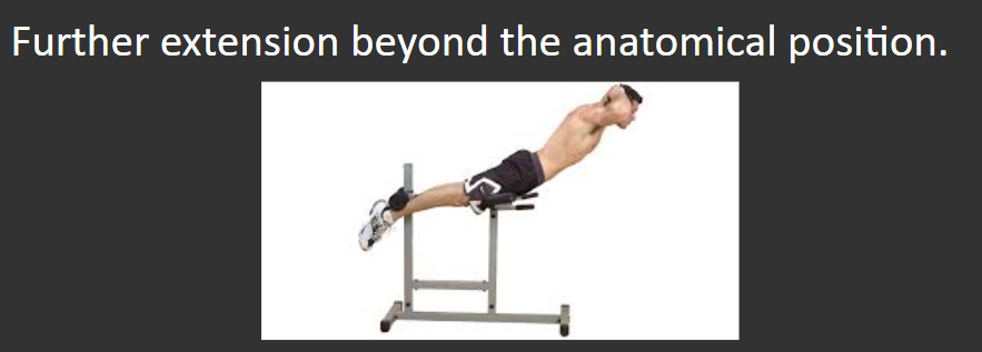
Define Hyperextension
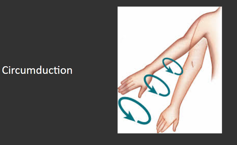
Joints capable of creating movement in two or three planes are also capable of ___________.
When performing the concentric phase (return to standing) of the squat exercise, your hips and knees perform _______.
Extension
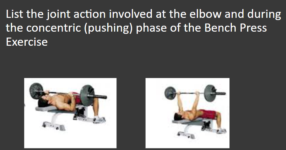
Elbow Extension
Smooth
Walls of veins and arteries, and organs
Cardiac
Heart muscle
Properties of muscle tissues
Extensibility: ability to stretch
Elasticity: ability to return to normal after being stretched
Contractility: ability to shorten and produce tension
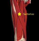
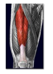
Longitudinal muscle
Fibers parallel to long axis of muscle
Ex: Sartorius
Fusiform muscle
Rounded muscle that tapers toward ends
Ex: Brachialis, brachioradialis, biceps brachii

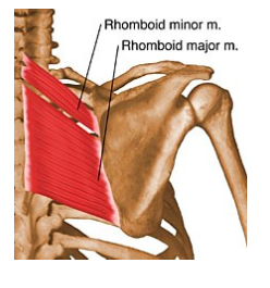
Quadrate muscle
Four-sided, generally flat, and have parallel fibers
Ex: Rhomboid, pronator quadratus
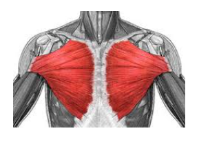
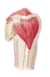
Triangular muscle
Fibers radiate from narrow to broad attachment
Ex: Pectoralis major
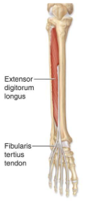
Unipenniform muscle
Fibers on one side of tendon
Ex: Extensor digitorum longus, tibialis posterior
Bipenniform muscle
Fibers on both sides of tendon
Ex: Flexor hallucis longus, Rectus femoris
Multipenniform muscle
Several tendons with fibers between them
Ex: Deltoid
Slow-twitch muscle fibers
Smaller, lots of blood, more myoglobin
Used in long duration/low intensity activities
Fast-twitch
Larger and less blood
Used in short duration/intense activities where strength and power are important
Forms Z line
Actin anchored to adjacent sarcomeres in series with each other
Sarcomere - from Z line to Z line
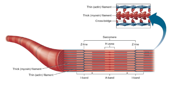
I band
Area of myofibril that contains Z line and actin filaments (no myosin)
Shortens in length during contraction
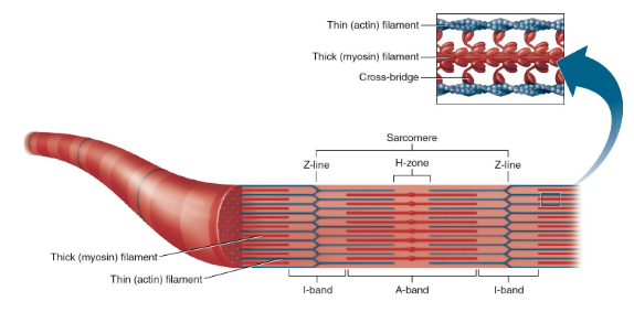
A band
whole length of myosin - overlap with actin filaments

H zone
within the A band where actin and myosin do not overlap

M band or line
connects adjacent myosin filaments with each other
(middle of H zone)

Axial skeleton
Skull, spine, sternum, ribs
Appendicular skeleton
Bones of the appendages: humerus, femur, phalanges, scapula, tibia, tarsals, etc.
Articular surface
connects bone to bone
Fossa
smooth, hollow surface
Facet
small, flat, smooth Notch - cut-out to allow passage
Foramen
hole in bone
Process
thinner projection
Condyle
large bony knob
Tubercle/tuberosity
small/medium bump in bone
Ossification (bone production) occurs
in diaphysis & epiphyses
Major growth continues until about, All epiphyses normally fused by
17-19 years of age, 25
The two major forms of articulations:
Diarthrodial: freely movable, possess an articular cavity
Synarthrodial: No separation or articular cavity
Hinge
-Movement in one plane
-Elbow, knee (modified)
Ball and socket
-Shoulder and hip joints
-Permits movements in all planes
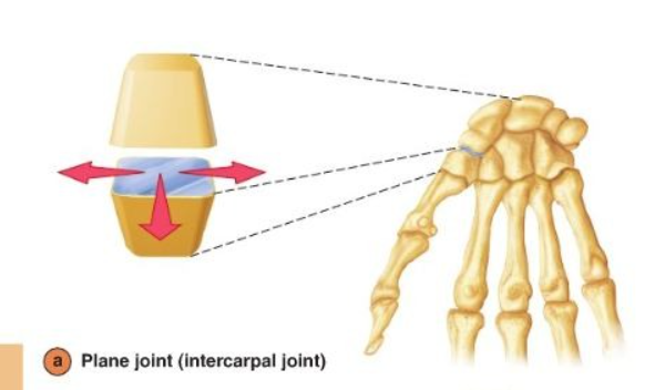
Irregular (flat-gliding) (plane)
Intercarpal, intertarsal, articular surfaces of vertebrae
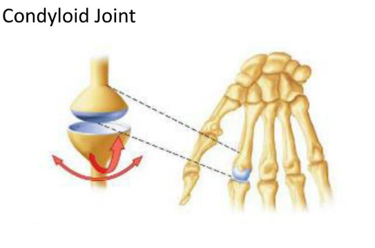
Condyloid
Radiocarpal and metacarpophalangeal
Movement in two planes
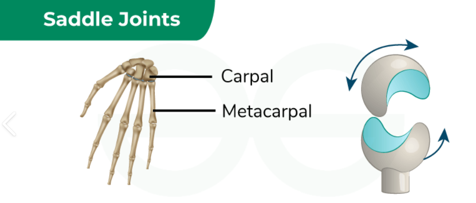
Saddle
Carpometacarpal joint of thumb (between 1st metacarpal/trapezium)
Movement in two planes
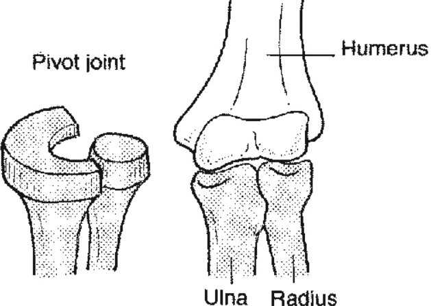
Pivot
Atlantoaxial (C1/C2), proximal radio-ulnar
Movement in one plane
Non-axial
carpals/tarsals
Uniaxial
elbow
Biaxial
wrist
Triaxial
shoulder/hip
Sutured
skull (no movement)
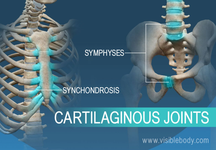
Cartilaginous
spinal column (some movement, fibrocartilage)
Ligamentous
forearm/leg and coracoacromial
(tie bones together)
Factors of Joint Stability
◼ Shape of bone structure
◼ Ligaments
◼ Muscle and tendons
◼ Fascia and skin
Joint range of motion is limited by:
-Elasticity of muscles, tendons, and ligaments
-Soft tissue near the joint
-Bone structure
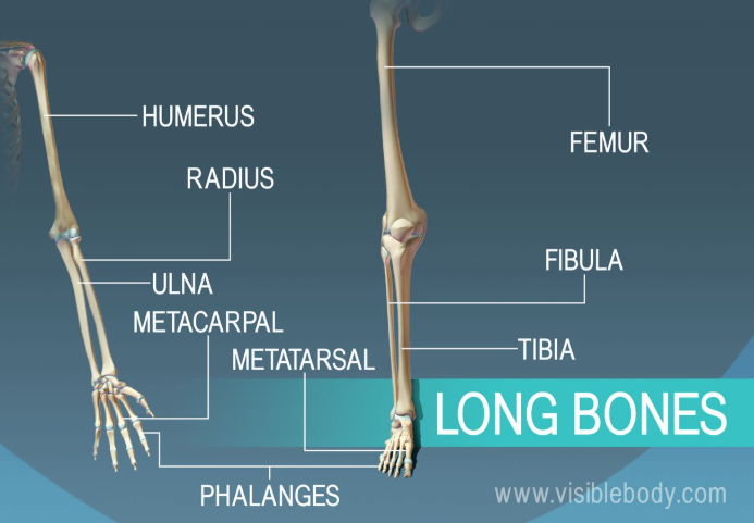
Long bone
Cylindrical shaft, contains a medullary canal
-Humerus, ulna, tibia, fibula, metacarpals, phalanges, femur
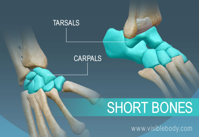
Short bone
small, chunky, solid
-Carpals and tarsals
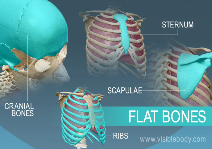
Flat bone
protect vital organs
-Sternum, ribs, skull, scapula, pelvis
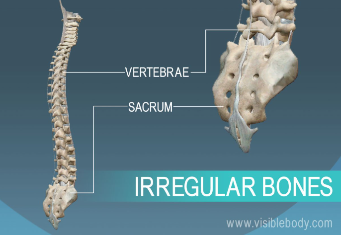
Irregular bone
-Vertebrae, sacrum, coccyx, ear bones
Sesamoid bone
-Small, nodular, enveloped in tendons (free floating)
-big toe, thumb, knee