Looks like no one added any tags here yet for you.
nervous system functions
regulate and control other systems of the body by communicating through electrochemical impulses
Neuron
nerve cell that responds to stimuli, conducts electrical activity and releases chemical regulators
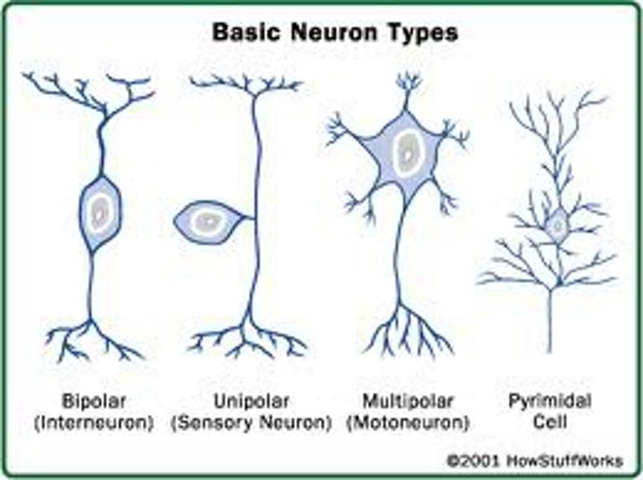
what are the structural classes of neurons based on?
the number of processes.
Parts of a neuron
cell body (soma)
dendrites
axon
axon hillock/initial segment
axon terminal
what are the functional classes of neurons based on?
the direction of impulse
Interneurons (association neurons)
located completely within the CNS and integrates functions of the nervous system (mulitpolar)
motor neurons
efferent neurons; conduct impulses from the CNS (brain & spinal cord) to target organs (muscles or glands)
sensory neurons
afferent neurons; conduct impulses from sensory receptors to the CNS
Glial cells in CNS
astrocytes, oligodendrocytes, microglia, ependymal cells
glial cells
constitute about half of the cells in the CNS, can divide by mitosis (unlike neurons), and provide physical and metabolic support
glial cells of PNS
Schwann cells and satellite cells
Oligodendrocytes
CNS; forms myelin sheaths which insulates and covers axons and speeds up the conduction of electrical signals along axon
Schwann cells
PNS; forms myelin sheath in PNS, same function as oligodendrocytes
membrane potential of neurons
resting potential = -70mV
established by large negative molecules inside the cell, Na+/K+ pumps, and permeability of the membrane
ions are constantly moving to maintain concentration gradients
channels in the membrane of a neuron
ligand-gated
voltage-gated
mechanical gated
ligand-gated channel
specified by ion, opening in response to the binding of a chemical ligand to its receptors
voltage-gated channels
protein channel that when stimulated, depolarizes the membrane to a threshold, specific to the ion
mechanical gated channels
open when physical deformation to membrane occurs (like stretching)
where are ion gated channels located on neuron?
on the receptive segment, dendrites & cell body
Where are voltage gated channels located on neuron?
axon hillock/initial segment
Where are mechanically gated channels found?
Found in sensory receptors (touch, pressure, vibration)
threshold
an approximate value needed for an action potential to occur. there is enough positive ions flowing in to move the membrane potential from -70mV to -55mV
does the strength of stimulus have an affect on action potential?
no, the stimulus strength doesn't matter after passing the approximate value. the strength of the stimulus affects the FREQUENCY of AP and may recruit more neurons to have an AP
why does the AP peak stop at +30mV?
this is where the K+ gates open and the Na+ gates close
changes in membrane potential are controlled by
changes in the flows of ions through channels such as Voltage gated K+ channels and voltage gated Na+ channels
action potential
all or nothing electrical event in a single cell where the membrane potential quickly becomes positive and returns to resting potential after
compound action potential
the sum of all the action potentials occurring in the individual neurons of the whole nerve
absolute refractory period
the period where a second stimulus will not produce an action potential
why does the absolute refractory period occur?
during this period, the neurons have to wait for the first AP to complete because since all Na+ channels are opening up another action potential cant start
- Na+ channels are inactivated
- As soon as inactivation is removed and Na+ are closed, the channel can reopen to the second stimulus
relative refractory period
a second action potential could happen only if the stimulus strength is greater than usual
Why can relative refractory period occur?
This occurs in a period of hyperpolarization (even more negative), have to bring it from a further negative point to +55
- This wavelength of action potential will have a lower amplitude
Why does the AP only travel in one direction down the axon?
because behind it is the refractory zone which is essentially a block that stops any other channels in that zone from opening. this forces channels in front of it to open ensuring the neuron operates forward
action potential conduction
- The depolarization of the first AP is the stimulus for the new action potential in the region just ahead of it and so on
- each AP is its own separate event and is said to be regenerated
-However, positive feedback of Na+ allows the action potential to travel. without decrement (decrease) thus reaching the end with the same amplitude.
saltatory conduction
Rapid transmission of a nerve impulse along an axon due to myelinated neurons.
myelin prevents Na+ and K+ from moving through the membrane causing the AP to move faster since its "leaping from node to node" compared to ion channels located ALL along the axon
Synapes
the functional junction between two neurons at which point the impulse is transmitted. use both chemical and electrical stimuli to pass info
synapses can either be _________ or _________ depending on the neurotransmitter (chemical signal) being transmitted
inhibitory or excitatory
presynaptic neuron
conducts impulses toward the synapse
postsynaptic neuron
conducts signals away from synapse
electrical synapses
Pre- and post-synaptic cells are connected by gap junctions. found in cardiac and smooth muscle to allow contraction as a single unit to occur.
chemical synapses
(the majority)
axon terminals hold synaptic vesicles. pre-synaptic neurons release neurotransmitters
Neurotransmitters
chemical messenger that travels across the synaptic cleft and binds to receptors on post-synaptic neurons
at the post-synapse
neurotransmitters come in large amounts across the synapses to ensure binding to a post synaptic receptor. unused neurotransmitters are transported away from the site
SNARE complex
set of proteins that allow the vesicle to attach to the plasma membrane, they loosely dock the vesicles
why are calcium ions important for the functioning of the SNARE complex?
Ca+2 ions trigger a change in the SNARE proteins that leads to the fusion and release of the neurotransmitter
Exitatory Postsynaptic Potentials (EPSPs)
opening Na+ or Ca2+ channels results in a graded depolarization. this brings the postsynaptic membrane closer to the threshold (more positive) and is a graded potential
inhibitory postsynaptic potential (IPSP)
opening K+ or Cl- channels results in a graded hyper-polarization. this brings the postsynaptic membrane further from the threshold (more negative). this decreases the likelihood of an action potential
ESPS and IPSPS are GRADED POTENTIALS:
amplitude decreases as the signal moves toward axon hillock. graded potentials MAY lead to action potentials
action potentials can begin at the hillock due to
a high amount of Na+ and K+ channels
characteristics of graded potentials
summation and lack of a refractory period
example of neurotransmitter at the synapse
acetylcholine
Nicotinic ACh receptors
Ach binds at post synaptic cell, ex. skeletal muscle cells (how muscles contract)
- agonist = nicotine
- antagonist = curare
binding of 2 acetylcholine molecules opens a channel
- due to electrochemical gradient, more Na+ flows in than K+ out, ESPS begins
Muscarine ACh receptors
Ach binds at post synaptic cell
ex: digestive cells or cardio cells
- agonist = muscarine
- antagonist = atropine (from plants)
binding at the receptor open sion channels indirectly using a G-protein
- dopamine and norepinephrine receptors do this too
Monoamines
synthesized from amino acids
ex: dopamine, norepinephrine, epinephrine (all 3 called catecholamines, and all use a second messenger system)
after activating their receptors, monoamines are _________________________
transported back into the axon terminal or are broken down by enzymes
neurons releasing catecholamines located in the CNS:
regulate mood, attention, hormone release, states of consciousness and more
Monoamine Oxidase Inhibitors (MAOIs)
enzyme breaking down monoamines
used to treat depression & other diseases/disorders. it inhibits the process of breaking down the catecholamine by enzymes, therefore increasing concentration of dopamine and norepinephrine at synapse
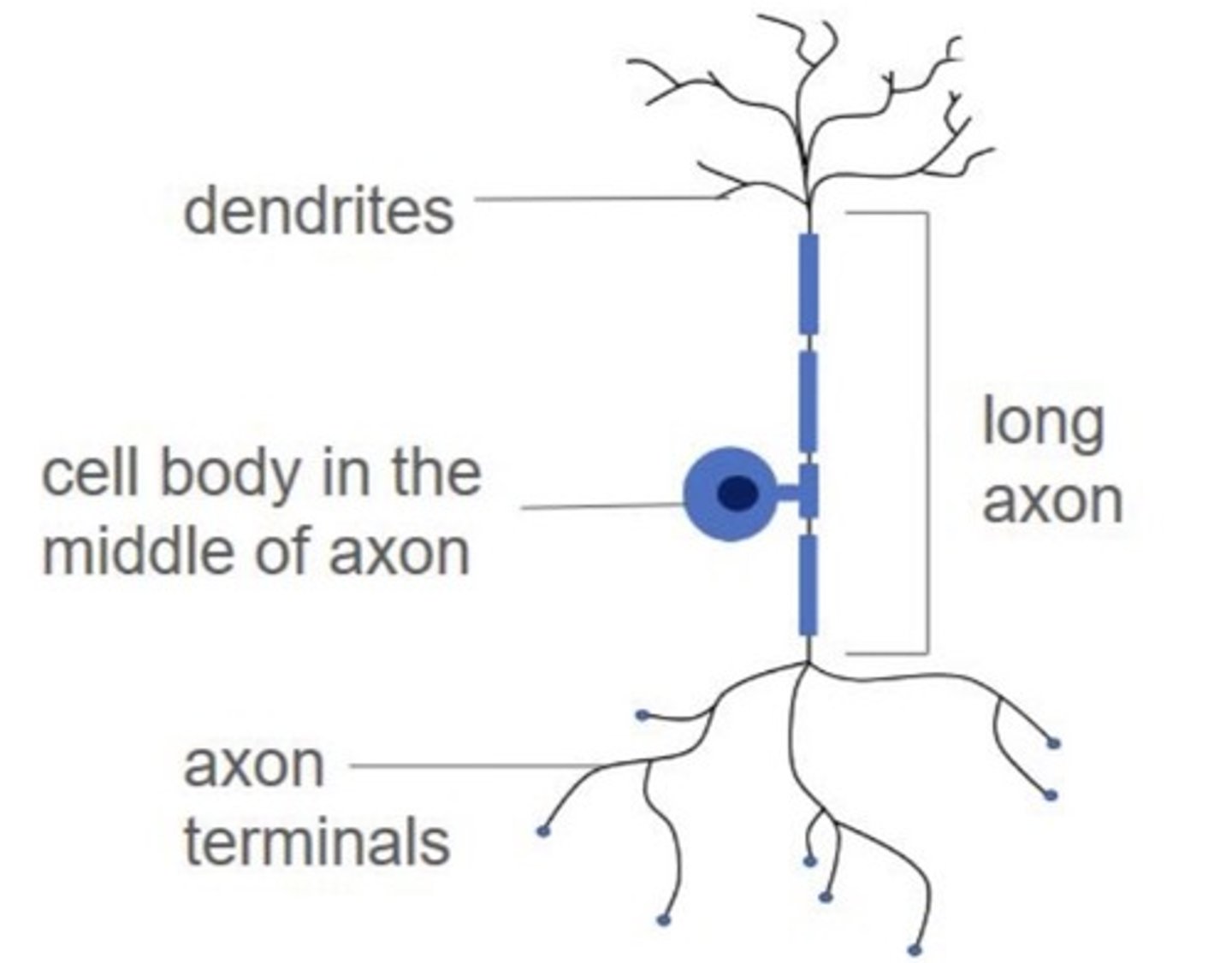
sensory neuron structure
sensory neurons have an end to receive sensory stimuli and produce the nerve impulse and the other end delivers impulses to synapse in the CNS. cell body located in the middle
Plasticity
the brain's ability to change, rewire/ build connections between neurons. associated with muscle memory, normal brain function during stress, during depression this is limited
sensory receptors
specialized cells that generate graded potentials called receptor potentials in response to a stimulus
receptor potentials
A slow, graded electrical potential produced by a receptor cell in response to a physical stimulus
Stimulus
energy or chemical activating a sensory receptor
Modalities
types of senses: arise from different receptors. every sensory neuron is specific to a sensation
major categories of sensory receptors:
mechanoreceptors = mechanical deformation (touch, pressure)
thermoreceptors = heat/cold
photoreceptors = light
chemoreceptors = chemical composition
nociceptors = pain
neural pathways in sensory systems
eyes: visual cortex
ears: auditory cortex
information (the coded action potential) then moves to the association area of the cerebral cortex.
complex integration occurs at
cortical association areas. this is where perception occurs along with emotional/varying factors that will affect perception
factors that affect perception
1. receptor adaptation and afferent processing
2. emotions and experiences
3. not all stimuli give rise to a conscious sensation
-ex: stretch receptors monitor blood pressure in several large arteries
4. lack of receptors for certain stimuli
-ex: radio waves
5. damaged neural pathways
-ex. phantom limbs
6. drugs- hallucinogens
7. mental illness
mechanoreceptors in the skin respond to
touch and pressure
phasic receptors
respond quickly but just as quickly adapt to stimulus
ex: vision, eye
fast-adapting, stimulus withdrawn quickly
tonic receptors
maintain response to stimulus
ex: pain. you stab yourself - that pain from the knife is going to continue to deliver a response
slow adapting
receptor potential (graded potential): coding potentials:
stimulus strength: increasing the frequency of AP
adaptation: a decrease in receptor frequency
Thermoreceptors
respond to thermal cues, some chemicals can open channels as well.
more receptors for cold (located close to surface) than warm
Nociceptors
respond to intense mechanical deformation, extreme temps & chemicals (PAIN)
transient receptor potential channels
Painful heat or painful cold stimulates a whole different set of channels to open in the membrane called: _________________________
How is pain different from other sensory information?
after initial AP, changes can occur that may increase/decrease sensitivity to pain
- pain can last after stimulus gone
- pain can be altered by pas experiences/emotions, and simultaneous activation of other senses (ex: phantom limb)
- pain reduction depends on endogenous opioids
eye: the shape of lens and thus degree of refraction is controlled by
muscles
as lens changes shape,
adjustments or accommodations to distance of objects occur
Where are photoreceptors located?
retina (back of the eye)
photoreceptors contain _________ for effective light trap called ______ and ______
pigment; rods, cones
rods
Retinal receptors that detect black, white, and gray, low levels of light
Cones
retinal receptor cells that function in daylight or in well-lit conditions. The cones detect fine detail and give rise to color sensations. (bright light signals = red, blue, green)
photoreceptors and bipolar cells ONLY undergo _________
graded responses, they lack the voltage-gated channels that mediate AP's
action potentials working with photoreceptors
start with photoreceptor, go through bipolar cell, and AP occurs at the ganglion cells (first cells in the pathway where AP can be initiated)
- glutamate is a neurotransmitter released here
neural pathways in the brain
optic nerve from each eye meet at the optic chiasm and project to many areas (mainly thalamus).
other inputs come from brainstem and visual cortex
some vidual pathway neurons project to areas other than the visual cortex like the hypothalamus
info is coded in spatial / temporal electrical activity - we perceive it all as a visual image consisting of lines, colors, contrast and movement
tympanic membrane (eardrum)
air molecules push against it at same frequency as sound wave. pressures and movement indicate pitch and volume
3 bones in ear
malleus, incus, stapes:
transduce sound by amplifying it through middle ear to the oval window
Organ of Corti
Center part of the cochlea, containing hair cells, canals, and membranes
organ of corti receptor cells
receptor cells called hair cells (mechanoreceptors)
- the hairs on the cell are called stereocilia, are bent back and forth as sound waves vibrate
- K+ channels open, Ca+ channels open
- bursts of neurotransmitters are then releases onto afferent neurons
- GLUTAMATE binds and causes AP in neurons that make up the vestibulocochlear nerve
fluid in the ear (endolymph) is
highly positive with more K+ ions, K+ moves down its concentration gradient
neural pathways in hearing
cochlear nerve fibers synapse with interneurons in the brainstem
- different arrival times from each ear and intensity determine sound source as well as the shape of outer ear and head movements
neural pathways in hearing travel path
action potentials carried by vestibular cochlear nerve ------> brainstem (medulla obloganta) -----> thalamus -----> auditory cortex
organization of CNS
association of interneurons form a network of communication from one area of brain to another
CNS is composed of
brain and spinal cord
CNS functions
1. receives input from sensory neurons and directs activity of motor neurons
2. association neurons integrate sensory info and help direct appropriate response to maintain homeostasis and respond to environment
3. humans capable of learning & memory adding a layer of modification to our behaviors
forebrain (cerebrum)
Higher mental functions - The largest portion of the brain
forebrain (cerebrum) consists of
cerebral hemispheres each divided into lobes
cerebral cortex
outer gray matter of cerebrum
corpus callosum
bundle of nerves that connects the cortex layers of the right and left hemispheres
limbic system
neural system associated with emotions. physically linked to thalamus, hypothalamus, and endocrine
Emotions controlled by the limbic system
Aggression: areas in the amygdala and hypothalamus
Fear: amygdala and hypothalamus, hippocampus
Hunger/satiety: hypothalamus
Sex drive: the whole system
Goal-directed behaviors: hypothalamus and other regions, reward/punishment system
there are few connections between the cerebral cortex and limbic system, which
limits conscious control over emotion