Looks like no one added any tags here yet for you.
reagent red cells are:
known red cell antigens
antisera are:
known red cell antibodies
Antiglobulin reagents are:
anti-IgG or anti-C3d or a combination of anti-IgG and anti-C3d
potentiators
Reagents added to the serum-cell mixture to enhance antibody uptake during the incubation phase of the indirect antiglobulin test
potency
strength of an Ag-Ab reaction
If reagents are produced for IN-HOUSE use (within the facility), a license is ___ required.
NOT
QC of reagents is performed ___ on commercial reagent red cells and antisera.
daily
Reagent product insert must include:
-description
-procedure for proper use
-interpretations
-performance characteristics
-limitations
-quality control
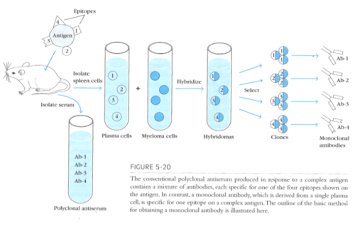
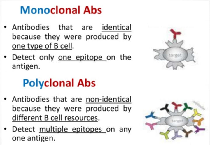
polyclonal antiserum
made from several different clones of B cells that secrete antibodies of different specificities
(AHG reagents)
Polyclonal vs. monoclonal antibodies
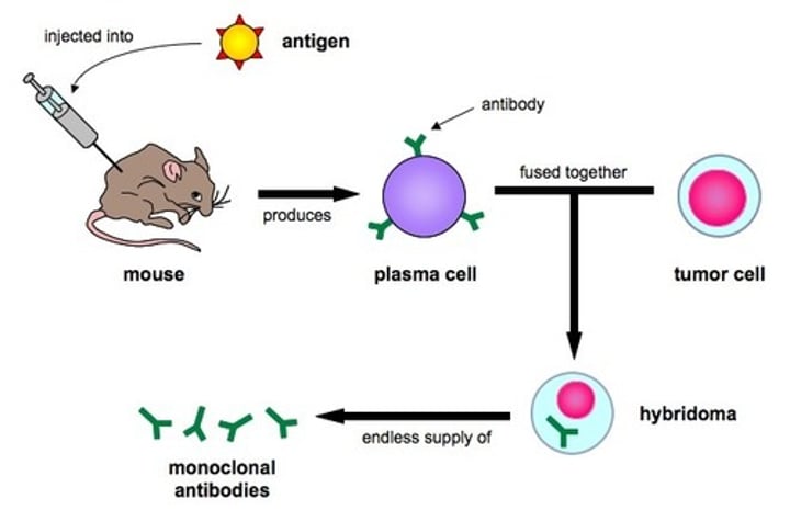
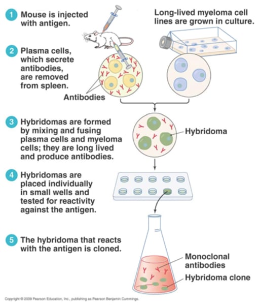
monoclonal antibody
made from single clone of B cells that secrete antibodies of the same specificity
monoclonal antibody production
hybridomas
hybrid cells formed by the fusion of myeloma cells and antibody-producing cells; used in the production of monoclonal antibodies
Epstein-Barr virus (EBV)
also called human herpesvirus 4 (HHV-4) and is one of eight viruses in the herpes family
heterohybridomas
hybrid cells formed by the fusion of lymphocyte of one species with the myeloma cell of a different species
phenotype
observable expression of inherited traits
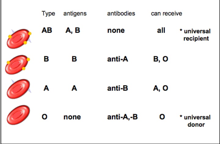
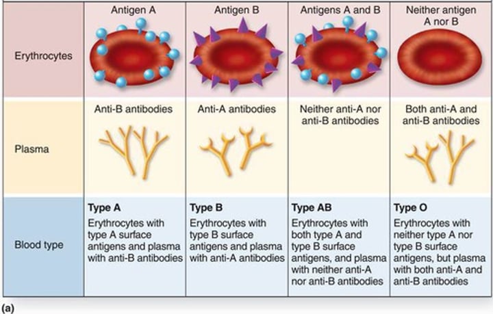
Four major blood phenotypes in the ABO blood group system:
A, B, AB, and O
Summary of monoclonal antibodies:
-secreted by a single clone of antibody-producing B cells
-one immunoglobulin class (IgG or IgM)
-unique specificity for a particular epitope
Summary of polyclonal antibodies:
-secreted by several different clones of antibody-producing B cells
-Mixture of IgM and IgG antibodies
-mixture of antibodies that may be directed at different epitopes of the same antigen
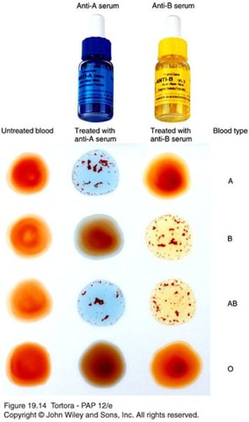
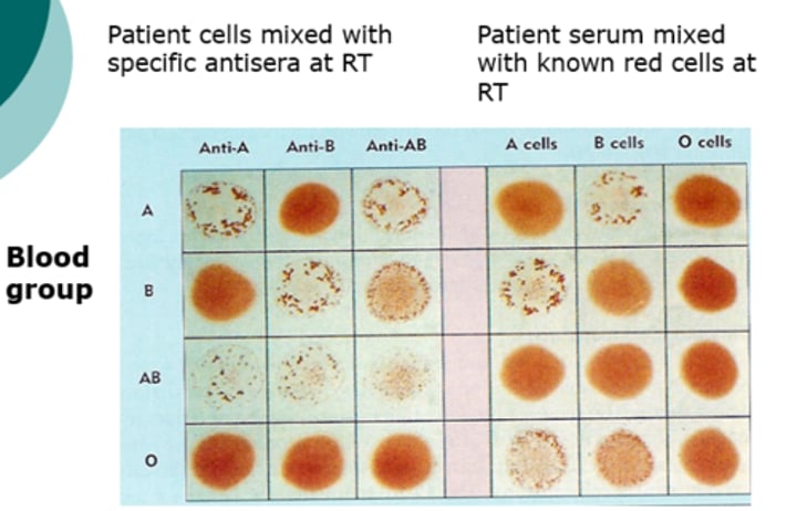
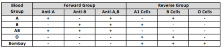
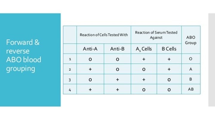
ABO Red Cell Testing (ABO forward grouping):
*ABO blood group antigen: A
-reaction with anti-A
*ABO blood group antigen: B
-reaction with anti-B
*ABO blood group reaction: AB
-reaction with anti-A and anti-B
*ABO blood group reaction: O
- no reaction with either
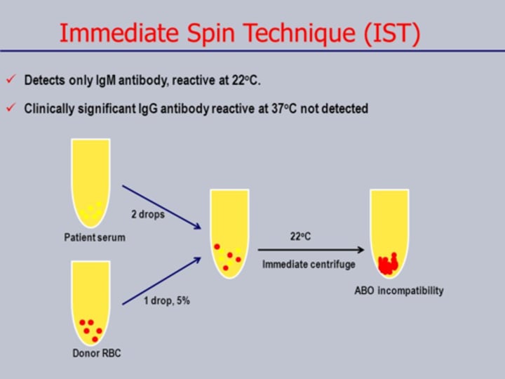
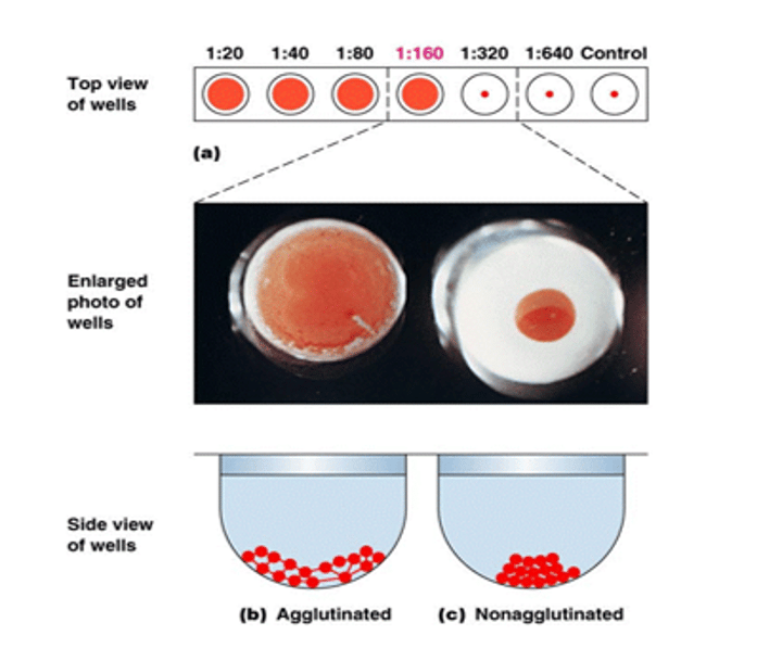
Immediate Spin Phase (IS)
source antigen and source antibody used in immunohematologic testing are combined, immediately centrifuged, and observed for agglutination
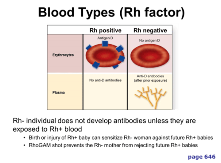
ABO antibodies
anti-A, anti-B, and anti-A,B; patients possess the ABO antibody to the ABO antigen lacking on their red cells (eg, group A individuals possess anti-B)
anti-A reagent color
always contains a blue dye
anti-B reagent color
always contains a yellow dye
autoantibodies
antibodies to self antigens
Typing for D antigen with patient or donor red cells
*D-positive: (D-antigen on RBC) reacts with anti-D and
*D-negative: (NO D-antigen on RBC) No reaction with anti-D
D typing procedure:
Commercial anti-D is combined with patient or donor red cells
-agglutination indicates presence of the D antigen on the red cells tested
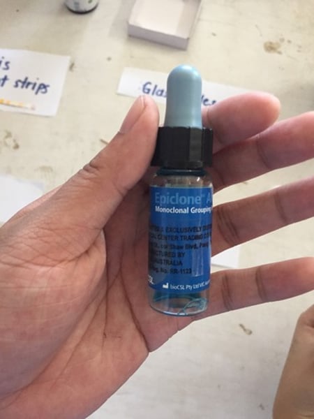
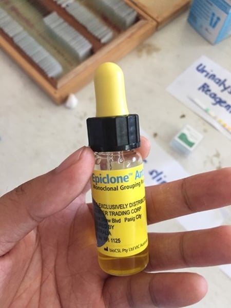
Summary of ABO and D typing reagents: Murine Monoclonal Anti-A and Anti-B
-for slide, tube, and microplate testing
-Anti-A=blue dye
-Anti-B=yellow dye
Summary of ABO and D typing reagents: Murine monoclonal Anti-A,B
-for slide, tube, and microplate testing
-blend of anti-A and anti-B clones
-Anti-A,B=clear
Summary of ABO and D typing reagents: Monoclonal Anti-D
-for slide, tube, and microplate testing
-monoclonal-polyclonal blend: IgM anti-D from human-murine heterohybridoma and polyclonal IgG anti-D
-monoclonal blend: IgM and IgG blending of human-murine heterohybridomas
-monoclonal: IgM from single clone
Examples of Low-protein controls in ABO and D typing:
1) reaction w/ anti-A and reaction w/ anti-D: A and D antigens present: reagent control present? yes; no agglutination w/ anti-B
2) reaction w/ anti-B: B antigens present: reagent control present? yes; no agglutination with anti-A
3) reaction w/ anti-B and anti-D: B and D antigens: reagent control present? yes; no agglutination with anti-A
4) reaction w/ anti-A and anti-B: antigens A and B: control present? yes; no agglutination w/ anti-D
5) reaction w/ anti-A, anti-B and anti-D: antigens: cannot interpret typing: control present? no; reagent control must be tested to determine ABO and D typing results
Reagent red cells
A1 and group B red cells: testing with patient serum or plasma confirms the ABO type
(known as reverse grouping or ABO serum testing)
ABO Reverse Grouping/Back type
Reagent red cells for serum testing are obtained from:
selected human donors and are manufactured in several optional packages (most common: two-vial set of A1 and B red cells)
All reagent red cells are ___ to remove blood group antibodies and are resuspended to a __-__% concentration in a buffered preservative solution to minimize hemolysis and loss of antigenicity during the dating period.
washed; 2-5%
recipient
patient receiving the transfusion
Antigram
profile of antigen phenotypes for each donor used in the manufacture of commercially supplied screening and panel cells
Screening cells are used in:
antibody screen tests
Antibody screening test
looks for antibodies with specificity to red cell antigens in patient and donor samples
An antibody identification panel is performed when the ___ ___ ___ is positive.
antibody screen test
Reagent red cell antibody identification panels:
are required to determine the specificity of a red cell antibody in a blood banking procedure called antibody identification
(patient or donor serum/plasma is tested with the reagent panel cells to identify an antibody to red cell antigens)
The antibody identification panel cells are individual group __ donors packaged in sets of __ or more, depending on the manufacturer.
O; 10
Three types of reagent red cells for routine testing include:
-A1 and B cells in ABO serum testing
-Screening cells to detect red cell antibodies
-panel cells to identify red cell antibodies
neutralization
blocking antibody sites, causing a negative reaction
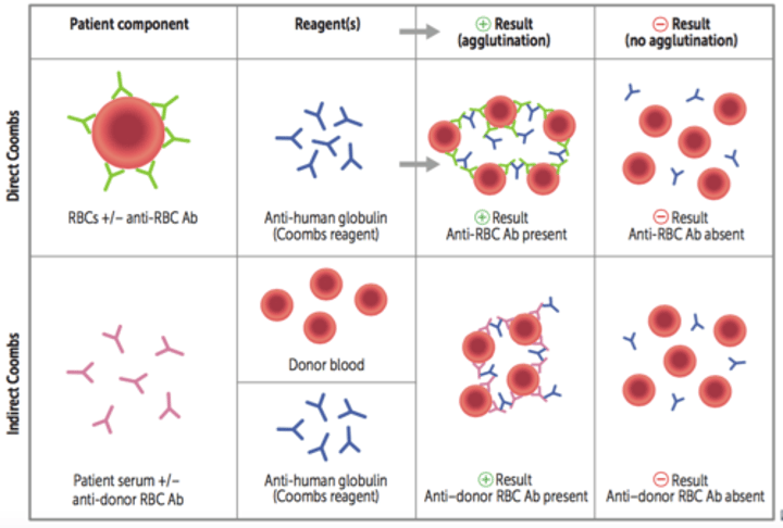
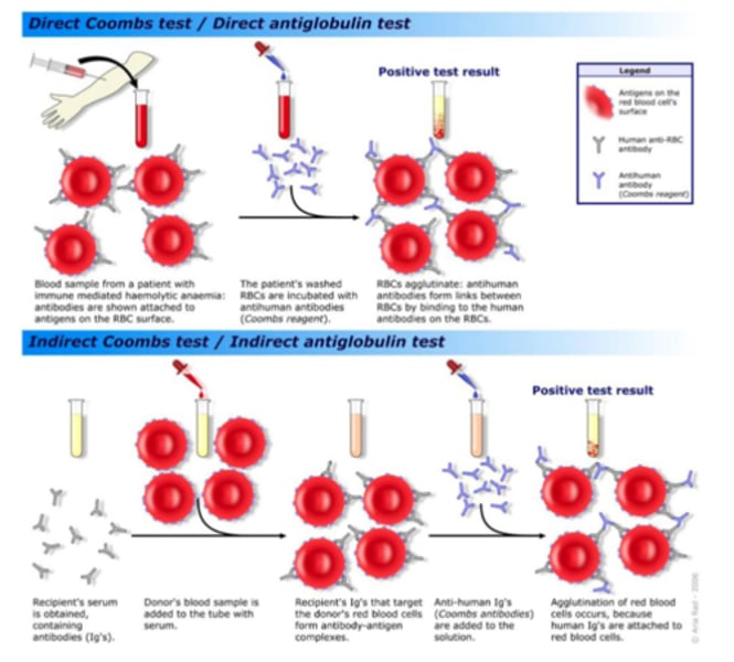
antiglobulin test (Coombs test)
detects IgG antibodies and complement proteins that have attached to red cells either in vitro or in vivo but do not produce visible agglutination
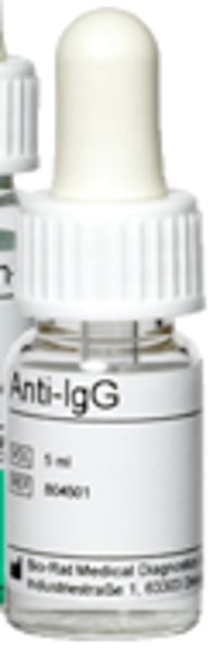
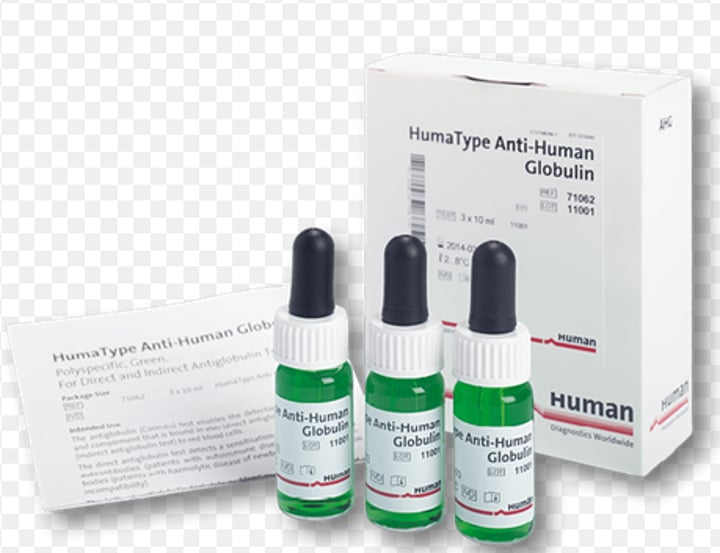
Polyspecific AHG reagent contains:
antibodies to IgG molecules (anti-IgG) and complement proteins (anti-C3d, anti-C3b)
Essential that red cells be washed with ___ ___ to remove any unbound molecules before the addition of the ___ reagent.
physiologic saline; AHG
AHG test washing steps:
-filling test tube with saline to mix with red cells already present in the tube
-saline-suspended red cells are centrifuged
-saline wash is decanted
-repeat for 2-3 cycles
-saline is removed, and the tube is blotted dry to remove most traces of the saline
to detect potential neutralization:
IgG-sensitized cells are added to tubes w/ negative reactions
(after centrifugation, a positive reaction should be observed to confirm that washing was adequate)
sensitized
Immunoglobulin or complement attached to the cells from the immune system (in vivo) or from a test procedure (in vitro).
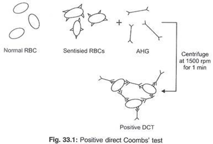
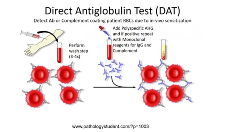
Direct Antiglobulin Test (DAT)
test used to detect antibody bound to red cells in vivo
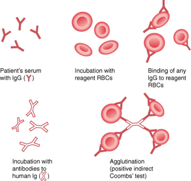
Indirect Antiglobulin Test (IAT)
test used to detect antibody bound to red cells in vitro
Autoimmune Hemolytic Anemia
immune destruction of autologous (self) red cells
DAT
-ordered to detect IgG or complement proteins bound to patient cells
-positive DAT is an important indicator of potential immune-mediated red cell destruction in the body
-b/c of IgG or complement attachment to red cells, macrophages are signaled to clear them
-this signals immune destruction of red cells and often leads to anemia
DAT procedure:
-A patient's RBCs are obtained and washed (3-4x) with physiologic saline
-Coomb's Reagent is added which will bind to any antibodies present on the RBCs
-Agglutination will occur if there are antibodies on the RBCs
polyspecific AHG reagent
contains both anti-IgG and anti-C3d antibodies and detects both IgG and C3d molecules on red cells
monospecific AHG reagent
reagents prepared by separating the specificities of the polyspecific AHG reagents into individual sources of anti-IgG and anti-C3d/anti-C3b
sample of choice for a DAT is collected in a:
EDTA tube
Clinical examples causing a positive DAT: Transfusion reaction
-caused by: donor cells coated with IgG
-source of IgG: recipient (patient) antibody
Clinical examples causing a positive DAT: Hemolytic disease of the fetus and newborn
-caused by: fetal red cells coated with IgG
-source of IgG: maternal antibody crossing the placenta
Clinical examples causing a positive DAT: Autoimmune hemolytic anemia
-caused by: IgG or C3 on patient red cells
-source of IgG: patient autoantibody
Clinical examples causing a positive DAT: Drug-related mechanism
-caused by: IgG-drug complex attached to cells
-source of IgG: immune complex formed with drug
IAT:
-detects in vitro sensitization of RBCs
-two stage procedure
1) antibodies first combine with red cell antigens in vitro during an incubation step
(plasma source is incubated at body temperature with a red cell source to allow the attachment of IgG antibodies to specific red cell antigens)
-red cell suspension is washed with physiologic saline to remove unbound antibody/complement
-after washing, AHG reagent is added to the test and centrifuged
reaction phase
observation of agglutination at certain temperatures, after incubation, or after addition of AHG
Polyspecific AHG reagents are used primarily in the ___ to determine that either IgG or complement molecules have attached to the red cells in vivo.
DAT
Comparison of DAT and IAT procedures: (DAT)
-detects IgG / complement-coated red cells
-IgG attachment to red cells has occurred within the PATIENTS BODY
-one-stage procedure
-patients red cells are tested with antiglobulin reagent withOUT an incubation step
-test for certain clinical conditions: HDFN, hemolytic transfusion reaction, and autoimmune hemolytic anemia
Comparison of DAT and IAT procedures: (IAT)
-detects IgG and complement-coated red cells
-IgG attchment to red cells occurred during the incubation step
-two-stage procedure
-test requires an incubation step before the addition of AHG reagent
-used as a reaction phase of several tests in immunohematology: antibody screen and antibody identification panel
Common sources of false-positive error in antiglobulin testing:
-Red cells are agglutinated before washing step and addition of antihuman globulin reagent
Possible explanation: potent cold reactive antibody of patient origin
Common sources of false-positive error in antiglobulin testing:
-Use of dirty glassware
Possible explanation:
particles or contaminants
Common sources of false-positive error in antiglobulin testing:
improper centrifugation-overcentrifugation
possible explanation: red cell button packed so tightly on centrifugation that nonspecific clumping cannot be dispersed
Common sources of false-negative error in antiglobulin testing:
Failure to wash cells adequately during the test procedure before the addition of AHG reagent
possible explanation: unbound human serum globulins neutralize AHG reagent
Common sources of false-negative error in antiglobulin testing:
testing is interrupted or delayed; AHG reagent is not added immediately after washing
possible explanation: Bound IgG or complement may detach from the coated red cells
Common sources of false-negative error in antiglobulin testing:
failure to identify weak positive reactions
possible explanation: technical error in testing
Common sources of false-negative error in antiglobulin testing:
loss of reagent activity
possible explanation: improper reagent storage, bacterial contamination, or contamination with human serum
Common sources of false-negative error in antiglobulin testing:
Failure to add AHG reagent
possible explanation:
technical error in testing
Common sources of false-negative error in antiglobulin testing:
improper centrifugation: undercentrifugation
possible explanations: conditions for promoting agglutination are not optimal
Common sources of false-negative error in antiglobulin testing:
inappropriate red cell concentrations-red cell suspensions fall outside the optimal 2%-5%
possible explanation: concentration of red cells influences the agglutination reaction
Differential DAT
test that uses monospecific anti-IgG and monospecific anti-C3d/anti-C3b reagents to determine the cause of a positive DAT with polyspecific antiglobulin reagents
IgG sensitized cells
-control system for antiglobulin tests interpreted as negative
-referred to as check cells or Coombs control cells
Potential reasons for a false-negative result detected by the use of IgG-sensitized red cells in an antiglobulin test:
-failure to add the antiglobulin reagent to the test
-failure of the added antiglobulin reagent to react
-failure to wash red cells adequately
Antihuman globulin reagents:
-Polyspecific: anti-IgG and anti-C3d; use in DAT
-Monospecific: either anti-IgG or anti-C3d/C3b; use in antibody screen/ identification, differential DAT
IgG sensitized red cells:
-added to negative AHG reactions
-should agglutinate after addition
-checks: sufficient washing, addition of AHG, AHG reagent was potent
-does NOT ensure that the AHG reaction is negative
antibody potentiators
(aka: enhancement media)
Reagents or methods that enhance or speed up the antibody-antigen reaction
enhancement media
reagents that enhance or speed up the antibody-antigen reaction
proteolytic enzymes
enzymes that denature certain proteins
Antibody potentiator: Low-ionic-strength saline (LISS)
-Increases rate of antibody uptake
Antibody Potentiator: Polyethylene glycol (PEG)
-Concentrates the antibody in the test environment in LISS
-removes water molecules
Antibody potentiator: Proteolytic enzymes (papain, ficin, and bromelin)
-Removes negative charges from the red cell membrane, which reduces the zeta potential; denatures some red cell antigens
-enhance warm and cold autoantibodies
-ficin comes from figs
-bromelin from pineapple
-papain from papaya
Antibody potentiators: Bovine serum albumin (BSA)
-Reduces the repulsion between cells but does not shorten the incubation time
-available in either 22% or 30% concentrations
-does NOT enhance warm autoantibodies
Lectins
plant extracts useful as blood banking reagents; they bind to carbohydrate portions of certain red cell antigens and agglutinate the red cells
Common lectins in Blood Bank: Dolichos biflorus
-origin: seed extract/plant
-antigen specificity: A1
Common lectins in Blood Bank: Ulex europaeus
-origin: gorse (flowering plant)
-antigen specificity: H
Common lectins in Blood Bank: Vicia graminea
-origin: seed extract/plant
-antigen specificity: N
Common lectins in Blood Bank: Iberis amara
-origin: plant
-antigen specificity: M
Gel test
- ID-MTS Gel card
-uses dextran acrylamide gel particles to trap agglutinated red cells
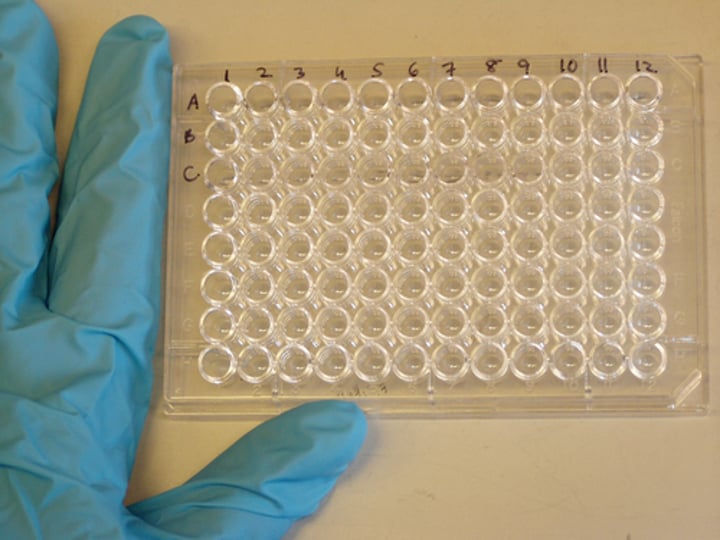
Microtiter plate
-96 wells serves as the substituted test tubes
-each well is considered a short test tube
-either U-shaped or V-shaped bottom in the microtiter plate well
-U-bottom well is more used
-small quantities of red cells and antisera are added to the microtiter wells, followed by centrifugation of the microtiter plates
-cell buttoms are resuspended by manually tapping the plate
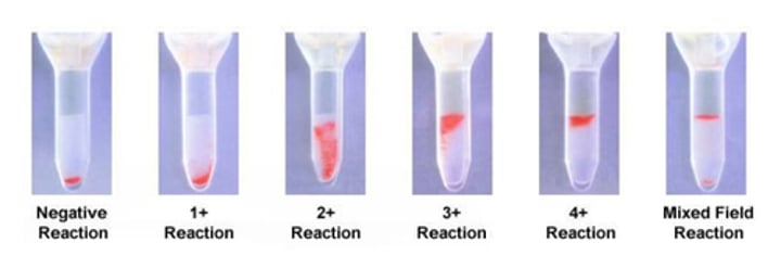
-concentrated button of red cells is indicative of Ag-Ab reactions, whereas the red cells in a negative result are dispersed throughout the well
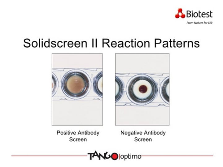
Solid-phase red cell adherence methods
-uses microplate test wells with immobilized reagent red cells
-for antibody screening, antibody identification, and compatibility testing
-suitable for automation
-antigen or antibody is immobilized to the bottom and sides of the wells
-In direct test: antibody is fixed to the wells. Ag-positive red cells from donor adhere to sides and bottom of well. Ag-negative red cells from donor settle to the bottom of the well and form a button after centrifugation
-In indirect test: red cell membranes are bound to wells. unknown patient serum is added and allowed to react. allows for the capture of IgG antibodies from patient serum to the rbc membrane. a washing step removes unbound IgG antibodies. In a positive indirect test: the indicator cells adhere to sides and bottom of wells. In negative indirect test: indicator cells settle to the bottom of wells and form button after centrifugation.
Microplate test method and interpretation: Microtiter Plate Assay
*Method:
-1 drop of anti-A and 1 drop anti-B are placed in separate wells of a U-bottom microplate
-1 drop of a 2-5% saline suspension of red cells is added to each well
-wells are mixed by gently tapping them
-the plate is centrifuged at an appropriate time and speed
-the cell button is resuspended by manually tapping the plate or using a mechanical shaker or placed at an angle for the tilt and stream method
-reactions are read, interpreted, and recorded
*Reactions of Microplate Testing:
-positive reaction: concentrated button of red blood cells
-negative reaction: smooth suspension of red blood cells or a streaming pattern of red blood cells when the plate is placed on an angle
Microplate technique apply the ___ principle of hemagglutination as the tube test. Solid-phase technology reactions are ____. A positive reaction is adherence to the ___; a negative reaction is a ___ ___ ______.
same; opposite; well; red cell button
What is the purpose of including a reagent control when interpreting group AB, D-positive red cells after testing with a low-protein anti-D reagent?
a. to detect false-positive agglutination reactions
b. to detect false-negative agglutination reactions
c. to identify a mix-up with a patients sample
d. to confirm ABO typing results
A. to detect false-positive agglutination reactions