[HISTOLOGY SLIDES] - Nervous Tissue (Laboratory)
1/36
There's no tags or description
Looks like no tags are added yet.
Name | Mastery | Learn | Test | Matching | Spaced |
|---|
No study sessions yet.
37 Terms
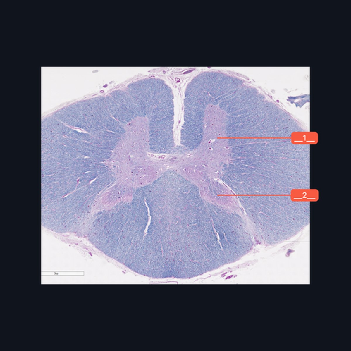
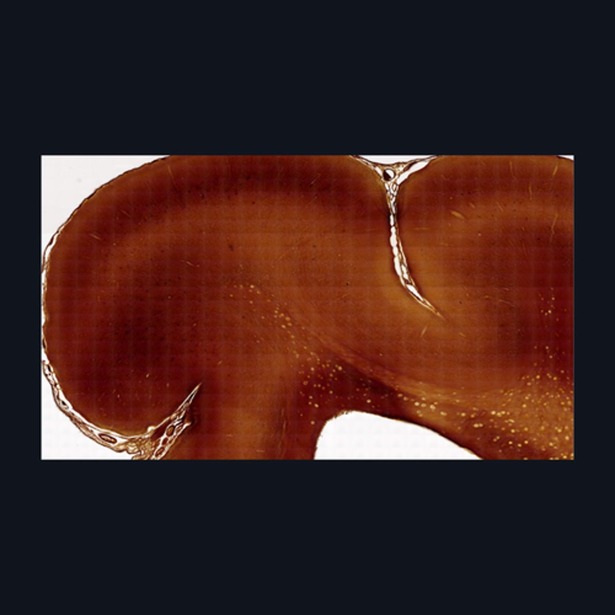
Gray Matter
Identify the pointed region in the spinal cord
(1) Ventral Horn, (2) Dorsal Horn
Identify the pointed regions of the gray matter of the spinal cord
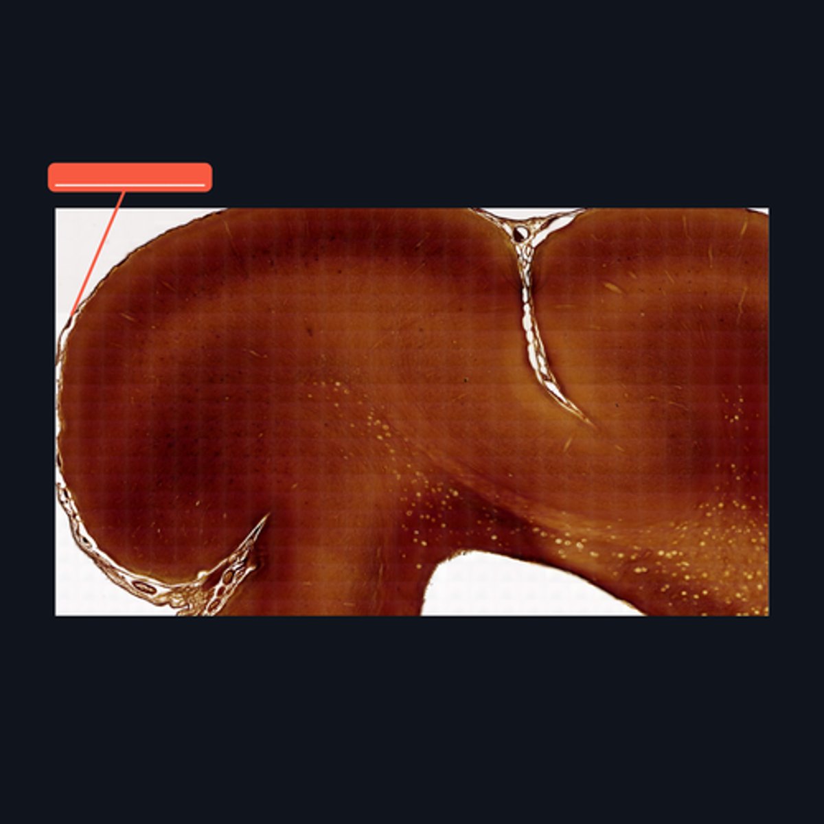
Gray Matter (When looking at speciemen from the brain, all areas in the periphery are considered Gray Matter)
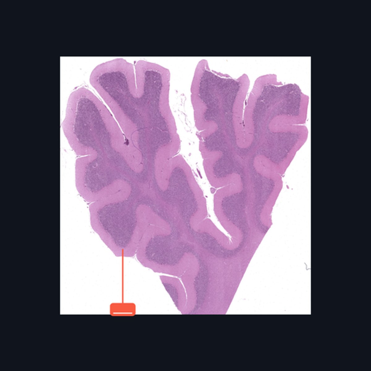
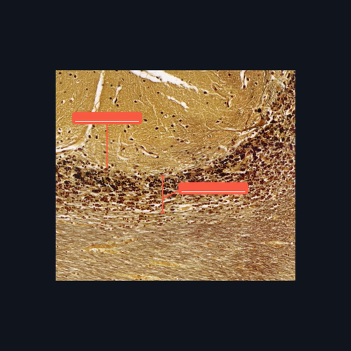
Identify the pointed region in the Cerebellum

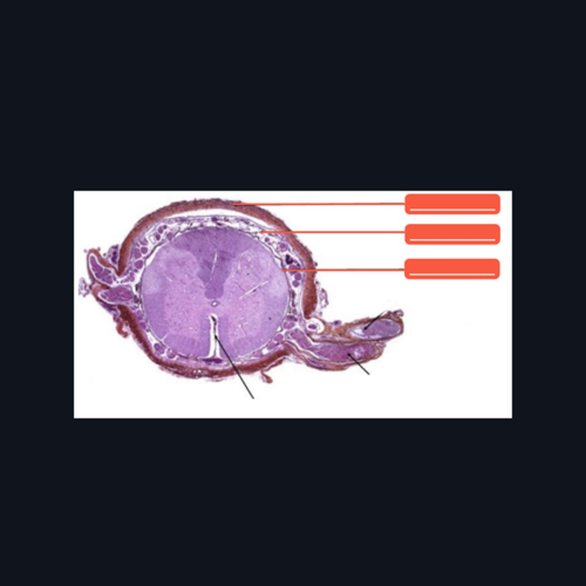
"cell bodies, dendrites, proximal portions of the axons, and neuroglial cells"
Enumerate the structures found in the pointed region
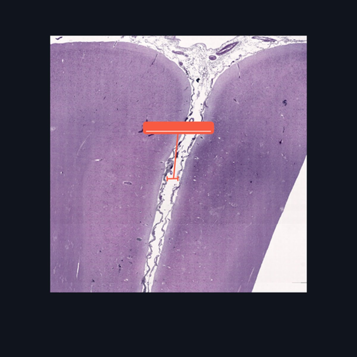
(top to bottom)
Dura mater
Subarachnoid space
Pia mater
Identify the pointed structures
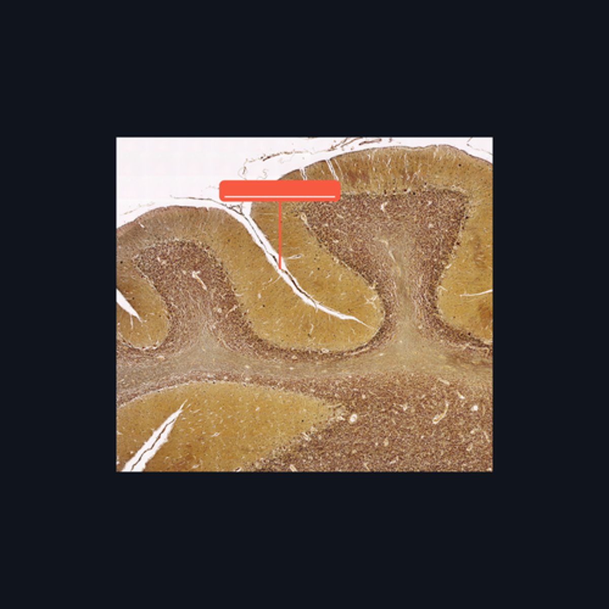
White matter
This type of cell/s is located mainly on what part of the brain?
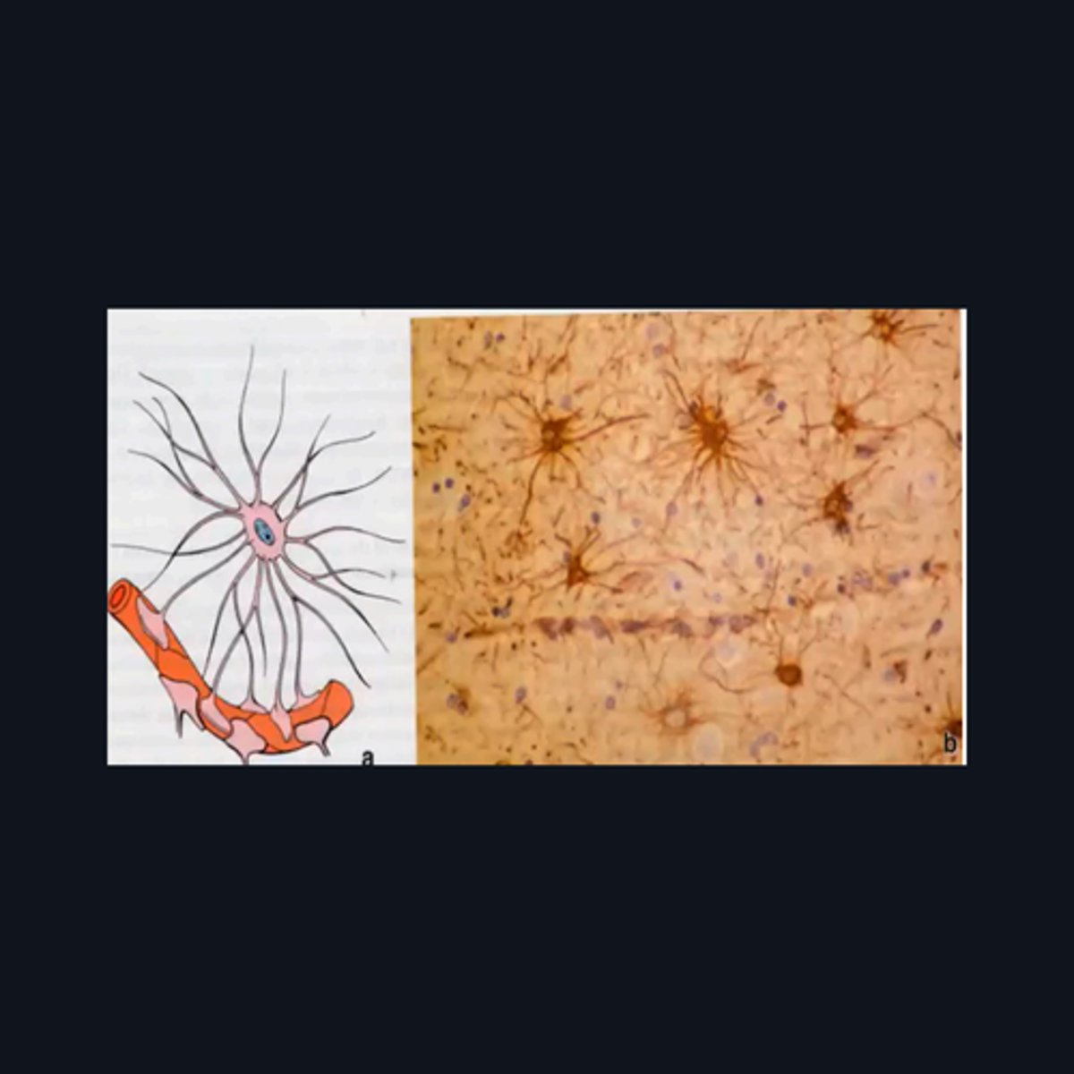
Sacral region
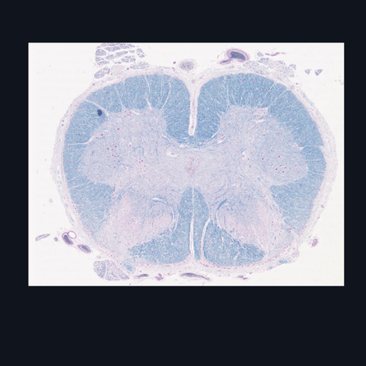
Identify the region of the spinal cord in the given image.
(top to bottom)
Ventral median fissure
White matter
Ventral horns
Central Canal
Central commissure
Dorsal midline sulcus
Dorsal horns
Identify the parts of the sacral region of the spinal cord.
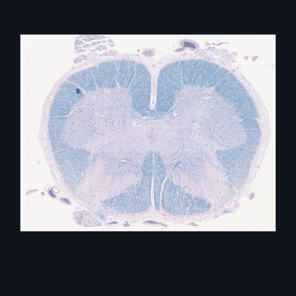
FALSE
T or F: The white matter in this region of the spinal cord is larger than the white matter in the thoracic region of the spinal cord.
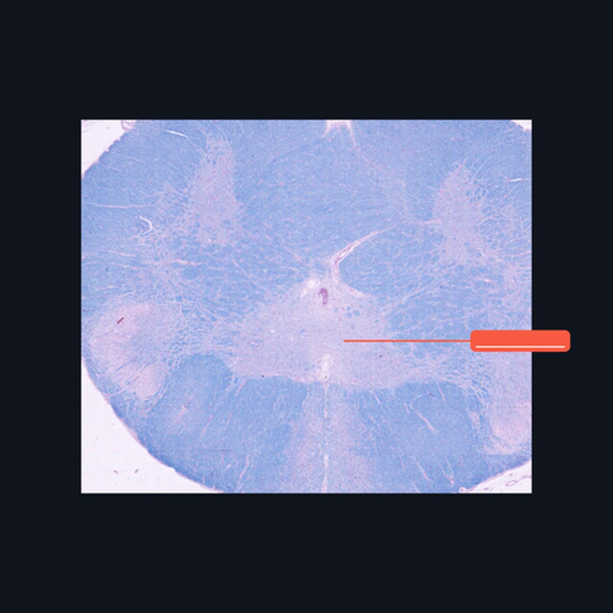
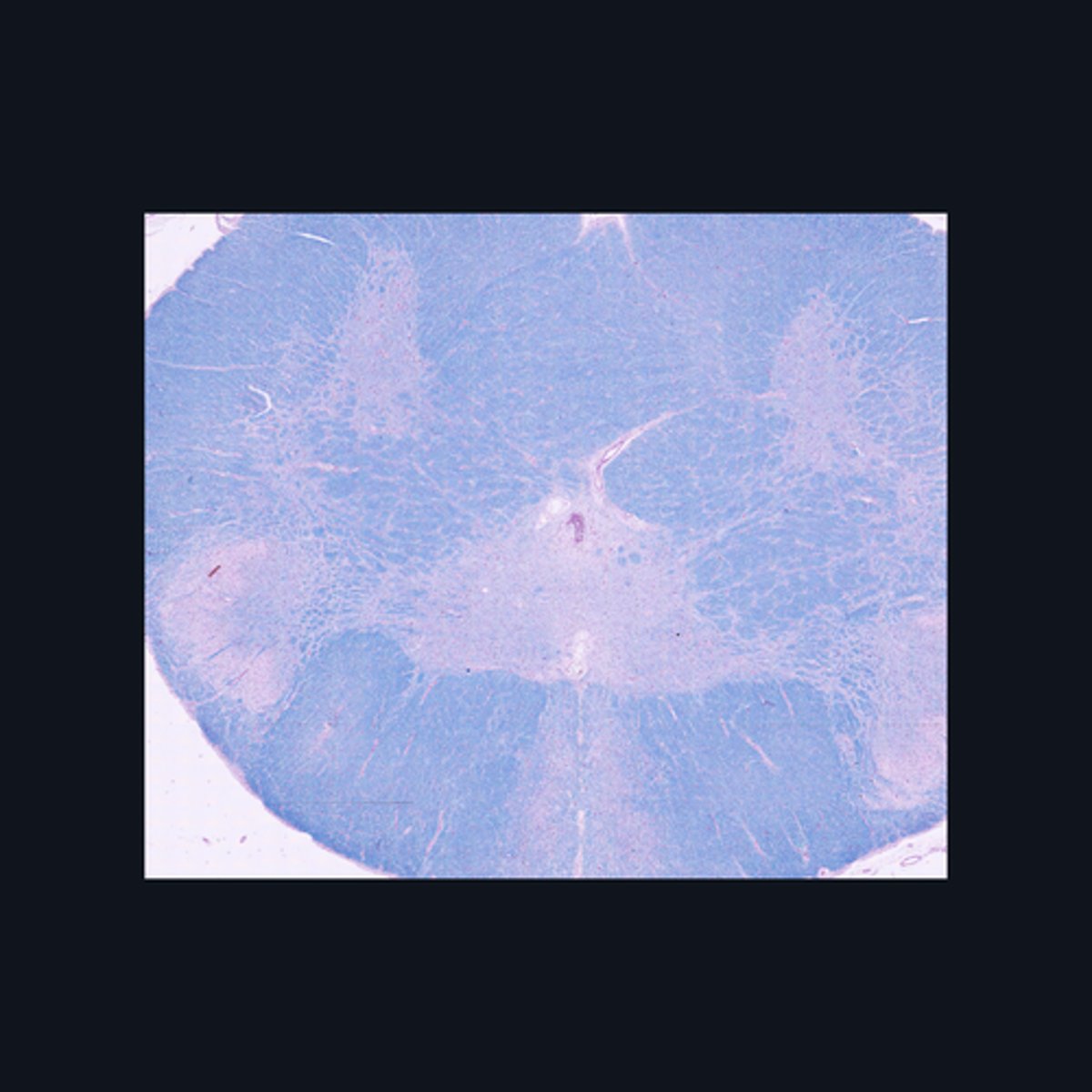
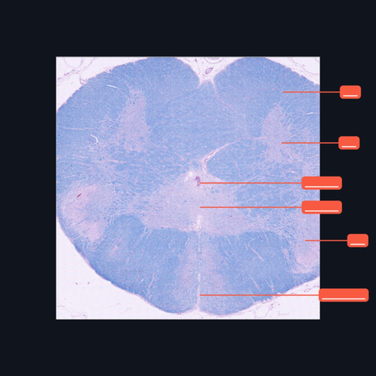
Upper Cervical region
Identify the region of the spinal cord in the given image.
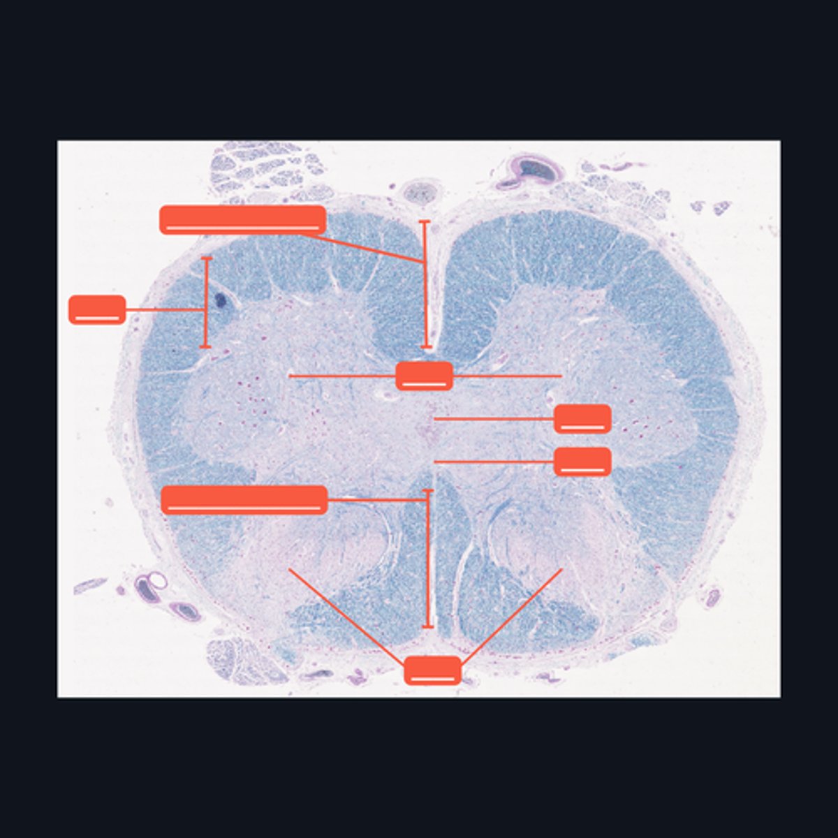
(top to bottom)
White matter
Ventral horn of gray matter
Central canal
Central Commissure
Dorsal horn of the gray matter
Dorsal Midline Sulcus
Identify the structures found in the upper cervical region of the spinal cord.
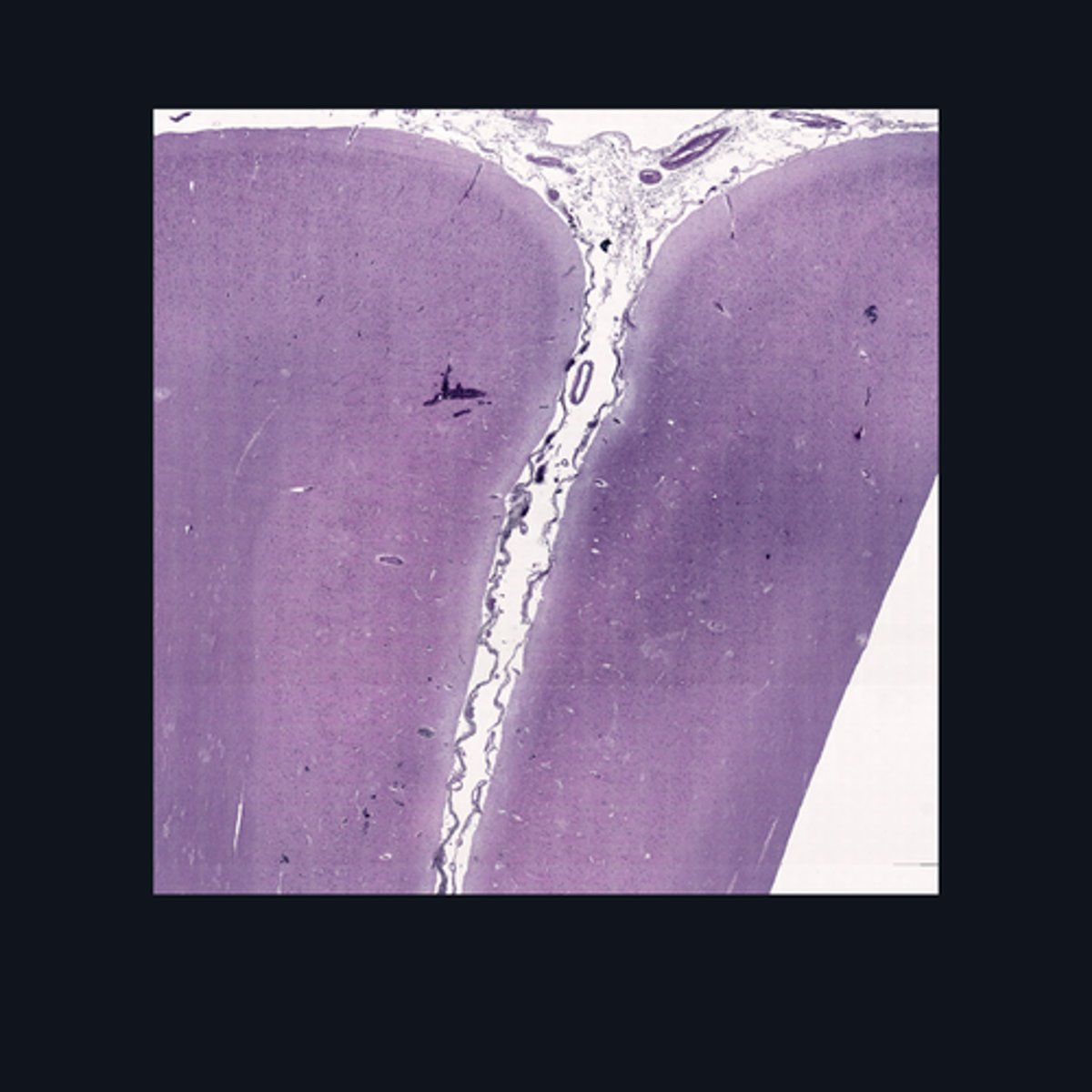
Brain, Cerebral cortex
Identify this structure.
Meninges
What covers this structure of the brain?
Sulci
Outer surface of cerebral hemisphere is highly folded to form convolutions known as
Plexiform (Molecular) Layer, Outer Granular Layer, Pyramidal Cell Layer, Inner Granular Layer, Ganglionic Layer, Multiform Cell Layer
What are the six poorly-designed layers of this structure?
Folia
The surface of the cerebellum exhibits transverse folds called the _____.
Purkinje cells
This middle layer of the cerebellar cortex consists of a single layer of pear-shaped multipolar neurons called _____.

Granule cells
The layer is densely populated by small, round to oval neurons called _____.
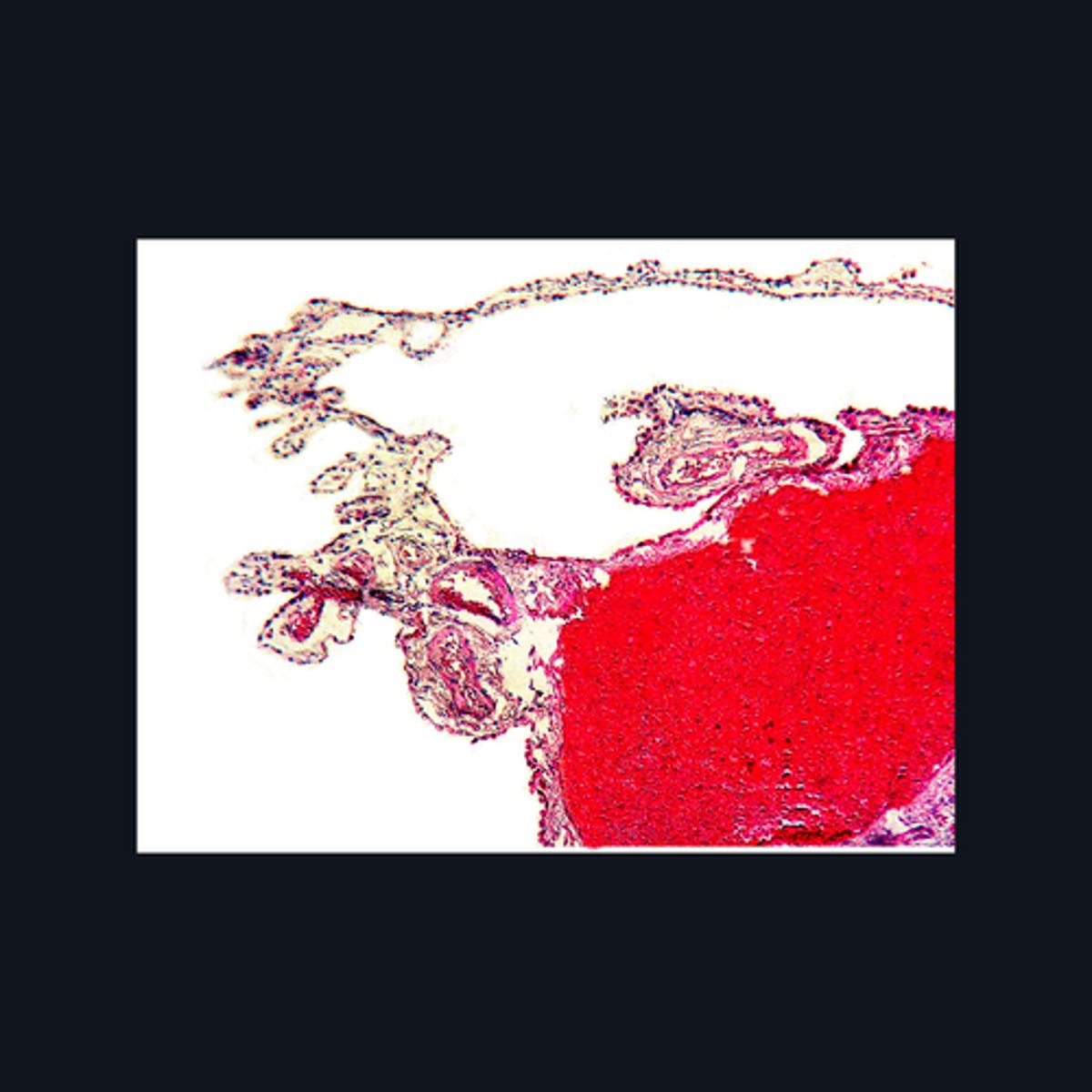
Choroid Plexus
Identify the region of the brain in the given image.
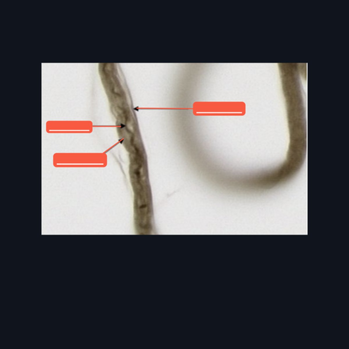
Peripheral Nerve
Identify where this structure is found
(top 3) from left to right
Perineurium
Axon
Myelin sheath
(bottom 2) from left to right
Endoneurium
Schwann cell
Identify the pointed structures.
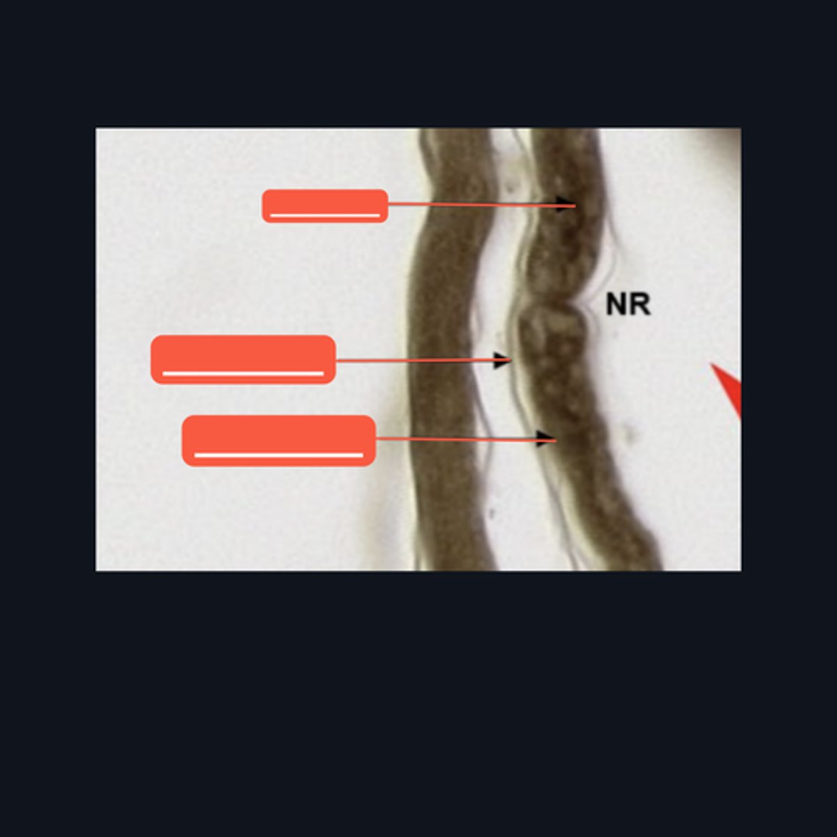
Peripheral Nerve
Identify the structure on the image.
(top to bottom)
Axon
Endoneurium
Myelin sheath
Identify the pointed structures.
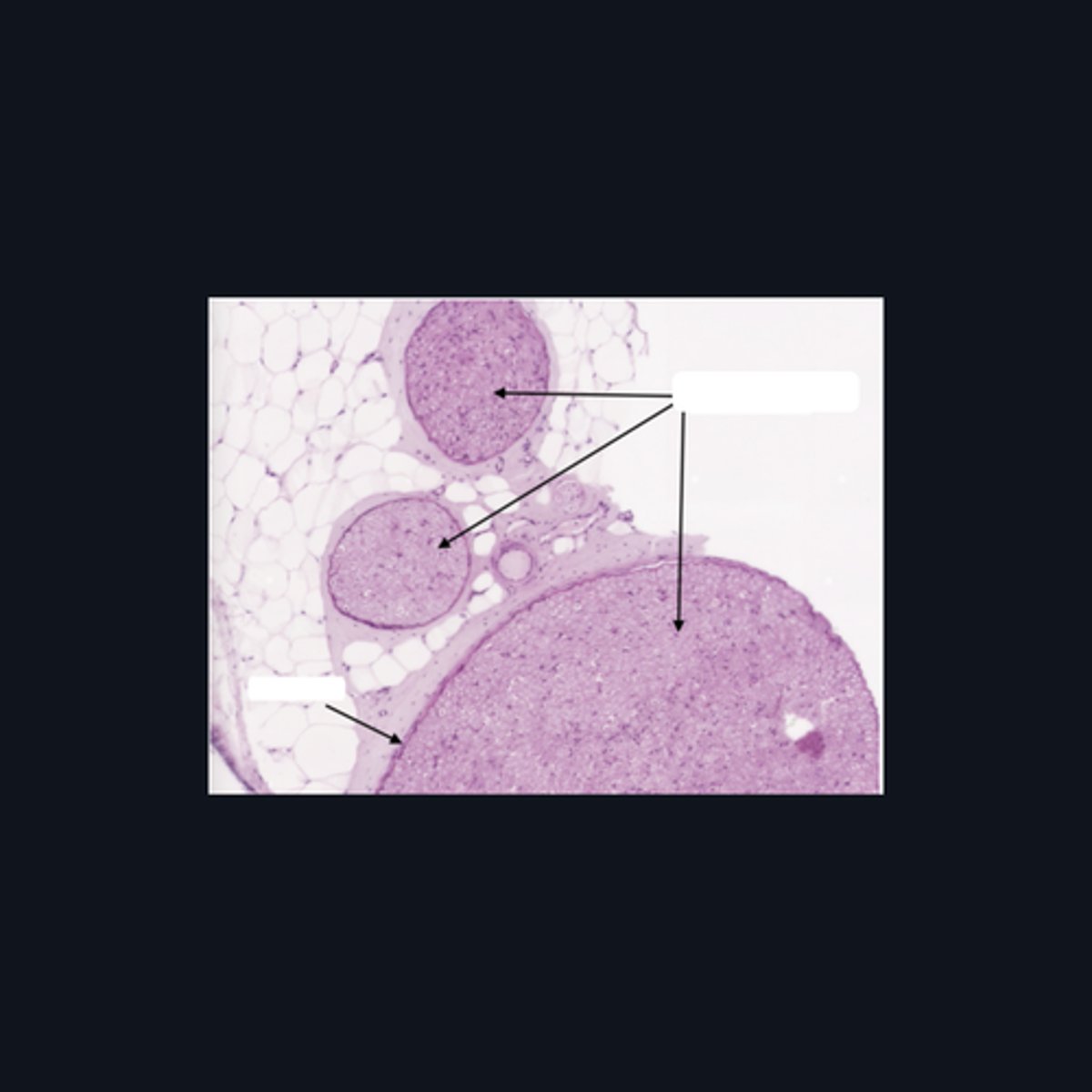
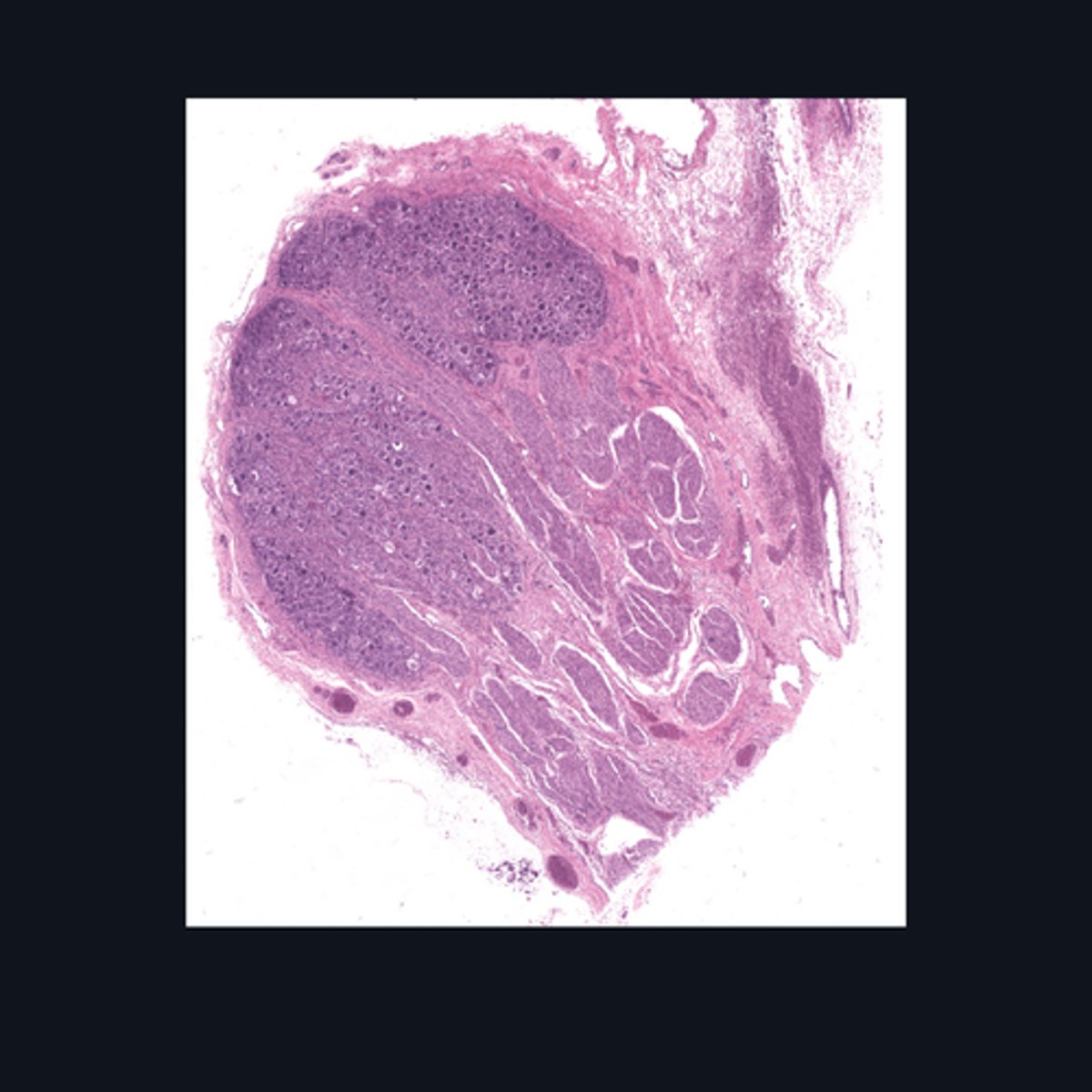
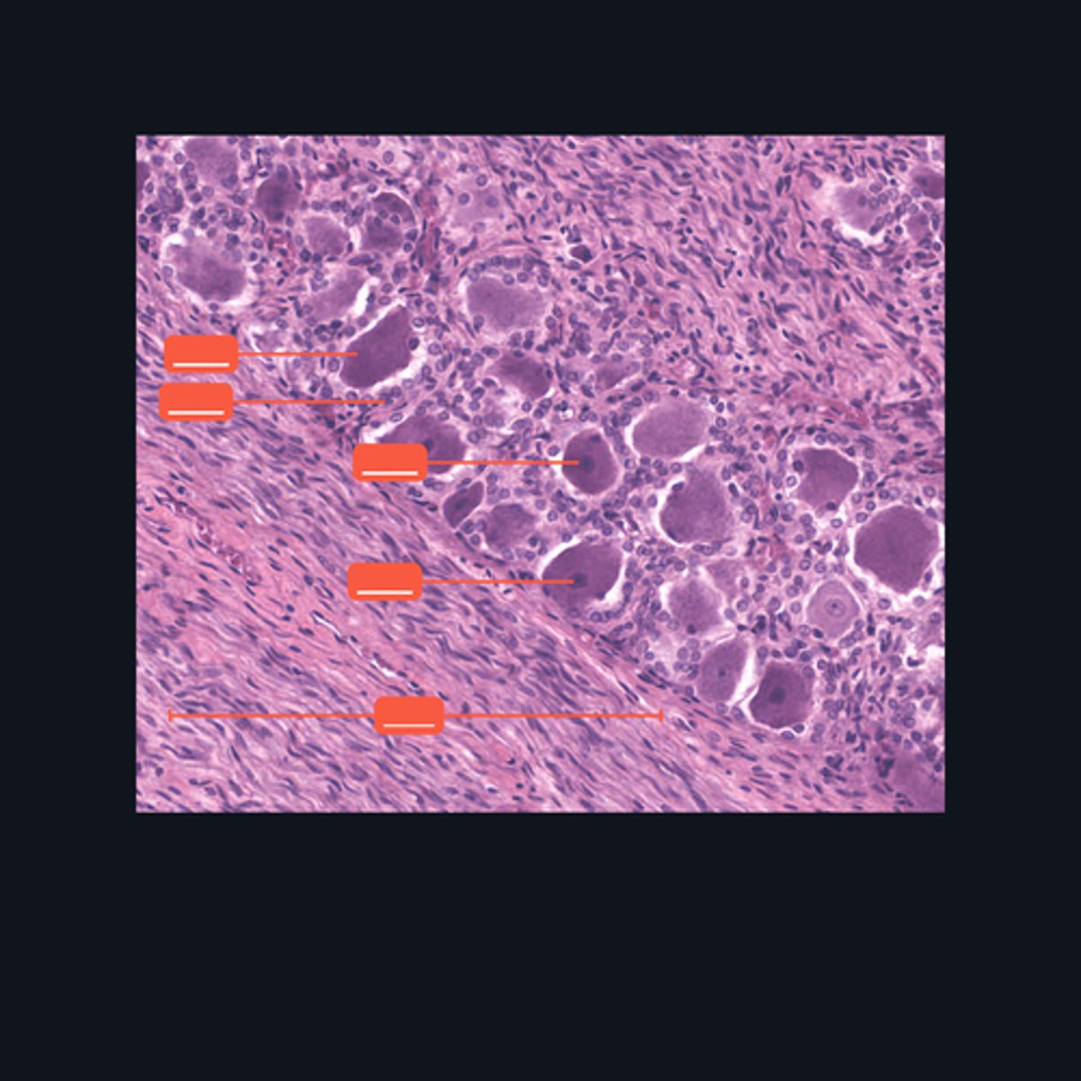
Dorsal root ganglion and nervous tissue
Identify the specific structure given in the image, as well as the type of tissue predominating in the structure.
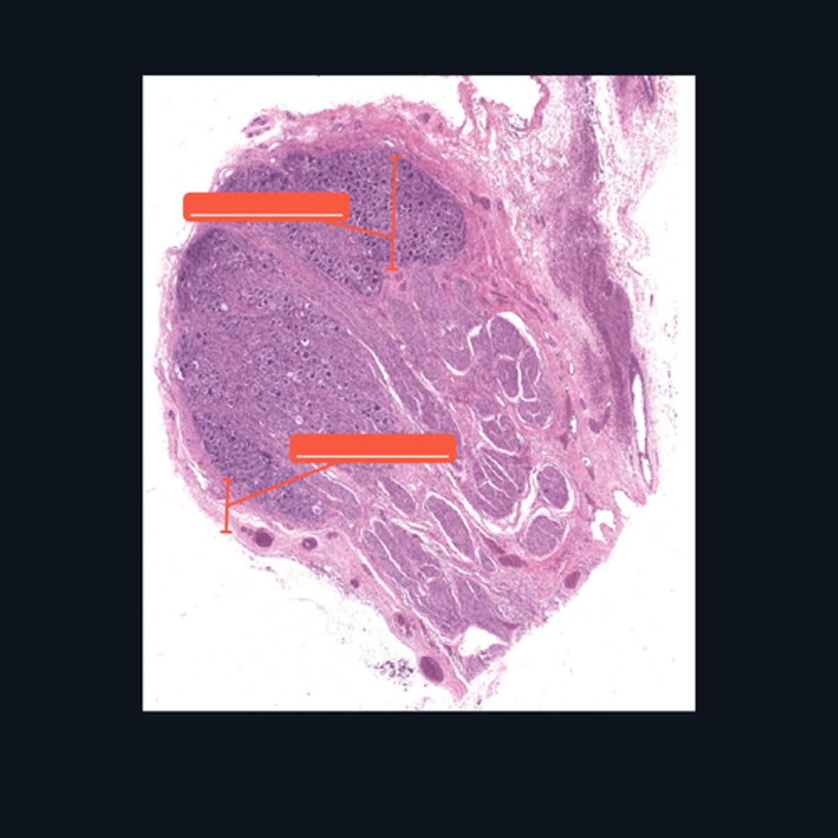
(top to bottom)
Somatic sensory neurons
Capsule
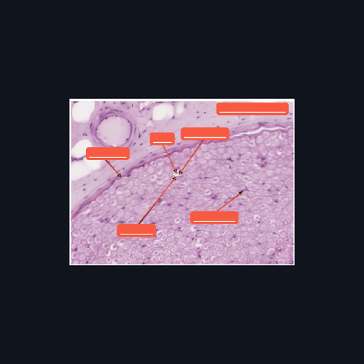
Identify the structures of the dorsal root ganglion. Be as specific as possible.
(top to bottom)
Cell body
Satellite cell
Nucleus
Nucleolus
Nerve fibers
Identify the parts of the spinal ganglion in high magnification.
False, pseudounipolar
T or F: The neurons that congregate in this structure is multipolar.

(bottom to top)
Capsule
Ganglion Cell
Satellite cell
Nerve fibers
Nucleolus
Nucleus
Nissl Granules
Identify the structures of a sympathetic ganglion.

Multipolar, autonomic motor neurons
What kind of motor neurons are present in this structure?

Dense Irregular Connective Tissue
What kind of tissue is the capsule of a spinal ganglion made up of? Be specific.
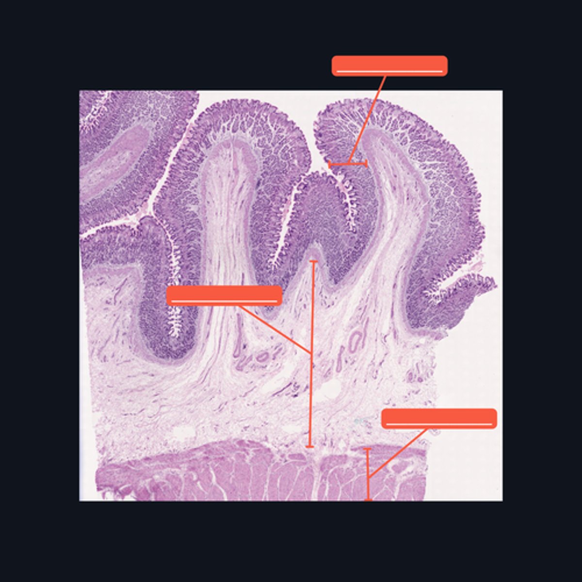
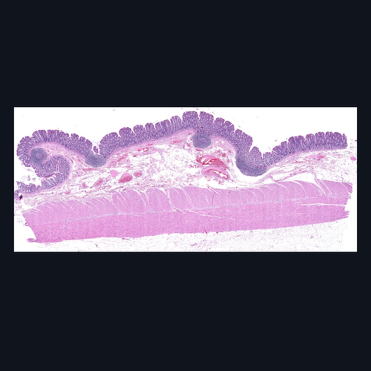
Mucosa, Submucosa, Muscularis Externa
Identify the histologic layers of the digestive tract
(1) Visceral Efferent Neuerons/ Autonomic Ganglion Cells
(2) Meissner's Plexus and Auerbach's Plexus
Cells from the division of the nervous system that innervates this specimen are made up of (1)_______, and whose cell bodies and fibers form (2) two ganglionated plexuses named: _______ and ________.
Submucosa
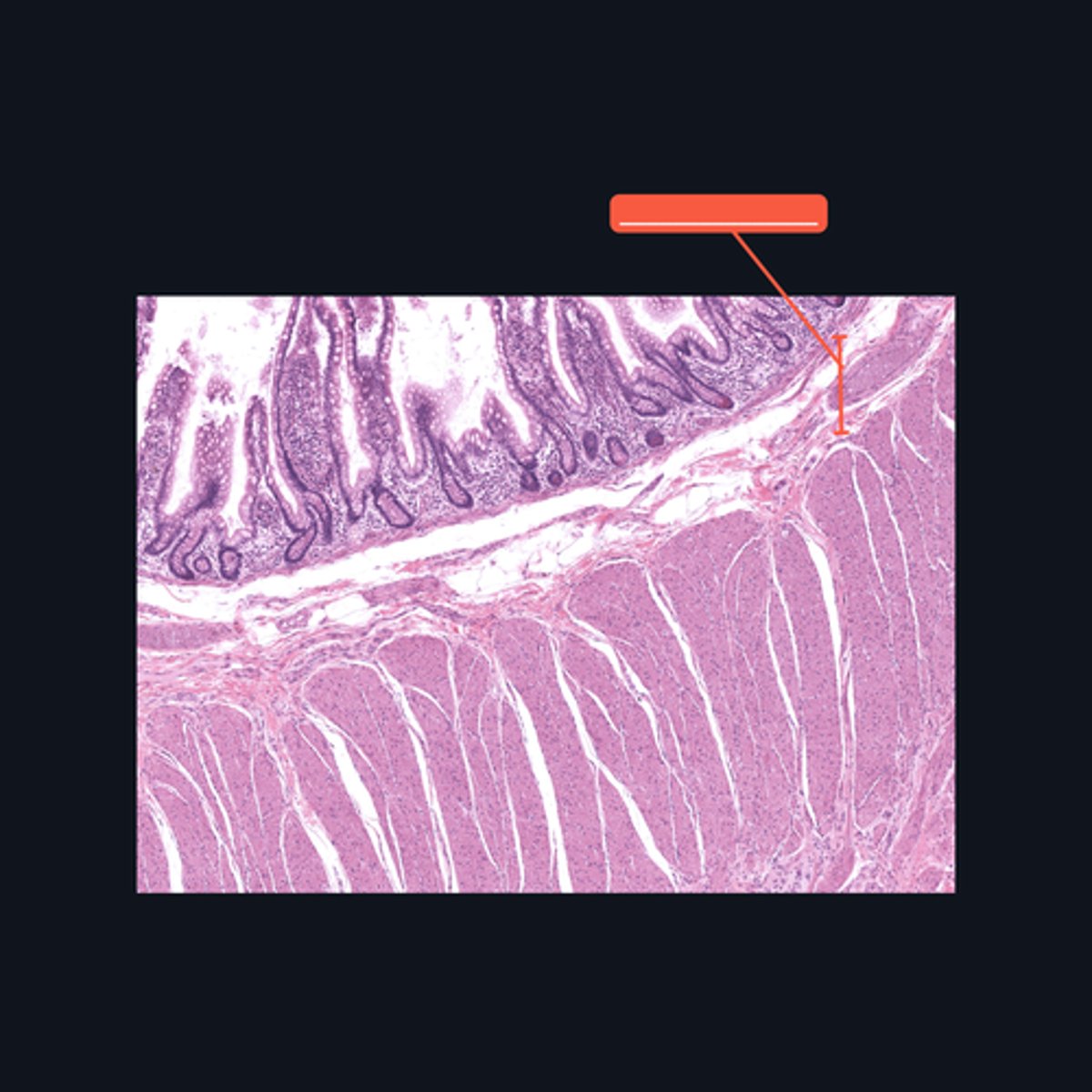
Identify the pointed histologic layer of the digestive tract
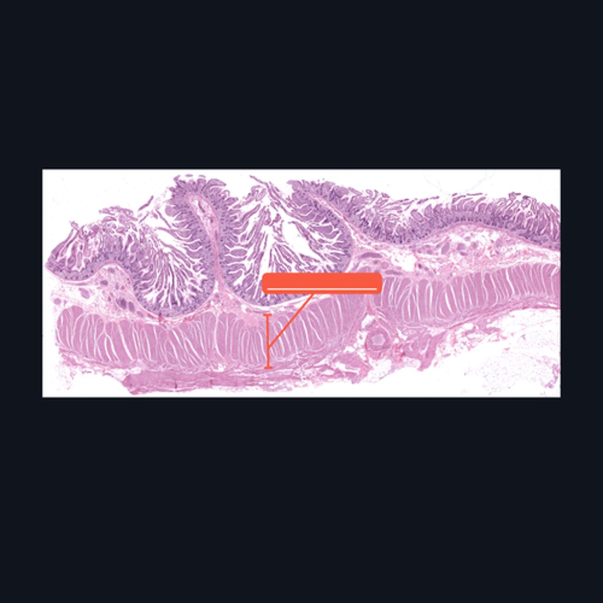
Muscularis Externa
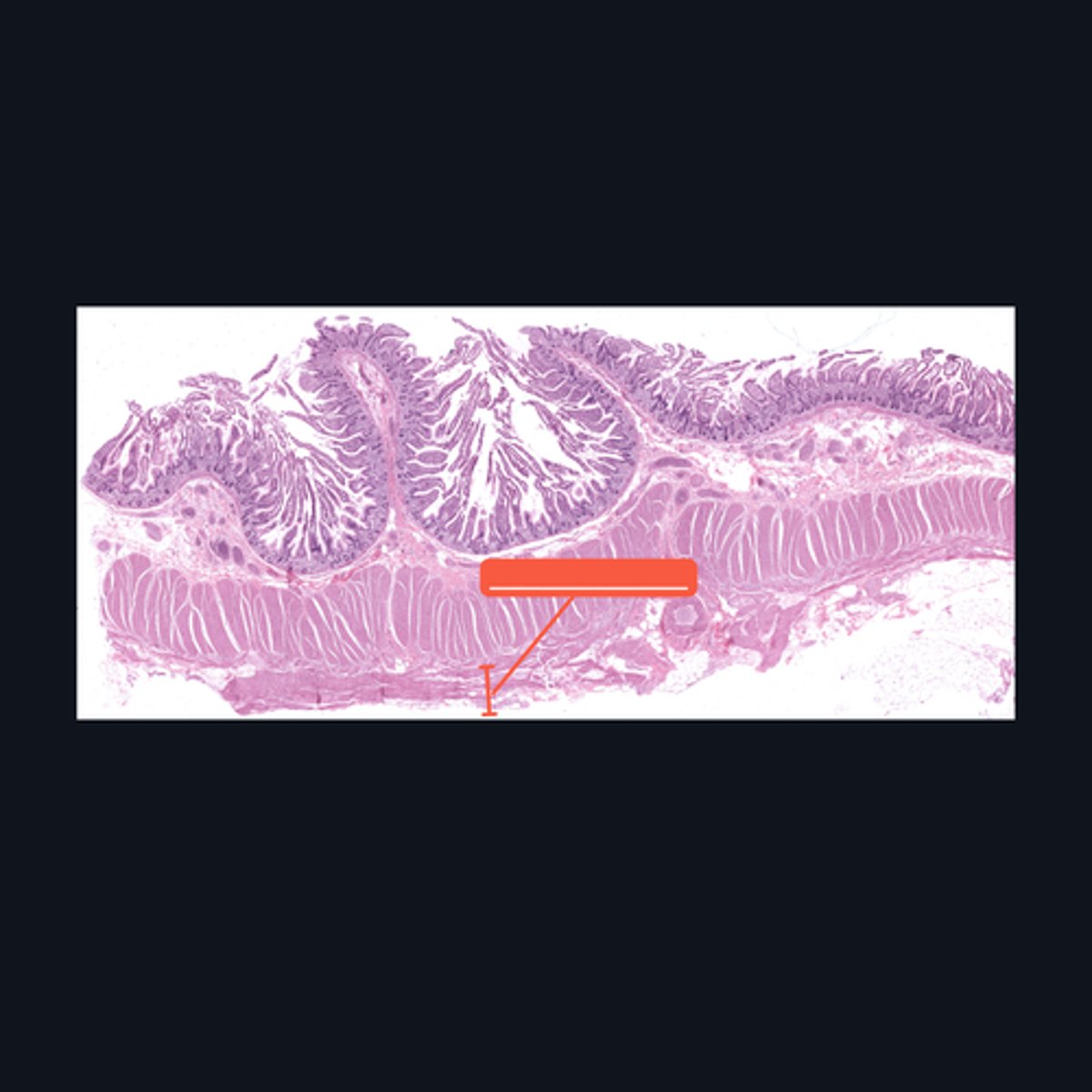
Identify the pointed histologic layer of the digestive tract
Serosa
Identify the pointed histologic layer of the digestive tract
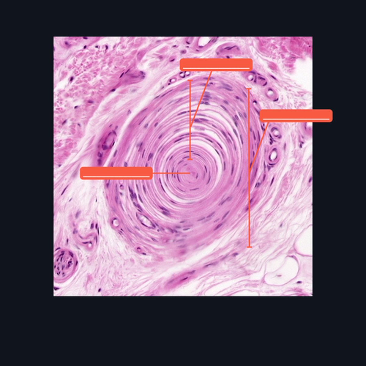
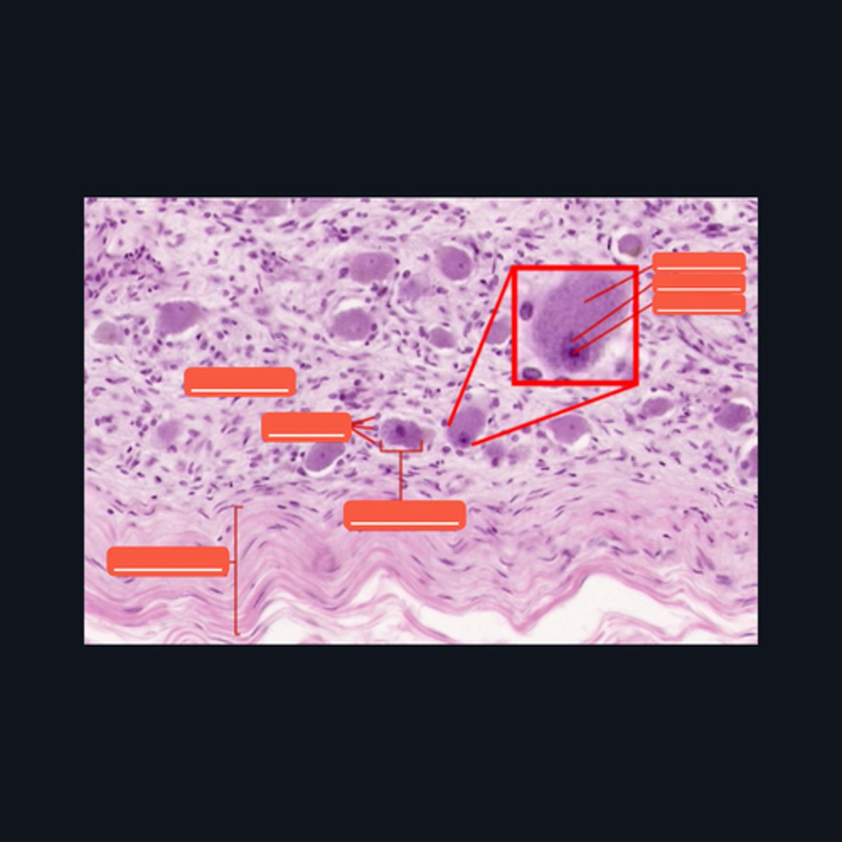
Meissner's corpuscle, Lamellar cells, and Capsule
Identify each pointed structures.
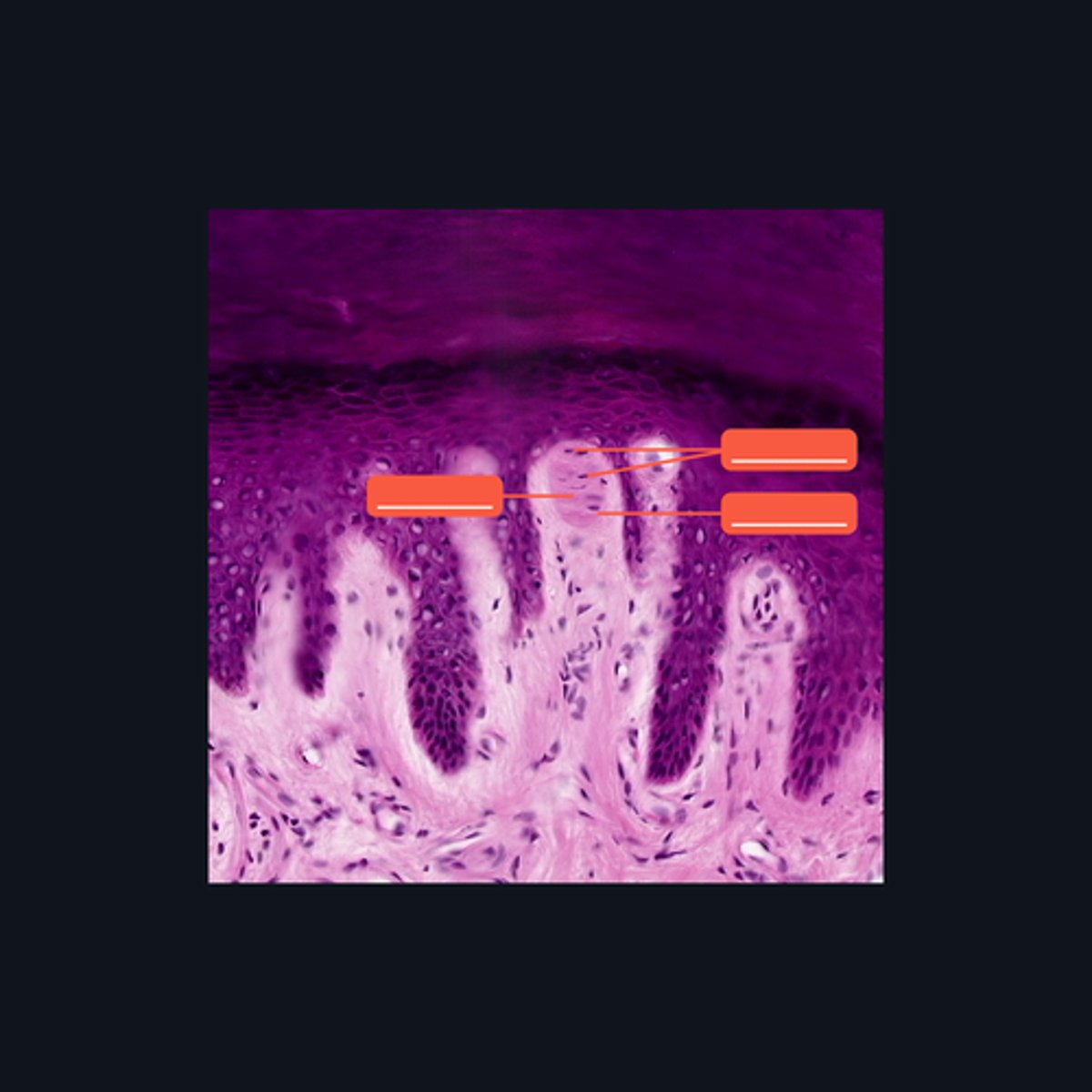
Pacinian corpuscle, Supporting cells, Axon
Identify the pointed structures.