General Human Anatomy Lecture Study Guide Unit 2 Lecture 6 free response
1/18
There's no tags or description
Looks like no tags are added yet.
Name | Mastery | Learn | Test | Matching | Spaced | Call with Kai |
|---|
No analytics yet
Send a link to your students to track their progress
19 Terms
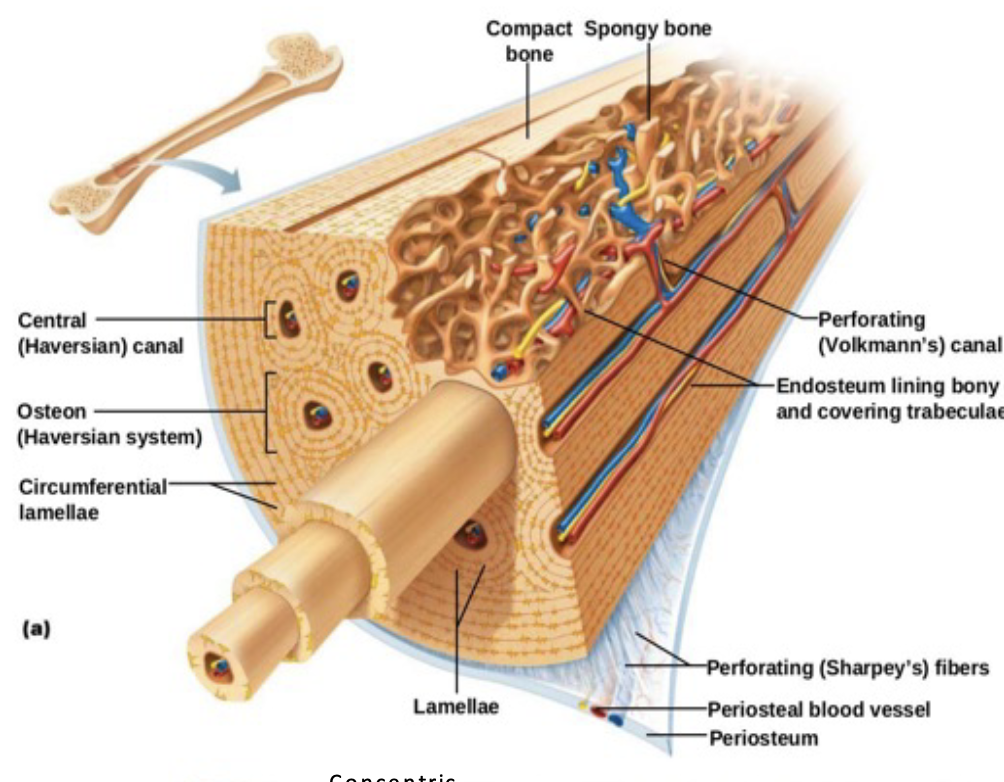
Be able to draw an osteon and label all components. Haversian canals (artery, vein, nerve), osteocytes, canaliculi, lamellae, lacunae
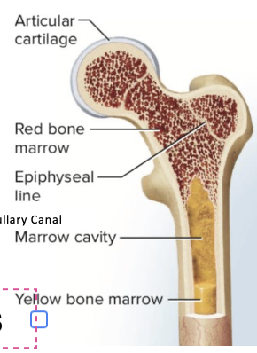
Be able to draw a long bone and label all components.
What are the types of marrow cavities? Red and yellow marrow
Red and yellow marrow
What is the composition of red marrow? What is its function? Location?
Blood connective tissue and produce red & white blood cells, found: epiphyses of long bones
What is the composition of yellow marrow? What is its function? Location?
Adipose connective tissue, function: long term energy storage, found: medullary canals of long bones
. Explain how intramembranous ossification occurs.
1. Osteoblasts cluster around blood vessels throughout sac = first bone 2. Osteoblasts secrete a collagen & calcium matrix 3. Calcium crystalize = becomes trabeculae (functional unit) 4. Trabeculae become denser creating compact bone connective tissue = hardening of surface 5. Remodeling occurs thru reabsorption of existing bone by osteoclasts 6. Original sac becomes the periosteum
. Explain how endochondral ossification occurs.
1. Hyaline cartilage connective tissue forms first 2 .primary ossification center forms in diaphysis 3. Osteoblasts create spongy bone connective tissue in primary ossification center 4. Osteoblasts create compact bone beneath periosteum 5. Osteoclasts break down spongy bone medullary canal. 6. Osteoblasts create spongy bone connective tissue in the epiphysis forming secondary ossification center. 7. Epiphyseal growth plate forms between the epiphysis and diaphysis
. Compare and contrast cartilage and bone tissue. Make sure to include location of tissue,
Cartilage: hyaline located: nose, fibro: menisci, elastic cartilage: epiglottis Cell: chondrocytes located in lacunae. Matrix: composed of collagen fiber, elastic fiber, chondrocytes in lacunae. High strength = resists compression. Flexible and avascular (lacks blood vessels)
Bone tissue:. Cell: osteocytes located in lacunae. Matrix: collagen fibers & hydroxyapatite crystal. These combined make bone tissue strong, nonflexible and avascular
18. Be able to draw a knee joint and label all components. Know the histology and function
6. What are the connective tissues that surround muscle?
Epimysium, perimysium, endomysium
What is the epimysium? What is its histology?
Superficial fascia that surrounds the whole muscle. Histology: dense irregular connective tissue
What is the perimysium? What is its histology?
Internal fascia that surrounds bundles of parallel fibers & form fascicles. Histology: dense irregular connective tissue
What is the endomysium? What is its histology?
Surrounds each individual fiber (muscle cell). Histology: areolar connective tissue
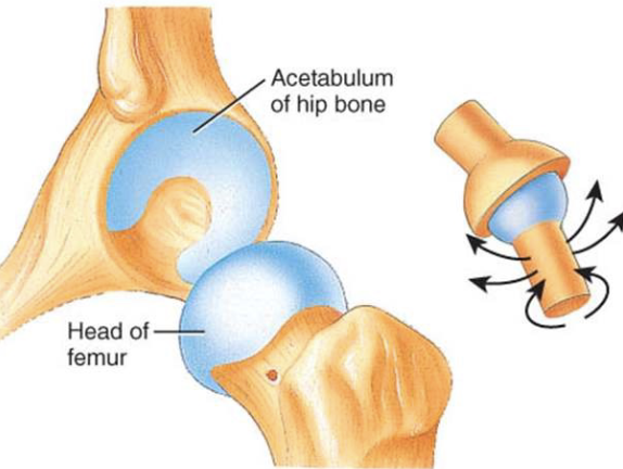
Ball and socket joint
multiaxial
all types of movement
hip
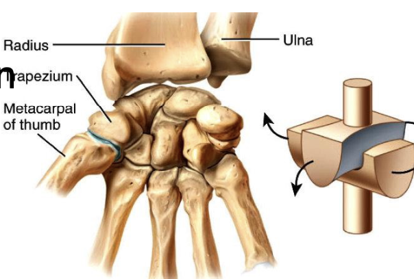
Saddle joint
biaxial
flex/extend
circumduction
first metacarpal/carpal
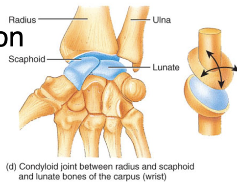
Ellipsoidal joint
biaxial
flex/extend
abduct/adduct
metacarpophalangeal
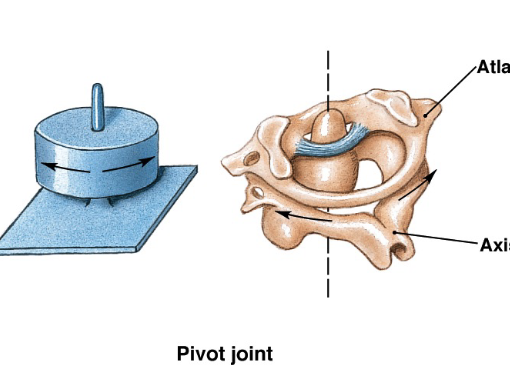
Pivot joint
uniaxial
rotation
C1/C2
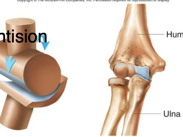
hinge joint
uniaxial
flex/extend
knee
Compare and contrast the three types of muscle tissue in regard to their characteristics,
Cardiac: Mono-nucleated, …, striated, multidirectional branching system
Smooth: