Looks like no one added any tags here yet for you.
Heart
Blood vesselsCirculatory System
Circulatory System
Cardiovascular System
Heart
Blood Vessels
Blood
Circulatory System
Approximately the size of a fist
Located
In the mediastinum between the second rib and fifth intercostal space
On the superior surface of the diaphragm
Two-thirds to the left of the midline
Anterior to the vertebral column, posterior to the sternum
Heart Anatomy
Location of Heart - Image
Heart - Image
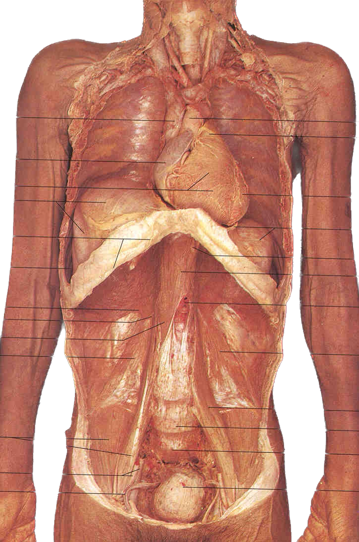
Right ventricle
Interventricular septum
Left ventricle
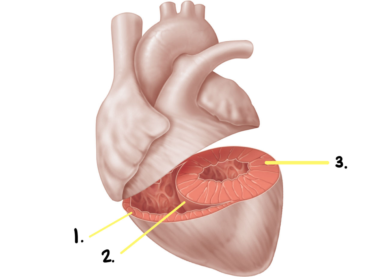
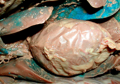
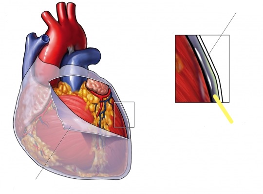
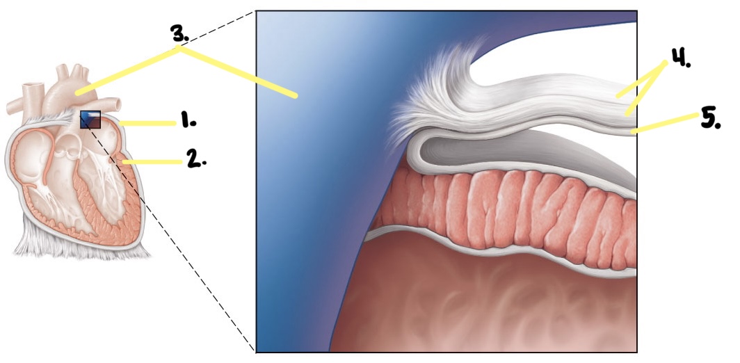
Double-Walled Sac
Superficial fibrous pericardium
Pericardium—Fibrous
Loosely surrounds heart
DICT sac – thick, tough white
Attached to the diaphragm and Great Vessels of the Heart
Protects, anchors, and prevents
overfilling
Superficial fibrous pericardium
Pericardium—Fibrous - Image
Pericardium - Image
Pericardial fluid - Image
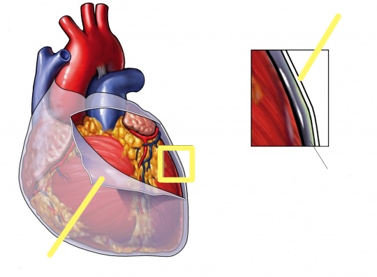
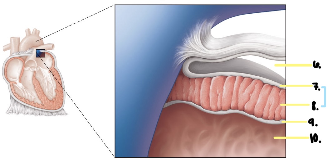
Deep two-layered serous pericardium
The parietal layer lines the internal
surface of the fibrous pericardium
Visceral layer (epicardium) on
the external surface of the heart
Separated by fluid-filled
pericardial cavity (decreases
friction)
Pericardium—Serous
Visceral and Parietal Pericardium - Image (A)
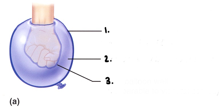
Outer balloon wall (comparable to parietal serosa)
Air (comparable to serous fluid)
Inner balloon wall (comparable to visceral serosa)
Visceral and Parietal Pericardium - Image (B)
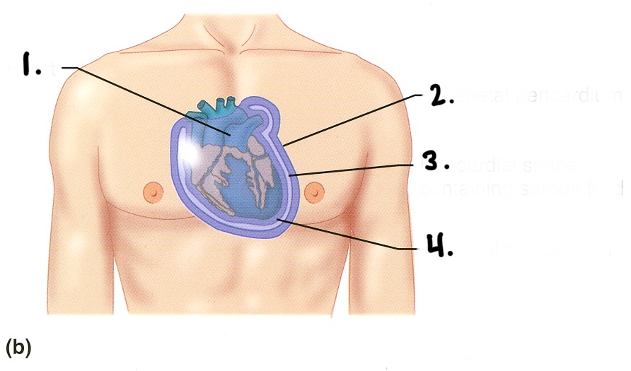
Heart
Parietal pericardium
Pericardial space (Has serous fluid)
Visceral pericardium
Heart - Image
Heart
Visceral pericardium
Parietal pericardium (Fibrous?)
Serous
Pericardial cavity
Heart - Image (Pt. 1)
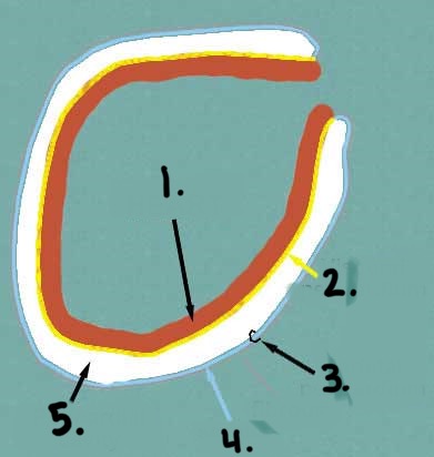
Pericardium
Myocardium
Pulmonary trunk
Fibrous pericardium
The parietal layer of the serous pericardium
Heart - Image (Pt. 2)
Pericardial cavity
Epicardium (visceral layer of serous pericardium) HEART
Myocardium WALL
Endocardium
Heart chamber
Epicardium
Myocardium
Endocardium
Heart Wall
Same thing as the visceral layer of the serous pericardium
Simple Squamous Epithelium
Shiny
Produces pericardial fluid
Epicardium
The cardiac muscle layer forms the bulk of the heart.
Spiral bundles of cardiac muscle cells
Fibrous skeleton of the heart: crisscrossing, the interlacing layer of connective tissue
Myocardium
Anchors cardiac muscle fibers
Supports great vessels and valves
Limits spread of action potentials to
specific paths
Fibrous skeleton (Heart)
Myocardium - Image
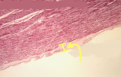
Endothelial layer of the inner myocardial surface
Lines the chambers of the heart
Simple Squamous Epithelium
Is in direct contact with Blood
It provides a smooth, slick surface for blood to “slide” against
Is continuous with endothelial lining of blood
vessels
Endocardium
Ventricular Endocardium - Image
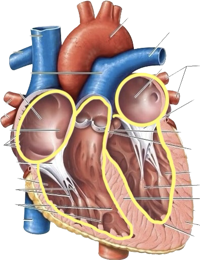
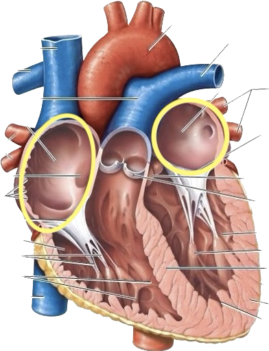
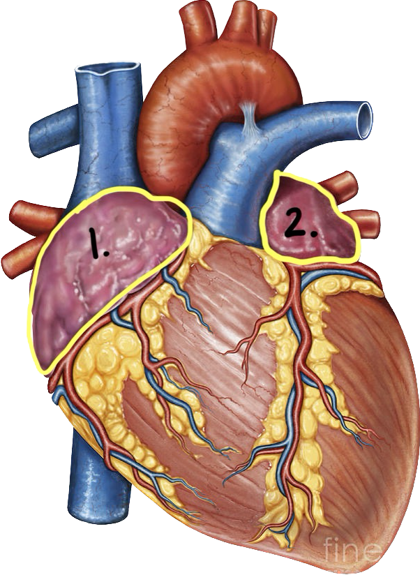
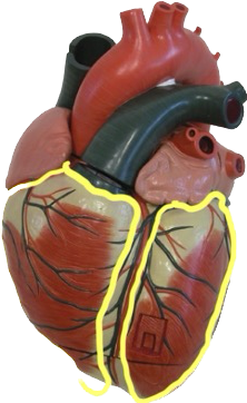
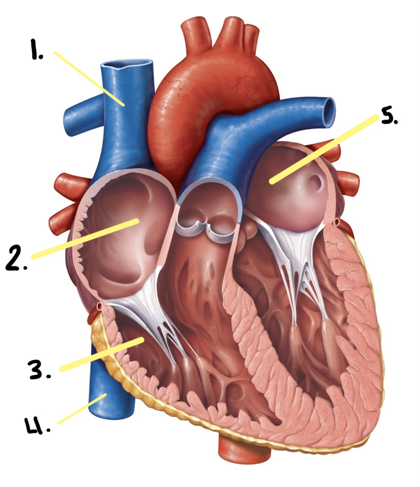
(2) Atria
(2) Ventricles
Chambers of Heart
Receiving Chambers
Thin-Walled
VERY little musculature
Low pressure
Atria )Pt. 1)
Pumping chambers
Thick-Walled
Heavily muscularized
High pressure
Ventricles (Pt. 1)
Chambers - Image
Left and Right
Separated internally by the interatrial septum (wall)
Auricles (ears) increase atrial volume
Atria (Pt. 2)
Atria - Image
Auricles
Right
Left
◦ Left and Right
◦ Separated by the interventricular
septum
◦ Anterior and posterior
interventricular sulci mark the
position of the septum external
Ventricles (Pt. 2)
Ventricles - Image
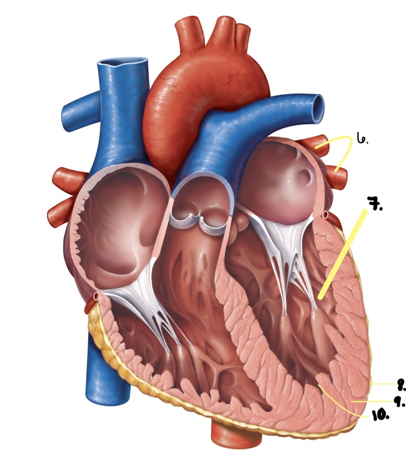
Thin walls ridged by pectinate muscles
Atria - The Receiving Chambers
◦ Superior vena cava
◦ Inferior vena cava
◦ Coronary sinus
Atria - Vessels emptying into the right atrium
Right and left pulmonary veins
Atria - Vessels emptying into the left atrium
Atria (Emptying vessels) - Image (Frontal section)
Heart - Image
Superior vena cava
Right pulmonary veins
Right atrium
Right ventricle
Heart - Image
Inferior vena cava
Left ventricle
Left atrium
Left pulmonary veins
Heart - Image
Superior vena cava
Right pulmonary veins
Inferior vena cava
Coronary Sinus
Left pulmonary veins
Heart - Image
Superior vena cava
Right atrium
Right ventricle
Inferior vena cava
Left atrium
Heart - Image
Left pulmonary veins
Left ventricle
Epicardium
Myocardium
Endocardium
Thick walls ridged by trabeculae carneae
Ventricles: The Pumping Chambers
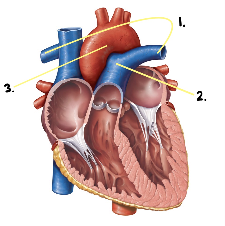
Pulmonary trunk
Ventricles: Vessel leaving the right ventricle
Aorta
Ventricles: Vessel leaving the left ventricle
Trabeculae carneae - Image
Heart - Image
Pulmonary Artery
Pulmonary Trunk
Aorta
Ensure unidirectional blood flow through the heart (prevent backflow of blood)
4 (2 AV & 2 SL)
Heart Valves
Heart Valves - Image
Lie between the atria and the ventricles
Prevent backflow into the atria when the ventricles contract
Atrioventricular (AV) Valves
AV Valves - Image
◦ Between the left atrium and left ventricle
◦ (2) flaps
◦ Bicuspid, Mitral
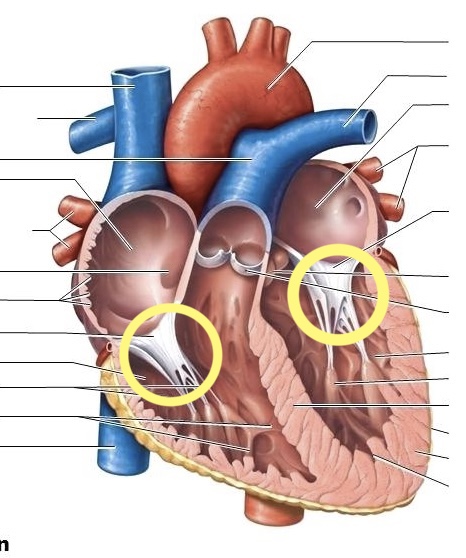
◦ Chordae tendons and papillary muscles
AV Valves - Left Valve
AV Valves (Left Valve) - Image
AV Valves (Left Valve) - Image
Heart - Image
◦ Between the right atrium and right ventricle
◦ (3) flaps
◦ Tricuspid
◦ Chordae tendons and papillary muscles
AV Valves - Right Valve
AV Valves (Right Valve) - Image
AV Valves (Right Valve) - Image
◦ Prevent backflow into the ventricles
when ventricles relax
◦ Aortic semilunar valve
◦ Pulmonary semilunar valve
Semilunar (SL) Valves (Pt. 1)
Semilunar (SL) Valves - Image (1)
Semilunar (SL) Valves - Image (2)
Heart valves ensure unidirectional blood flow through the heart
Semi-lunar valves are found in the large arteries that leave the ventricles
◦ Pulmonary SL valve
◦ Aortic SL valve
Semilunar (SL) Valves (Pt. 2)
Prevent backflow into the
ventricles
Each has three flaps
Semilunar (SL) Valves (Pt. 3)
Pulmonary and aortic valves from above - Image
Image
Pulmonary valve
Aortic valve
Area of cutaway
Mitral valve
Tricuspid valve
Image (Anterior)
Fibrous skeleton
Myocardium
Tricuspid (right atrioventricular) valve
Mitral (left atrioventricular) valve
Aortic valve
Pulmonary valve
Semilunar valves open - Image
Semilunar valves closed - Image
(Lub-dup) are associated with the closing of heart valves
The first sound occurs as AV valves close
The second sound occurs when SL valves close
Heart Sounds
Heart Sounds - Image
Vessels returning blood to the heart include
Superior and inferior vena cavae
Coronary Sinus
Right and left pulmonary veins
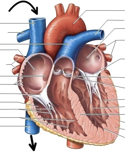
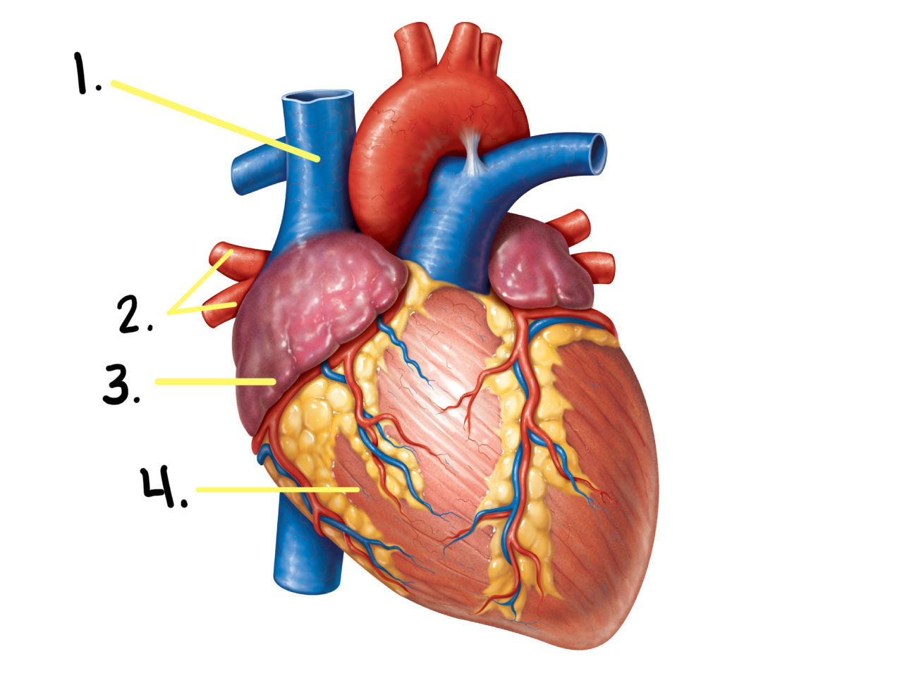
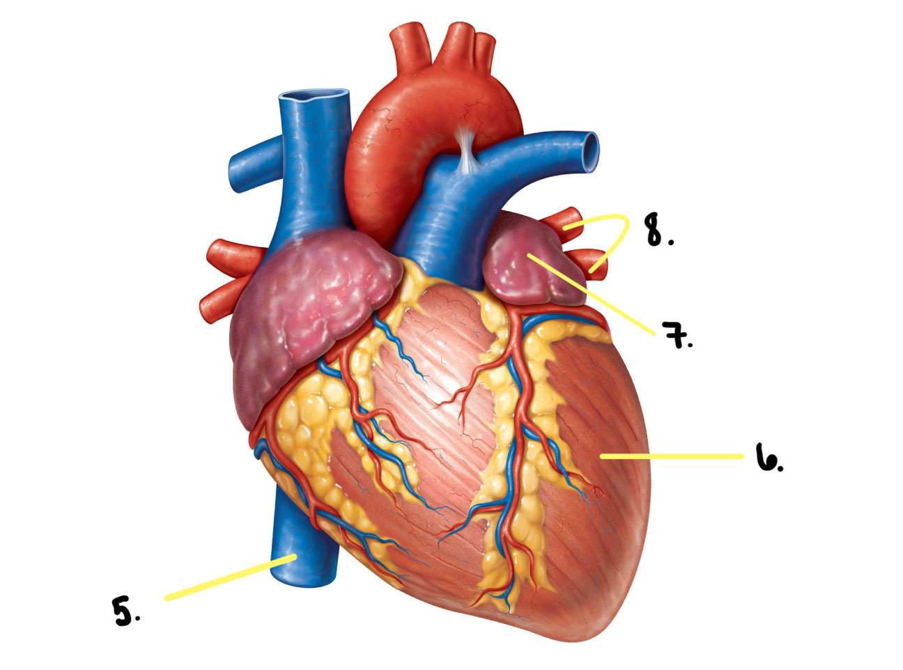
External Heart: Major Vessels of the Heart (Anterior View)w)
External Heart: Major Vessels of the Heart (Anterior View) - Image
Vessels returning blood to the heart include:
Right and left pulmonary veins
Coronary Sinus
Superior and inferior venae cavae
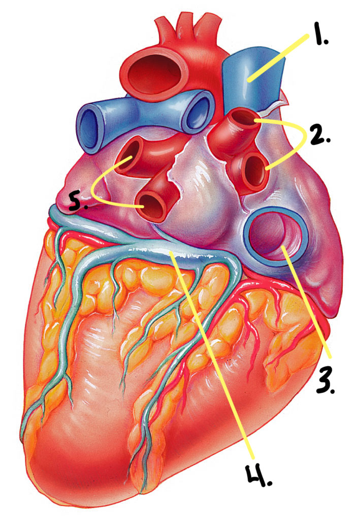
External Heart: Major Vessels of the Heart (Posterior View)
Vessels conveying blood away from the heart
The pulmonary trunk, which splits into the right and left pulmonary arteries
Ascending aorta (three branches) -brachiocephalic, left common carotid, and subclavian arteries
External Heart: Major Vessels of the Heart
Circulation - Image
Circulation - Image
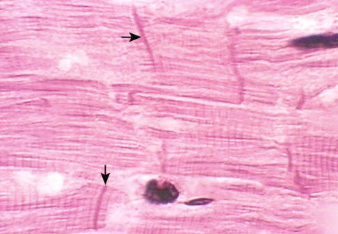
• Cardiac muscle tissue
• Cardiac cycle
• Cardiac conduction system
?
Image
Nucleus
Intercalated discs
Cardiac muscle cell
Gap junctions
Desmosomes
Cardiac muscle is striated, short, fat, branched, and interconnected
The connective tissue endomysium acts as both the origin and insertion
Microscopic Anatomy: Heart Muscle (Pt. 1)
Intercalated discs anchor cardiac cells together and allow free passage of ions
Heart muscle behaves as a functional syncytium
Microscopic Anatomy: Heart Muscle (Pt. 2)
Excitability
Contractility
Extensibility
Elasticity
Properties of Muscle
•Automaticity
•Auto rhythmicity
Cardiac Muscle (+2) & Contraction
• Contracts automatically—no nervous stimulation required
Automaticity
• Contracts in a rhythm—very consistent over time
Auto rhythmicity
Pulmonary Circuit (circulation)
Systemic Circuit (circulation)
Coronary Circuit (circulation)
Circuits
• Receives blood from organs (deoxygenated)
• Sends blood to lungs
Pulmonary Circuit (circulation) (Pt. 1)
• Receives blood from lungs (oxygenated)
• Sends blood to organs
Systemic Circuit (circulation)
Nourishes the myocardium
Coronary Circuit (circulation)
The right side of the heart is the pump for the pulmonary circuit
Short-distance
Low resistance through lungs
Low-pressure pump
Thinner walls (ventricles)
Pulmonary Circuit (circulation) (Pt. 2)
Pulmonary Circuit (circulation) - Image
Image
Right ventricle
Interventricular septum
Left ventricle
Systole
Diastole
Contraction & Relaxation
Contraction of the heart muscle
Systole
Relaxation of the heart muscle
Diastole
Diastole (filling) - Image
Systole (pumping) - Image
Diastole & Systole - Image
Cardiac cycle - Image
When one cell undergoes an action potential, the action potential spreads to all connecting cells (they all contract together)
Action potentials are all or nothing in the heart.
Functional Syncytium (Pt. 1)
• One heartbeat spreads to the whole heart
• Muscular contraction is followed after depolarization
Functional Syncytium (Pt. 2)
• Atria group/syncytium
• Ventricle group/syncytium
Functional Syncytium: Types (2)