Looks like no one added any tags here yet for you.
Central Nervous System (CNS)
Processes sensory input and motor output; includes brain and spinal cord.
Peripheral Nervous System (PNS)
Connects CNS to limbs; includes cranial and spinal nerves.
Sensory (Afferent) Division
Carries signals to the CNS from sensory receptors.
Motor (Efferent) Division
Carries signals away from the CNS to effectors.
Somatic Nervous System
Controls voluntary skeletal muscle movements.
Visceral Nervous System
Controls involuntary functions of cardiac and smooth muscles.
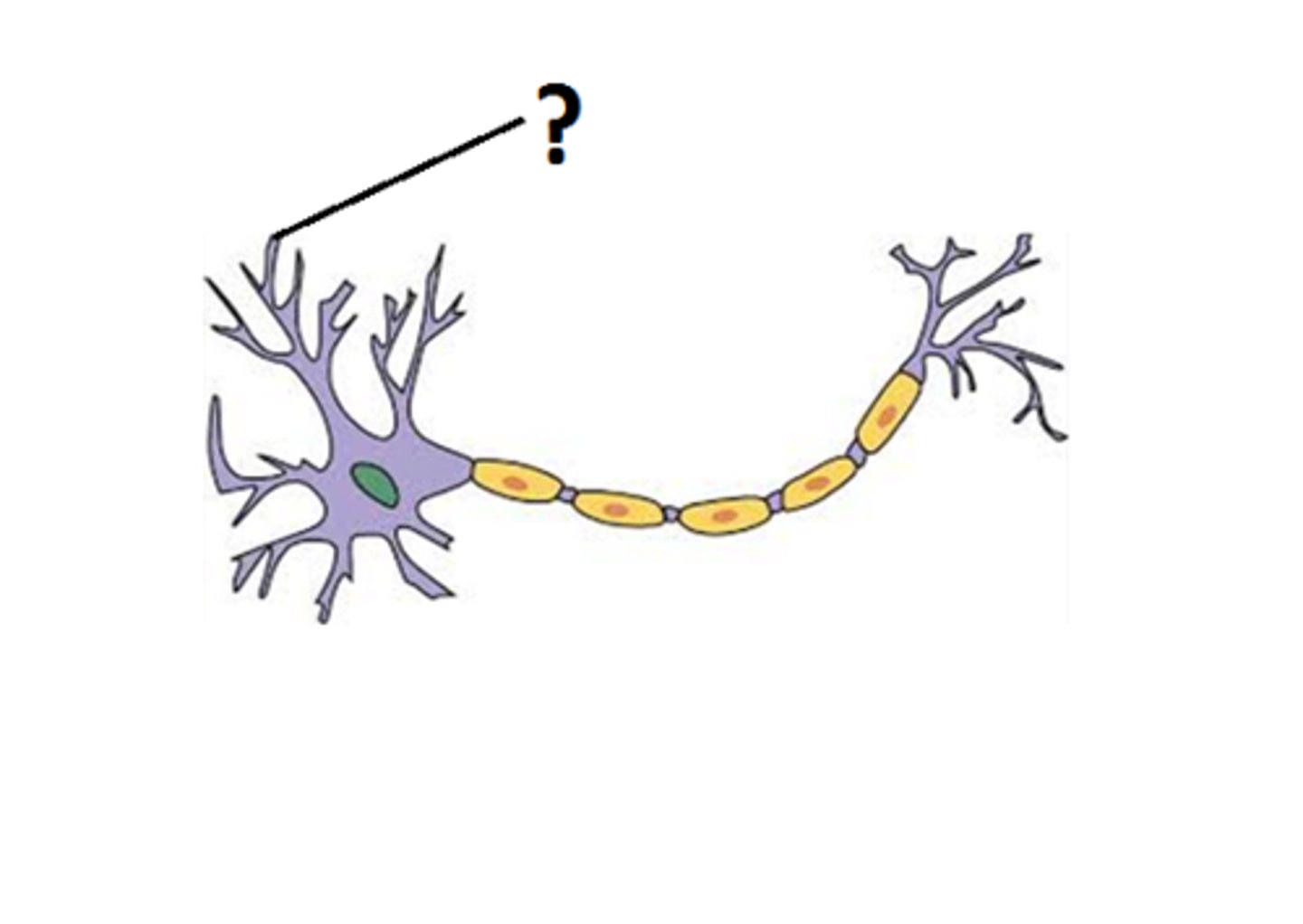
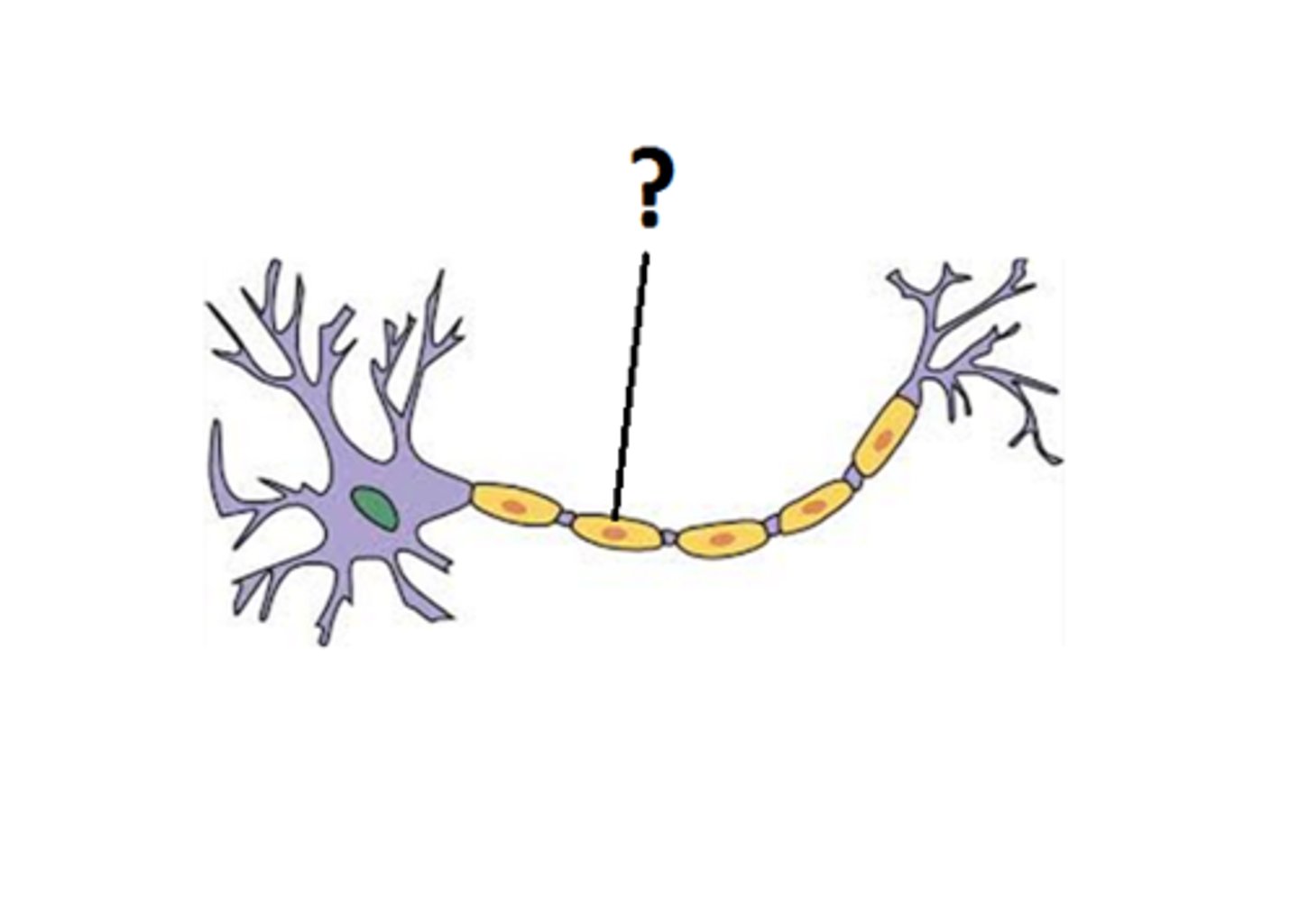
Dendrites
Receive signals from other neurons.
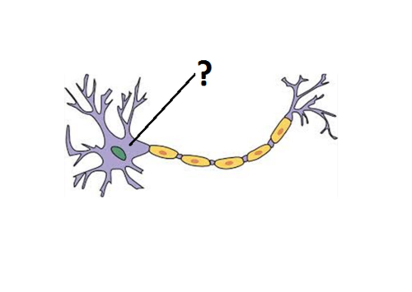
Cell Body (Soma)
Processes information and contains the nucleus.
Axon
Transmits impulses away from the neuron cell body.
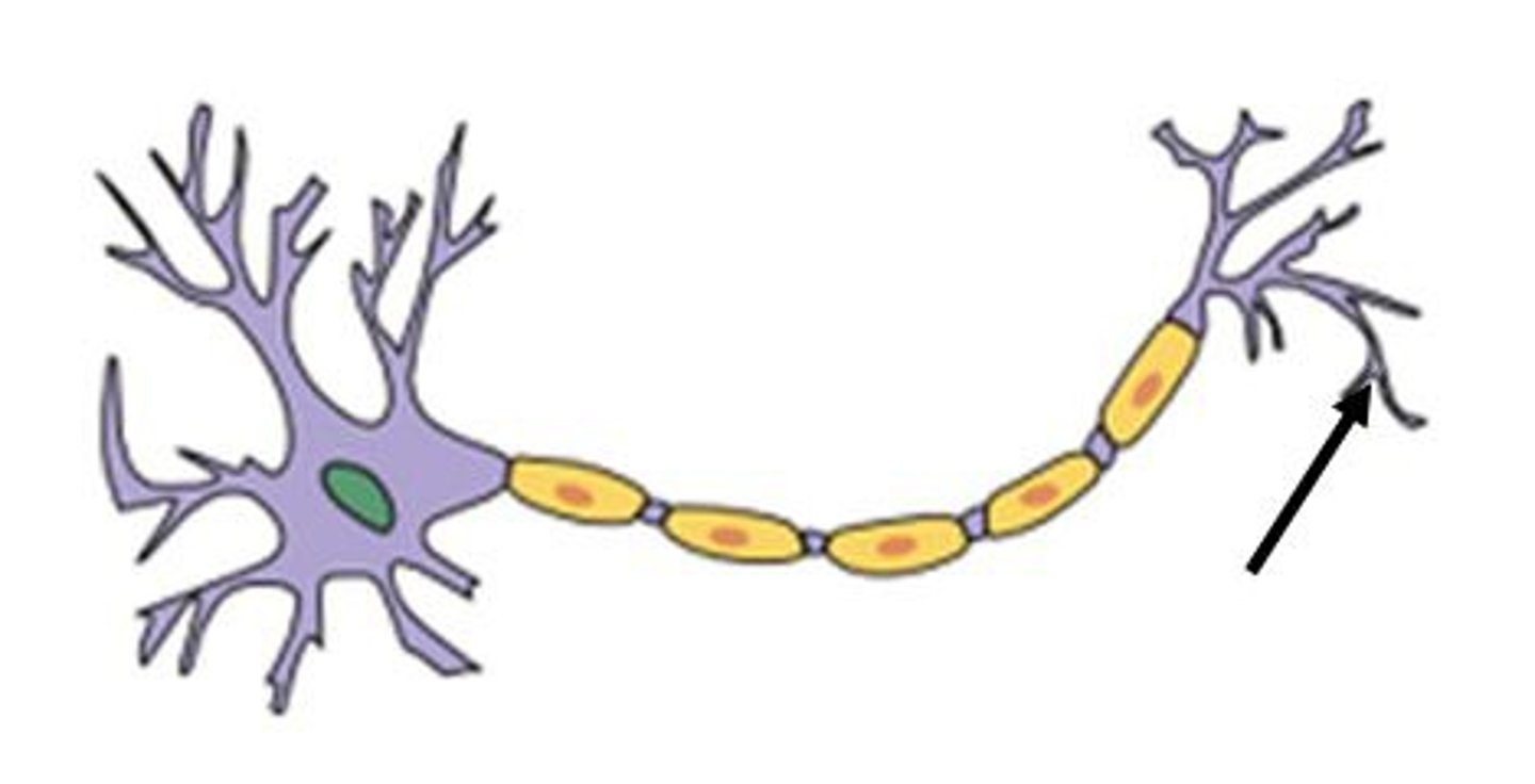
Axon Terminals
Release neurotransmitters at synapses.
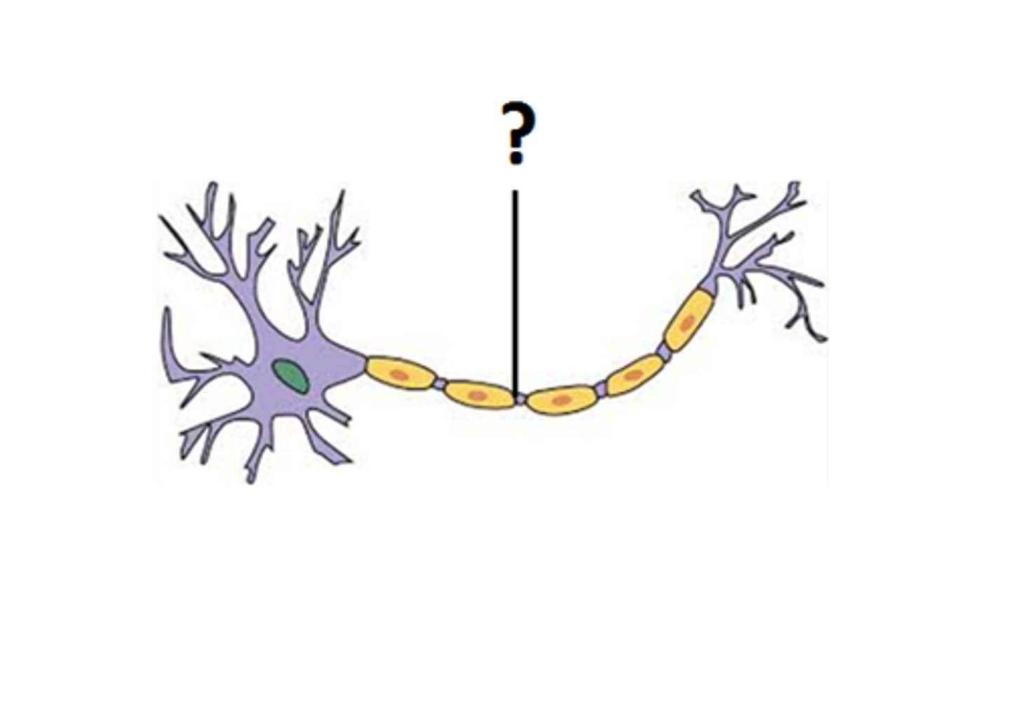
Myelin Sheath
Insulates axons and speeds up nerve impulses.
Multipolar Neuron
One axon, multiple dendrites; common in CNS.
Bipolar Neuron
One axon, one dendrite; found in special senses.
Unipolar Neuron
Single process that splits; found in sensory neurons.
Astrocytes
Support neurons and maintain blood-brain barrier.
Microglia
Act as immune cells in the CNS.
Ependymal Cells
Line ventricles and produce cerebrospinal fluid (CSF).
Oligodendrocytes
Form myelin sheath around CNS neurons.
Schwann Cells
Form myelin sheath around PNS neurons.
Satellite Cells
Support neuron cell bodies in ganglia.
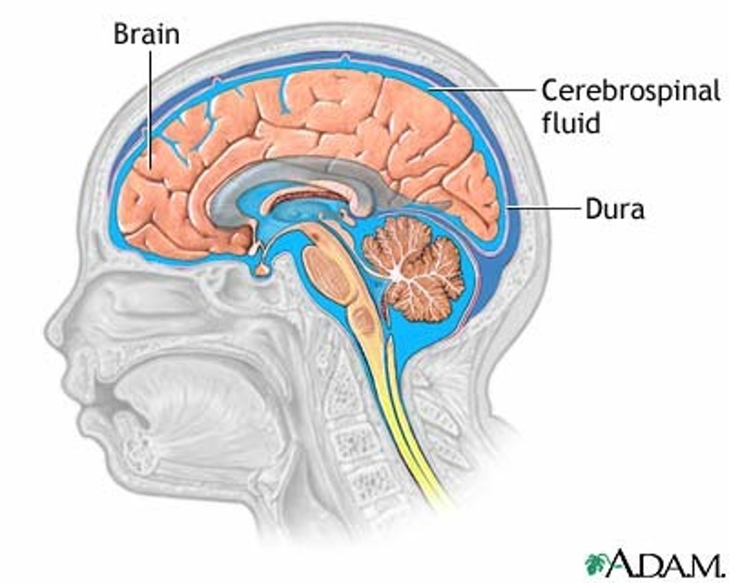
Cerebrospinal Fluid (CSF)
Fluid that cushions and nourishes the brain and spinal cord.
Neural Pathway
Sequence of neurons transmitting signals from stimulus to response.
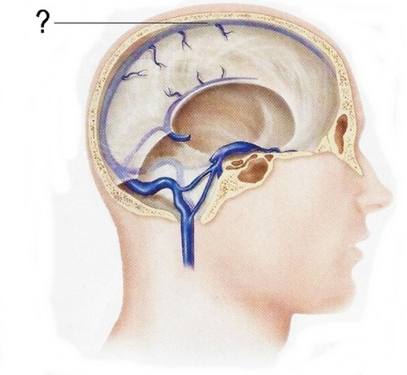
Superior Sagittal Sinus
CSF reabsorption into the bloodstream.
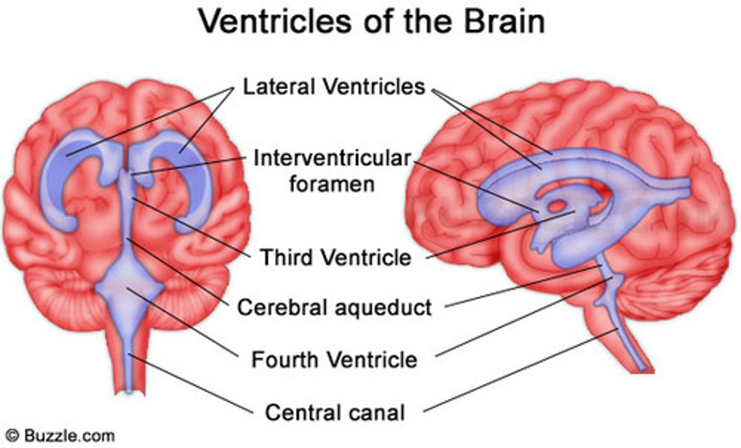
Lateral Ventricles
Largest ventricles, one in each hemisphere.
Third Ventricle
Located in the middle of the brain.
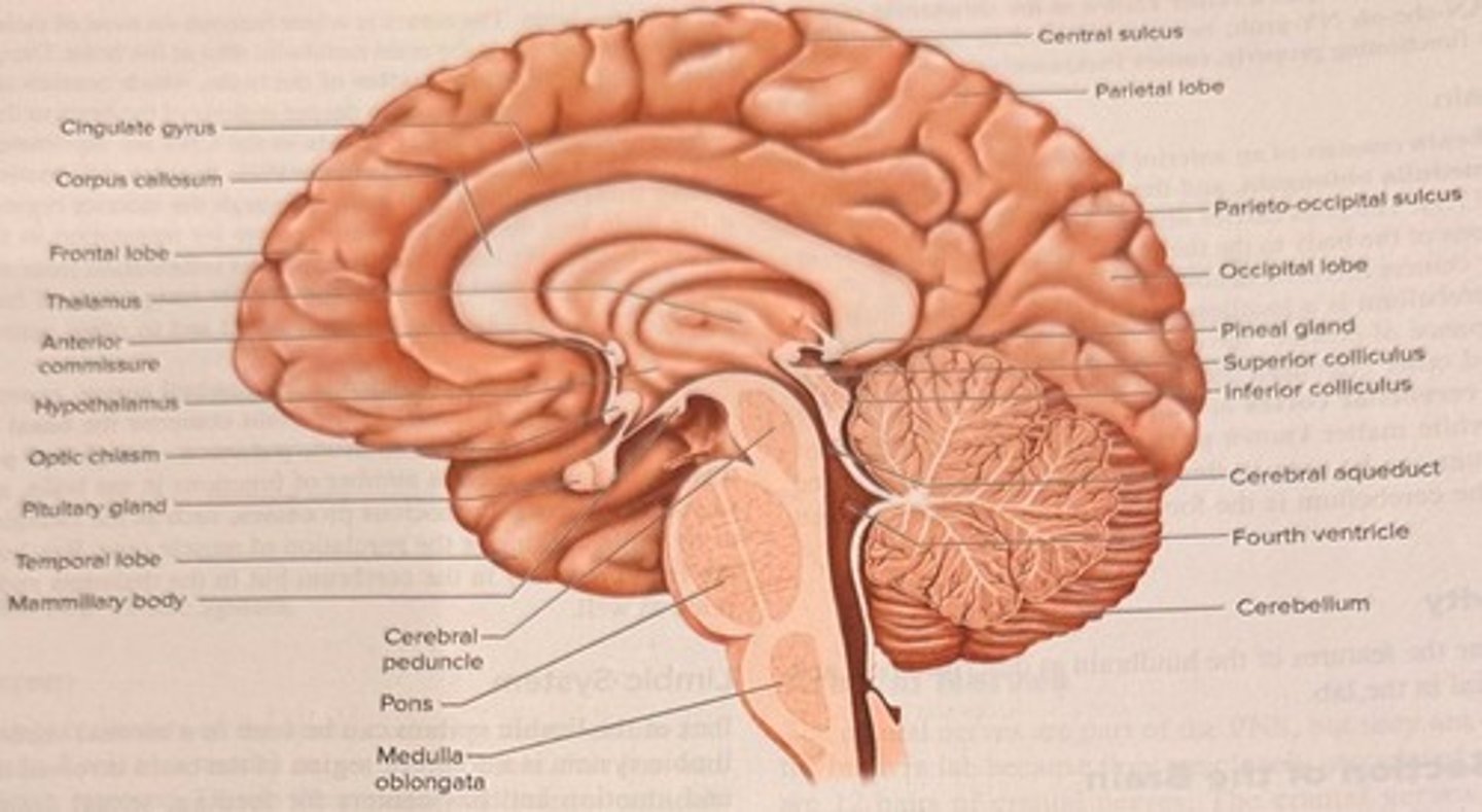
Cerebral Aqueduct
Narrow passageway for cerebrospinal fluid.
Fourth Ventricle
Located near the brainstem and cerebellum.
Subarachnoid Space
CSF flows around the brain and spinal cord.
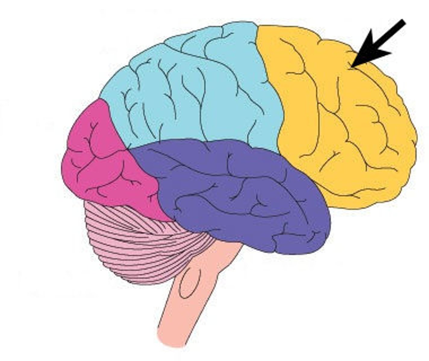
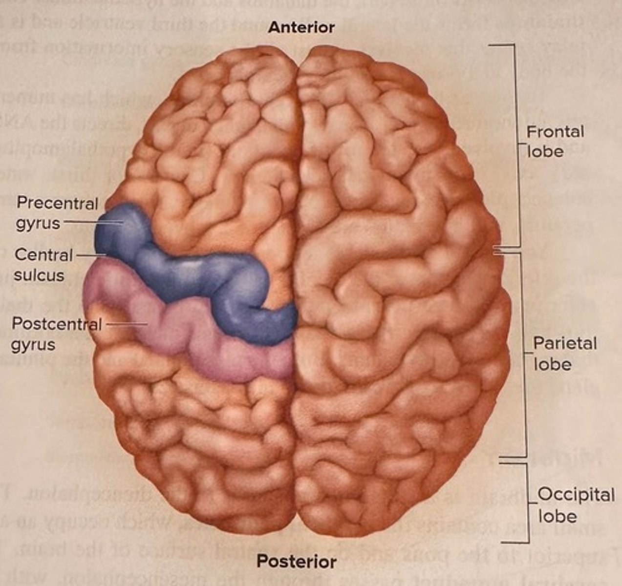
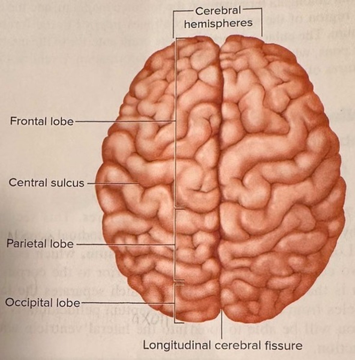
Frontal Lobe
Controls reasoning, movement, and decision-making.
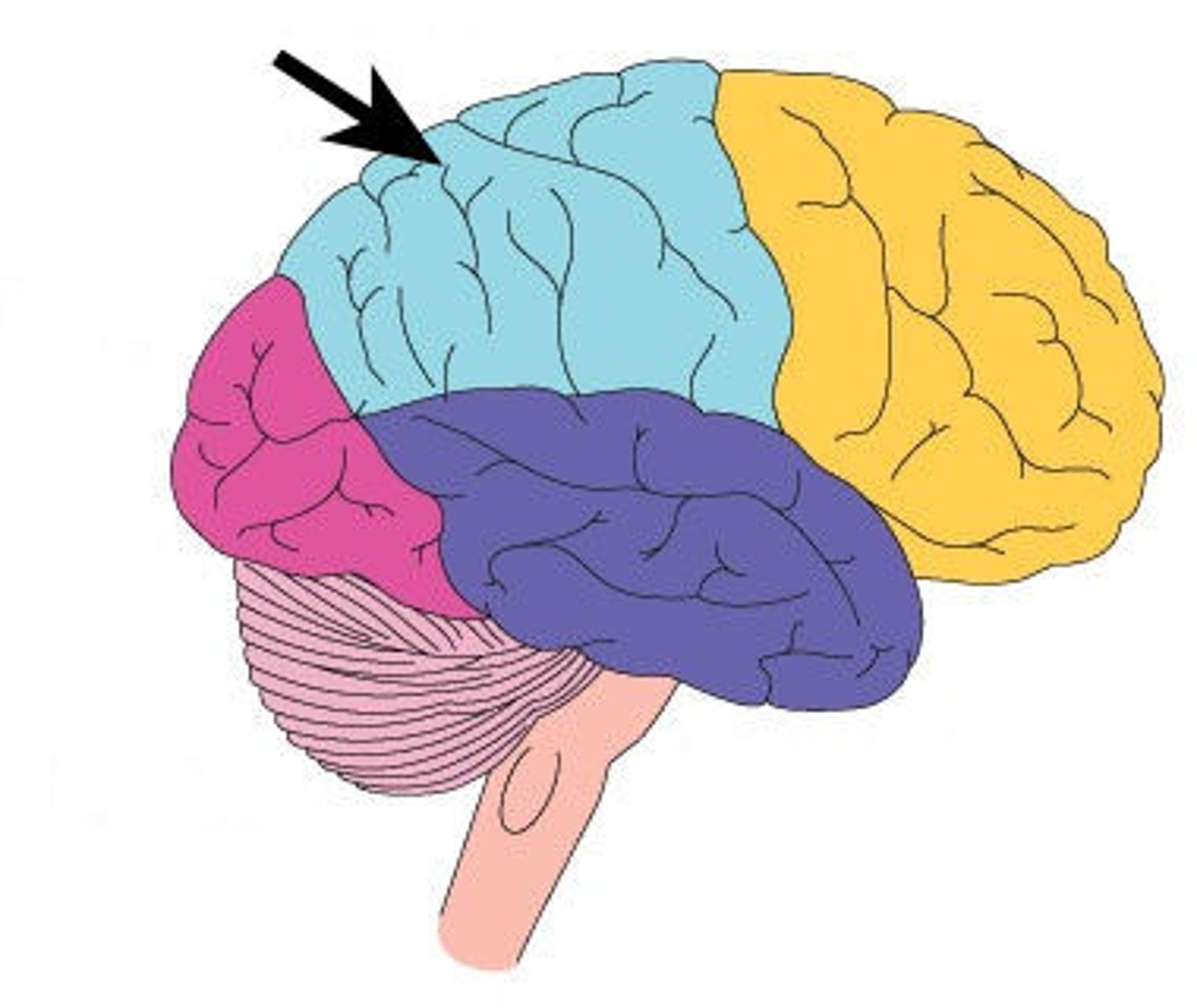
Parietal Lobe
Processes sensory information like touch and pain.
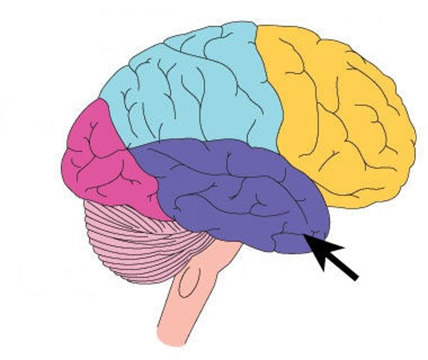
Temporal Lobe
Processes hearing, memory, and language functions.
Gyri
Raised ridges or folds on the brain's surface.
Sulci
Shallow grooves between the gyri.
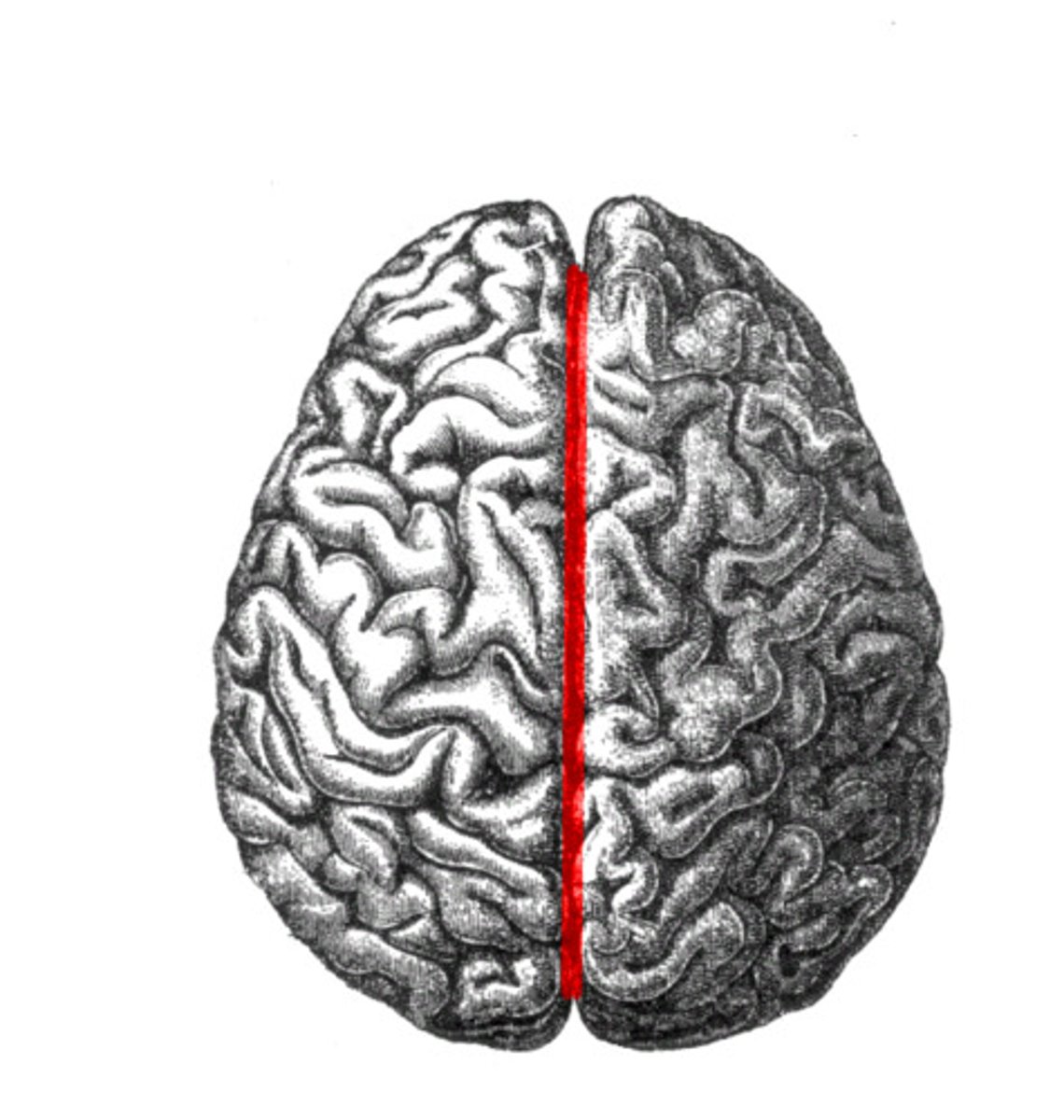
Fissure
Deep groove, e.g., longitudinal fissure.
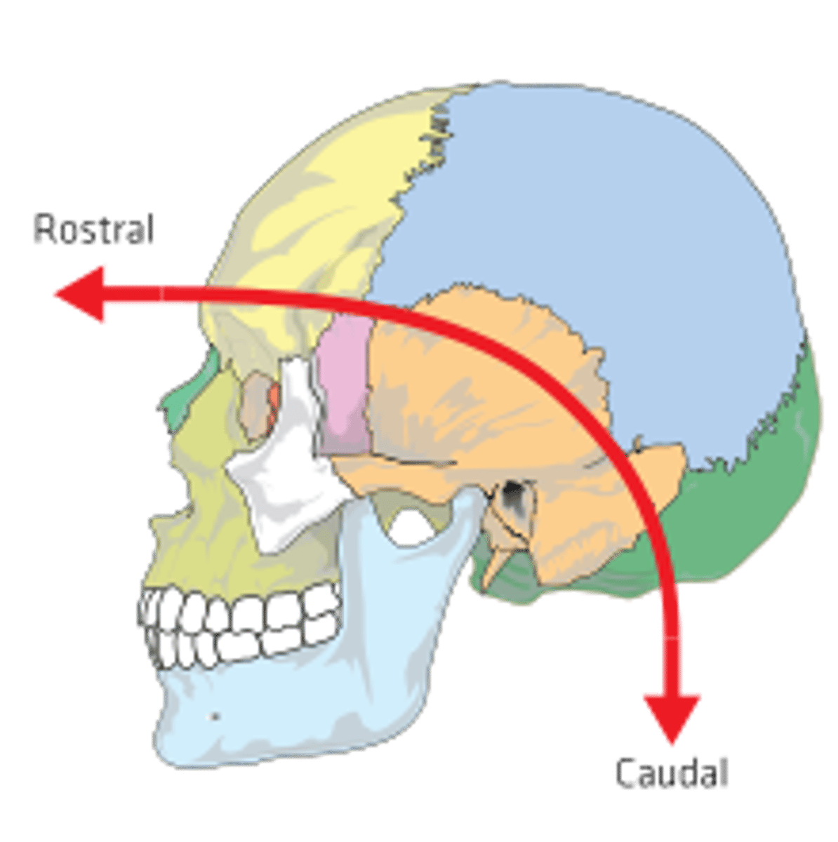
Rostral
Direction toward the front of the brain.
Caudal
Direction toward the back of the brain.
Longitudinal Fissure
Separates the left and right cerebral hemispheres.
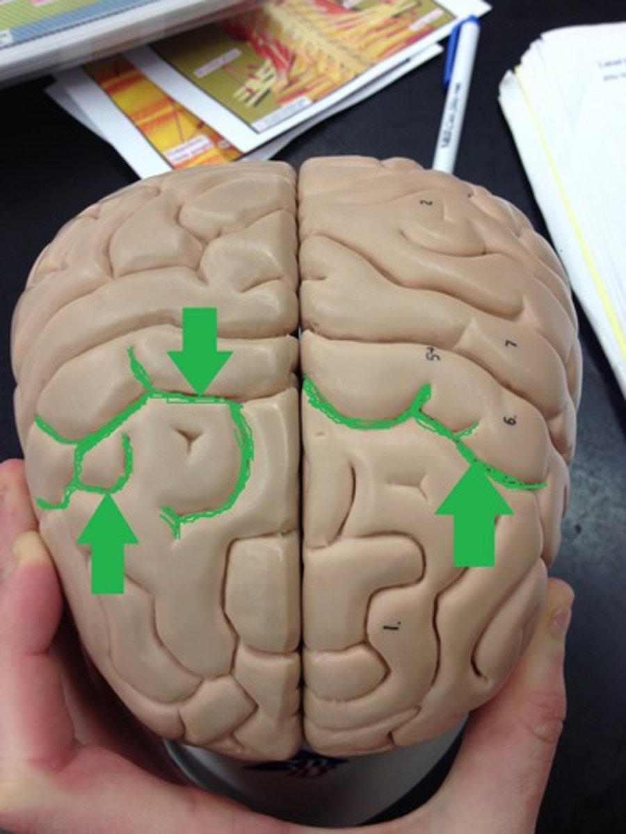
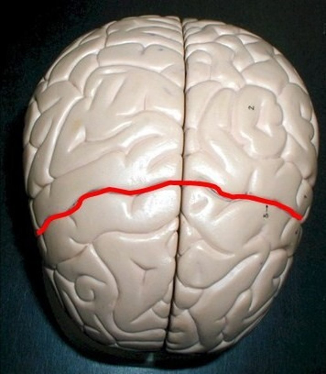
Central Sulcus
Separates frontal lobe from parietal lobe.
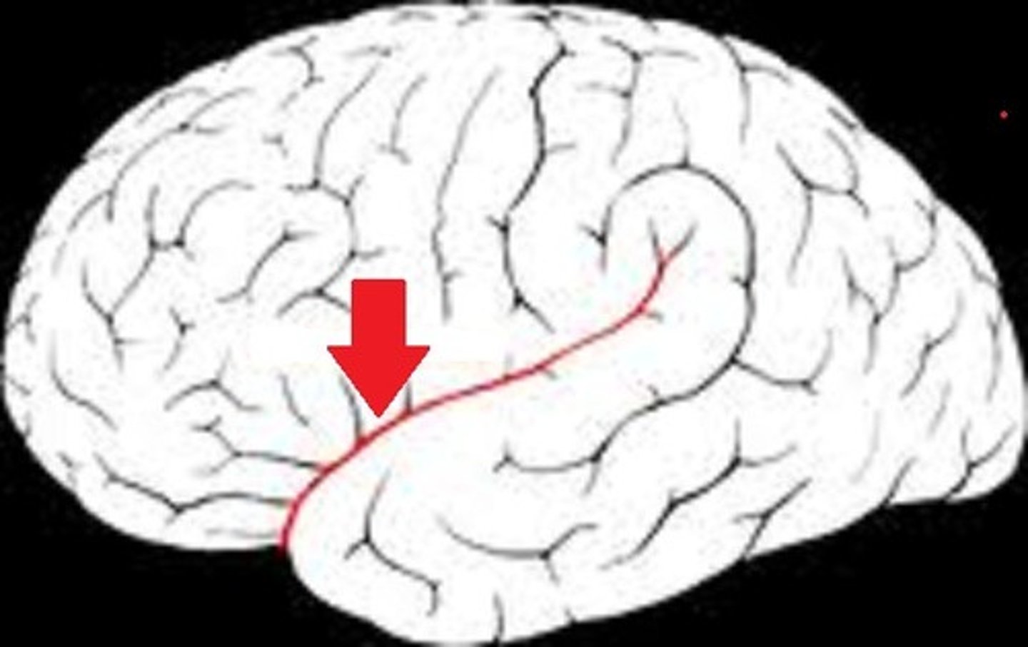
Lateral Sulcus
Separates temporal lobe from frontal and parietal lobes.
Human Brain
Larger, more complex, and vertically positioned.
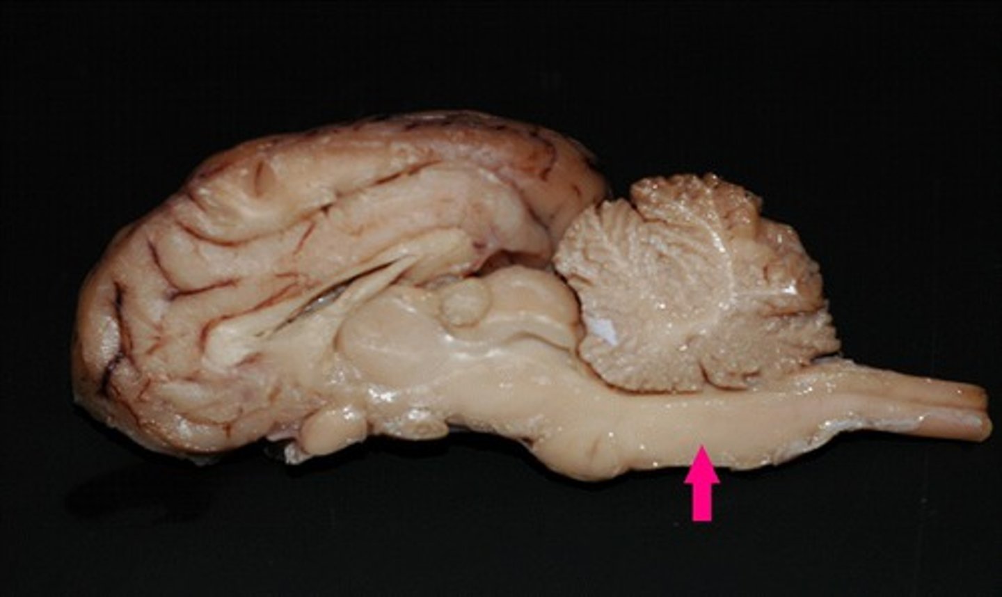
Sheep Brain
Smaller, elongated, and horizontally positioned.
Finger-to-Nose Test
Assesses coordination by touching nose and target.
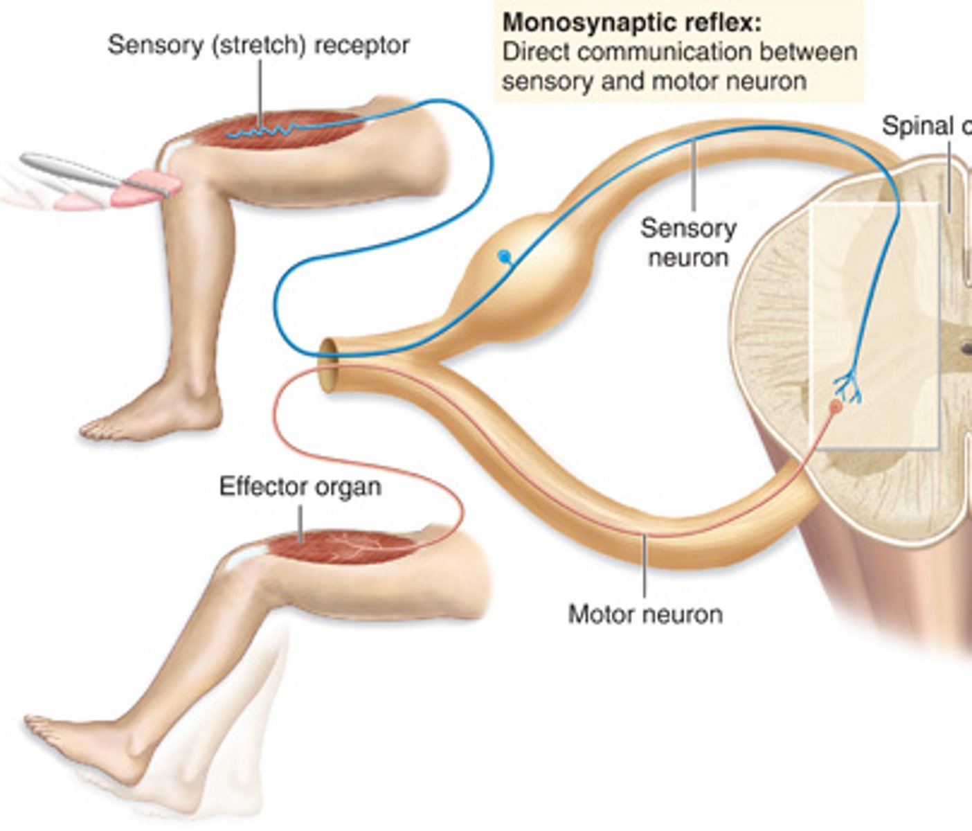
Monosynaptic Reflex
One synapse, fast response, e.g., knee-jerk reflex.
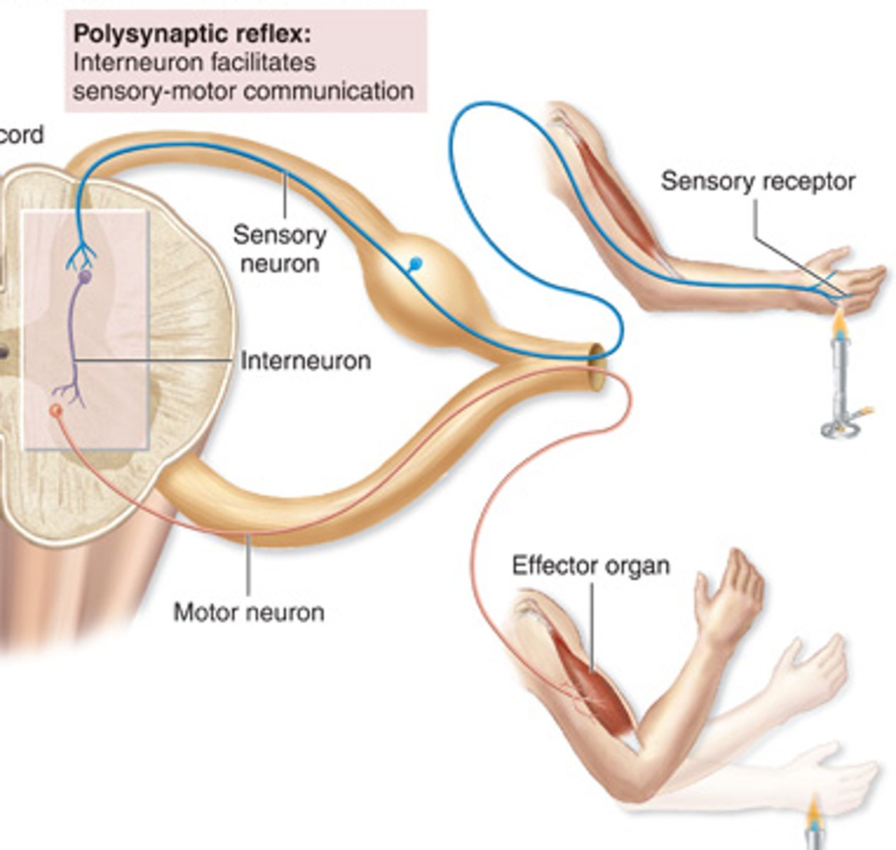
Polysynaptic Reflex
Multiple synapses, slower, more complex reflex.
Muscle Spindles
Detect muscle stretch, trigger reflex to resist.
Golgi Tendon Organs
Detect tension in tendons, prevent muscle overuse.
Patellar Reflex
Leg kicks forward, tests L2-L4 spinal segments.
Biceps Brachii Reflex
Forearm flexes, tests C5-C6 spinal segments.
Calcaneal Reflex
Foot points downward, tests S1-S2 spinal segments.
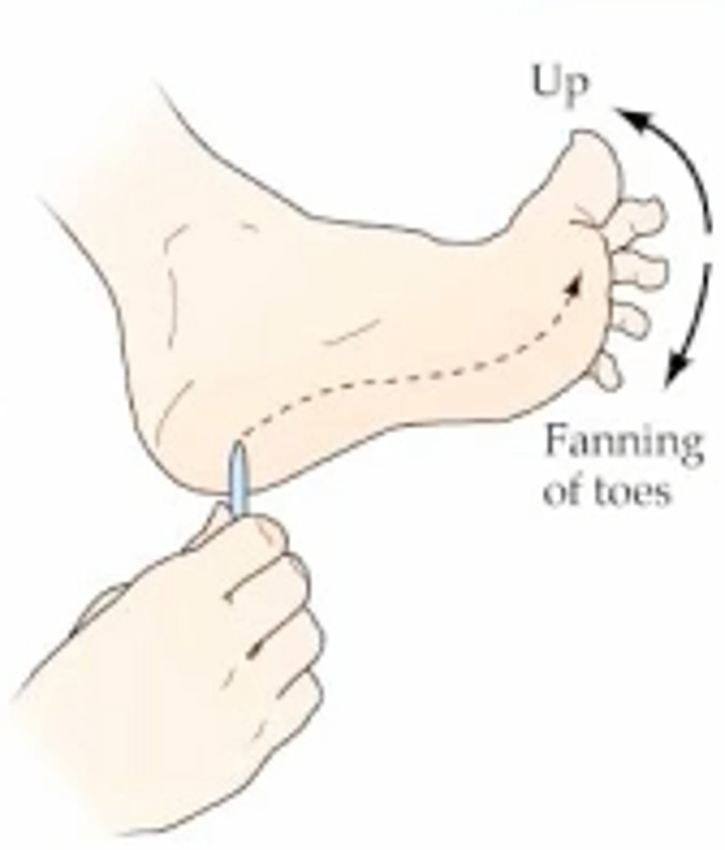
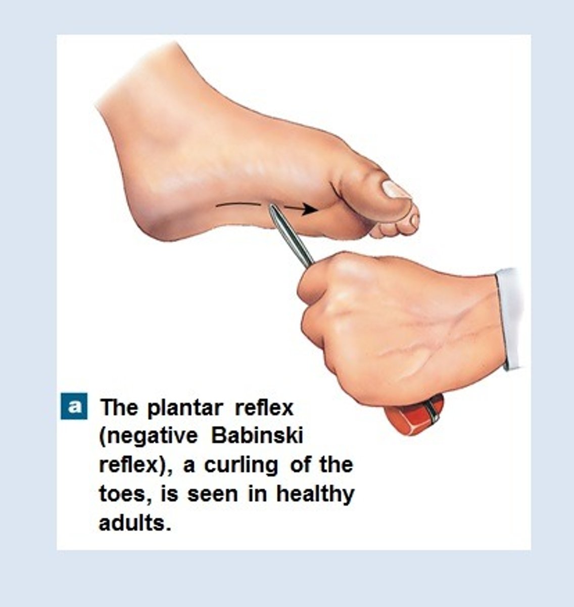
Babinski Reflex
Plantar reflex test, checks neurological function.
Positive Babinski
Toes fan out; normal in newborns.
Negative Babinski
Toes curl inward; normal in adults.
Mechanoreceptors
Detect touch, pressure, vibration.
Nociceptors
Detect pain via free nerve endings.
Thermoreceptors
Detect temperature changes, hot and cold.
Chemoreceptors
Detect chemical changes like taste and smell.
Proprioceptors
Detect body position and movement.
Punctate Distribution
Receptors unevenly distributed; some areas more sensitive.
Absolute Temperature
Actual temperature measurement of an object.
Relative Temperature
Temperature perception compared to a previous stimulus.
Sensory Adaptation
Receptors cease responding to constant stimuli.
Phasic Receptors
Adapt quickly to stimuli changes.
Tonic Receptors
Adapt slowly, responding to ongoing stimuli.
Two-Point Discrimination Test
Measures touch receptor density via distance felt.
Mapping Warm, Cool, and Fine Touch Receptors
Determines location and density of temperature receptors.
Temperature Judgment
Perception changes based on prior temperature exposure.
Proprioceptive Judgment
Tests body awareness without visual input.
Merkel's Discs
Detect light touch and pressure, slow adapting.
Meissner's Corpuscles
Detect fine touch and vibration, rapid adapting.
Free Nerve Endings
Detect pain, temperature, and crude touch.
Referred Pain
Pain felt in different location due to nerve pathways.
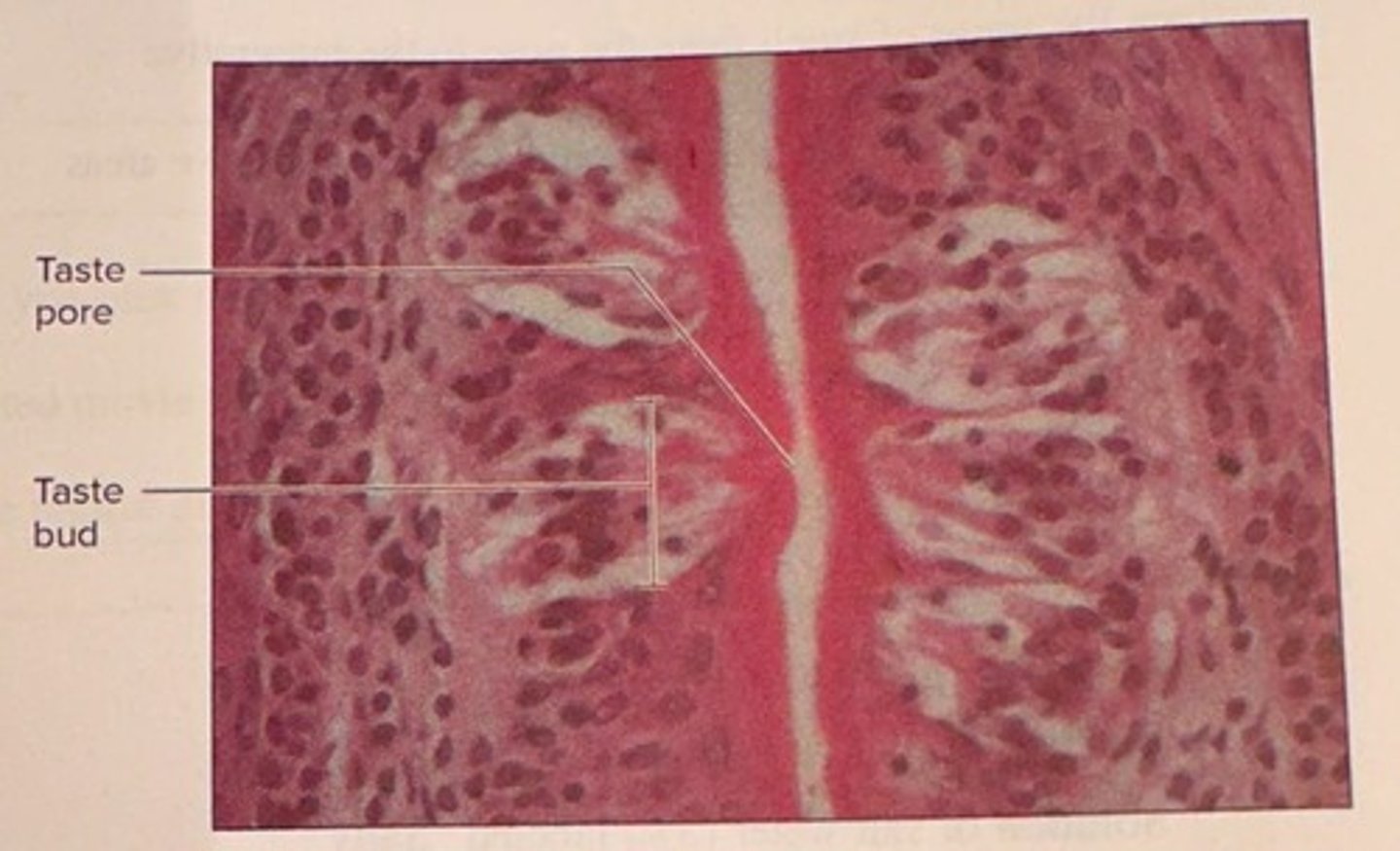
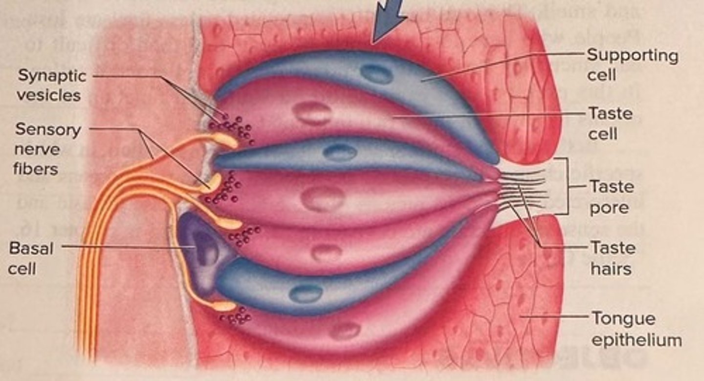
Tastebud
Contains taste hair cells detecting taste molecules.
Taste Buds
Contain taste hair cells detecting taste molecules.
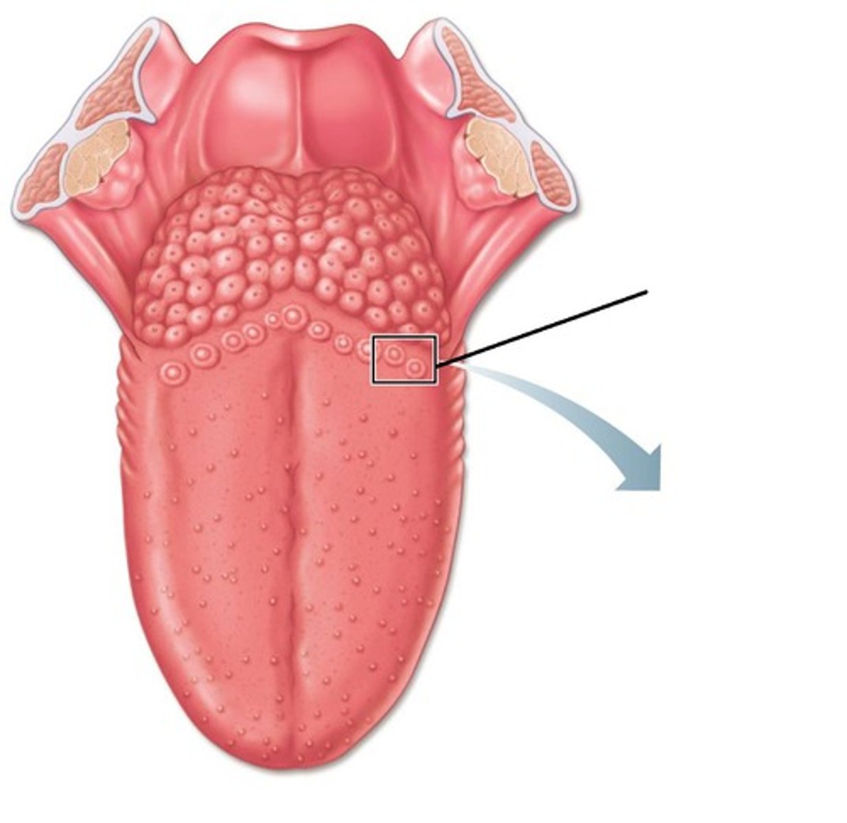
Papillae
Bumps on the tongue housing taste buds.
Olfactory Epithelium
Nasal cavity tissue aiding flavor perception.
Cranial Nerves
12 pairs of nerves that come directly from the brain and brainstem. They control senses (sight, smell, taste, hearing, touch) and movements (eye, face, tongue, swallowing, neck, and internal organs).
Facial Nerve (VII)
Facial expressions, taste (front 2/3 of tongue) (sensory & motor)
Glossopharyngeal Nerve (IX)
Taste (back 1/3 of tongue), swallowing (sensory & motor)
Vagus Nerve (X)
Controls heart, digestion, and autonomic functions (sensory & motor)
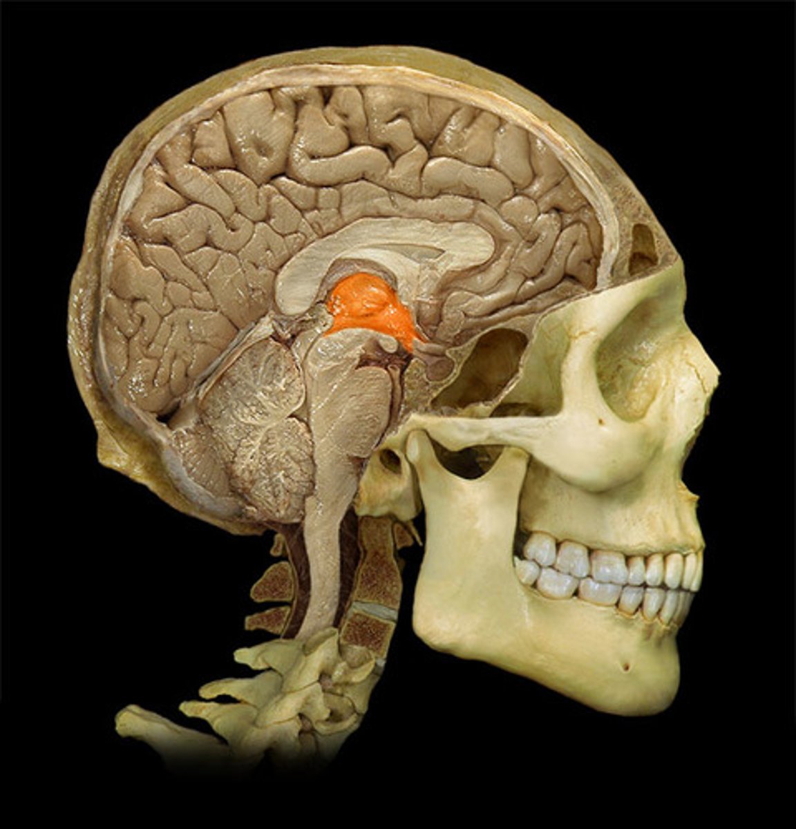
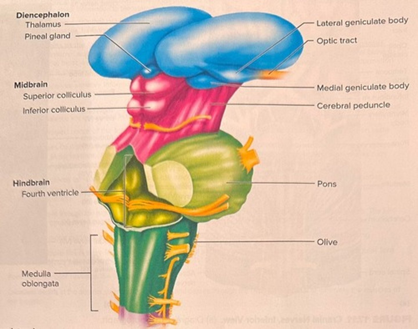
Medulla Oblongata
Brainstem area processing taste signals.
Thalamus
Sensory processing center for taste information.
Gustatory Cortex
Brain region for taste perception in insula.
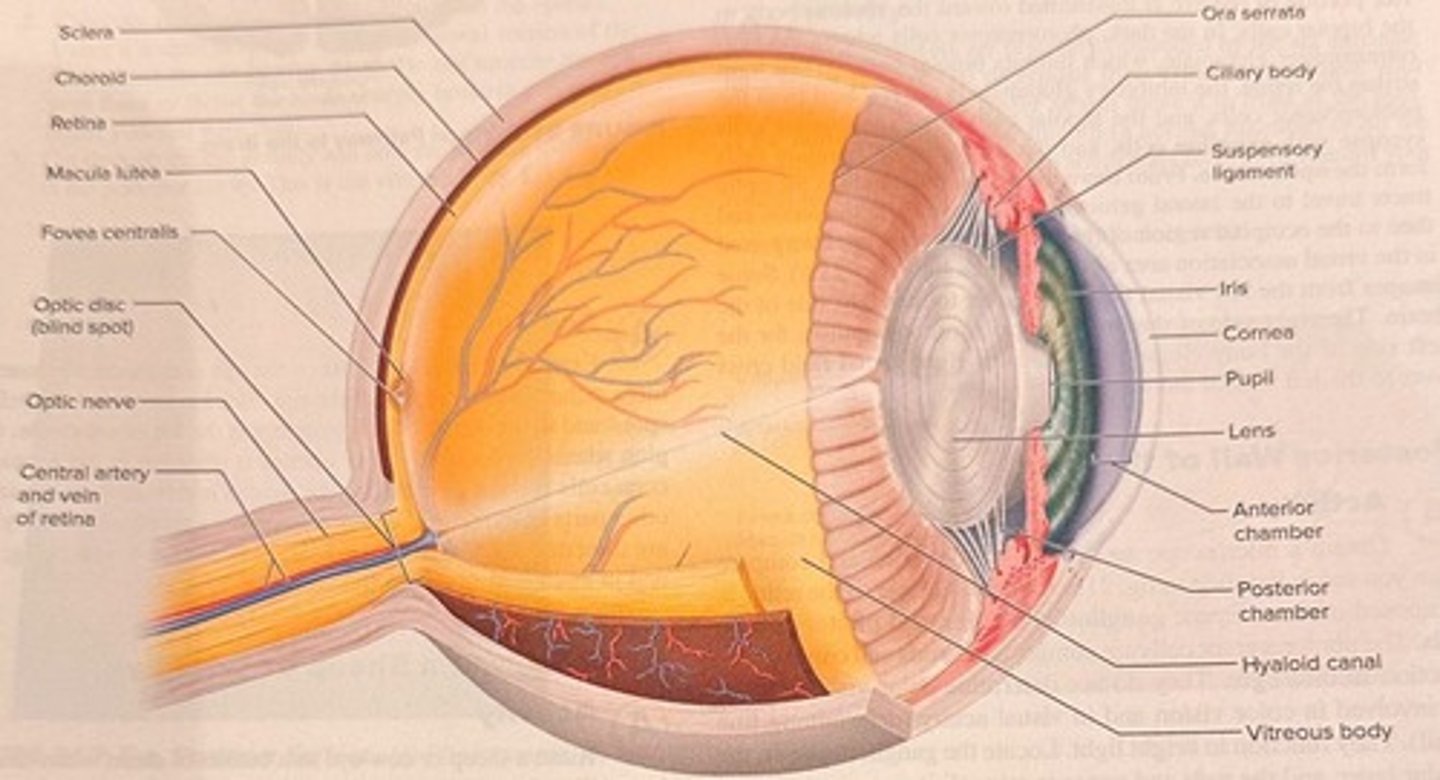
Sclera
Outer layer maintaining eye shape and protection.
Choroid
Middle layer with blood vessels nourishing the eye.
Retina
Inner layer with photoreceptors detecting light.
Anterior Chamber
Space filled with aqueous humor in front of lens.
Posterior Chamber
Space filled with aqueous humor behind the lens.
Vitreous Chamber
Largest chamber filled with vitreous humor.
Optic Nerve (Cranial Nerve II)
Nerve that carries impulses for sense of sight (sensory)
Optic Chiasm
Point where some optic nerve fibers cross.
Optic Tract
Carries visual signals to the thalamus.
Cornea
Transparent layer focusing light onto the retina.
Fovea Centralis
Area with highest cone concentration for sharp vision.
Optic Disc
Blind spot where optic nerve exits the eye.
Optic Nerve
Carries visual signals to the brain.
Occipital Lobe
Processes visual information from the eyes.
Astigmatism Test
Detects uneven cornea/lens causing blurred vision.
Visual Acuity
Measures sharpness of vision (20/20 standard).
Pupillary Reflex Test
Tests pupil reaction to varying light conditions.
Ishihara Test
Identifies color blindness using colored dot plates.