Chapter 12 Nervous Tissue - Part B
1/54
There's no tags or description
Looks like no tags are added yet.
Name | Mastery | Learn | Test | Matching | Spaced |
|---|
No study sessions yet.
55 Terms
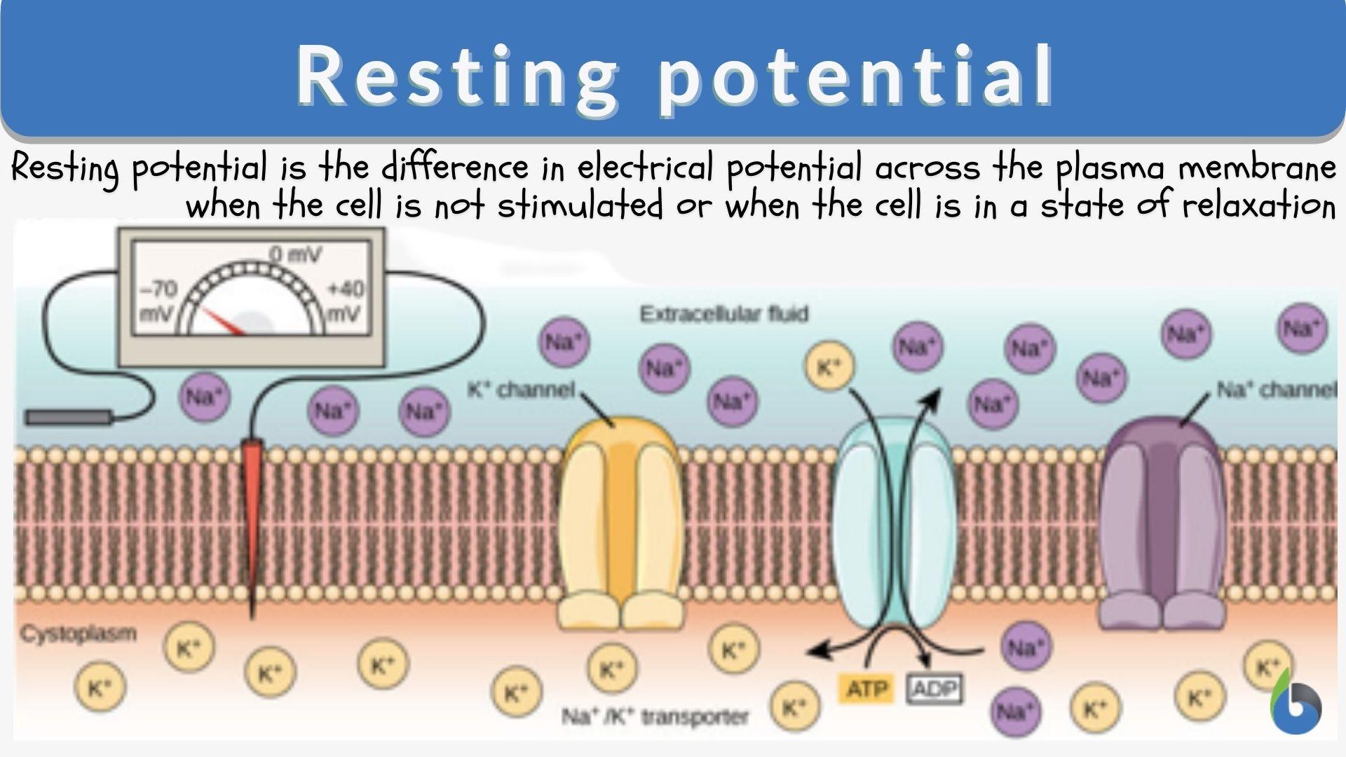
Resting Membrane Potential
Electrical difference between the inside of the neuron and the outside.
The inside is -70 mV, a negative charge.
The outside is 0 mV, a relatively positive charge.

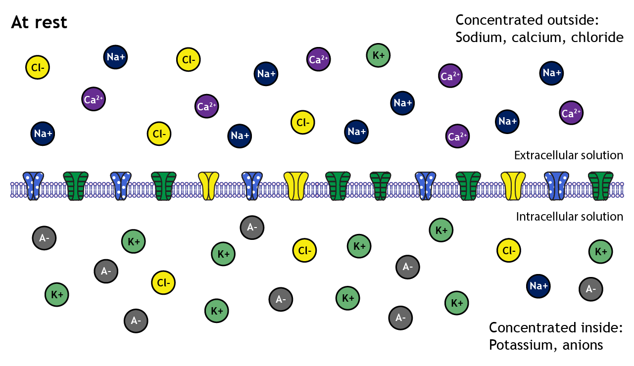
Ion concentrations relative to the cell membrane:
There is more k+ inside.
There is more OA- inside.
There is more Na+ outside.
There is more Ca+ outside.
There is more Cl- outside.
Electrical force
Ions will always travel to the area of opposite charge. Electrical force has less influence compared to chemical force.

Chemical force
Ions travel from an area of low concentration to an area of high concentration. Chemical force has more influence compared to electrical force.
K+ Forces
Electrically K+ wants to go in.
Chemically K+ wants to go out.
OA- Forces
Electrically OA- wants to go out.
Chemically OA- wants to go out.

Na+ Forces
Electrically Na+ wants to go in.
Chemically Na+ wants to go in.

Cl- Forces
Electrically Cl- wants to go out.
Chemically Cl- wants to go in.

Ca+ Forces
Electrically Ca+ wants to go in.
Chemically Ca+ wants to go in.
Resting Membrane Potential Mechanism
Who up charging they membrane??????
The method by which a neuron retains a -70mV charge inside, and a +0 charge outside.

Steps of the Resting Membrane Potential Mechanism
(All of these technically happen at once).
The rough ER in the cell body produces negatively charged organic proteins. These proteins are too big to flow down their concentration gradient, so the inside of the cell gains a -5 mV charge.
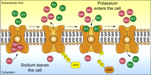
The sodium potassium pump pumps 3 Na+ out of the cell and 2 K+ into the cell. Since the pump removes 3 positive species and takes in only 2 positive species, the charge of the inside of the cell jumps to -10mV. (Note that this pump does not effect extracellular fluid cause it big and spacious)
K+ exits the cell via leakage channels, further decreasing the membrane potential to -70 mV. Once at -70mV, the electrical force and chemical force acting on potassium balance out.
Cl- is removed via K+/Cl- symports.
Ca2+ is removed via Na/Ca+2 exchangers (antiports).

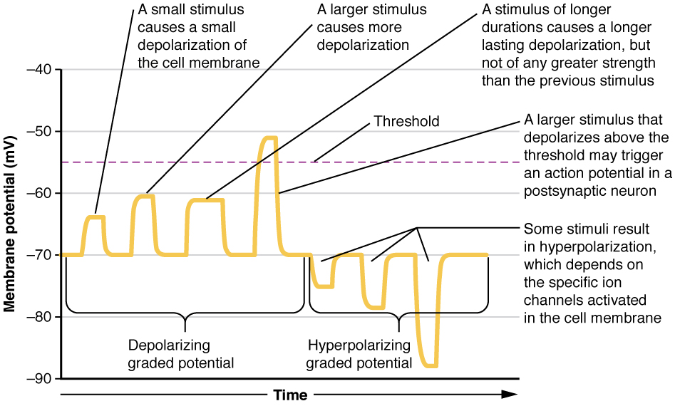
Graded Potential
A small, localized change in a cells membrane potential, usually hovering around the resting membrane potential of -70 mV.

Depolarization
An positive increase in charge of the membrane potential.

Hyperpolarization
A decrease in charge away from -70 mV of the membrane potential.

Repolarization
A decrease in charge back towards -70 mV of the membrane potential.

Potential threshold
-55 mV. Once the trigger zone (axon hillock) reaches a charge of -55 mV the action potential will spread through the axon and the neuron will fire an action potential.

Excitatory Action
A different axon sending neurotransmitters to the neuron, causing the charge to increase.

Inhibitory Action
A different axon sending neurotransmitters to the neuron, causing the charge to decrease.

Action strength
A neuron can send:
A short burst of charged particles.
A large burst of charged particles
A sustained burst of charged particles.
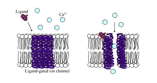
Ligand Gated Channel
A specific kind of channel that requires a particular neurotransmitter to open, like fitting a key into a lock. Once the key is inserted, a specific kind of ion will either enter or exit the cell, leading to depolarization/repolarization/hyperpolarization.
How do charges dissipate after entering?
Some exit through pumps, like Na+.
Others simply diffuse through the cell, becoming so spread out that their addition has a negligable effect on the charge.
Remember, the charge must be -55 mV AND reach the axon hillock (Trigger zone) to fire an action potential.
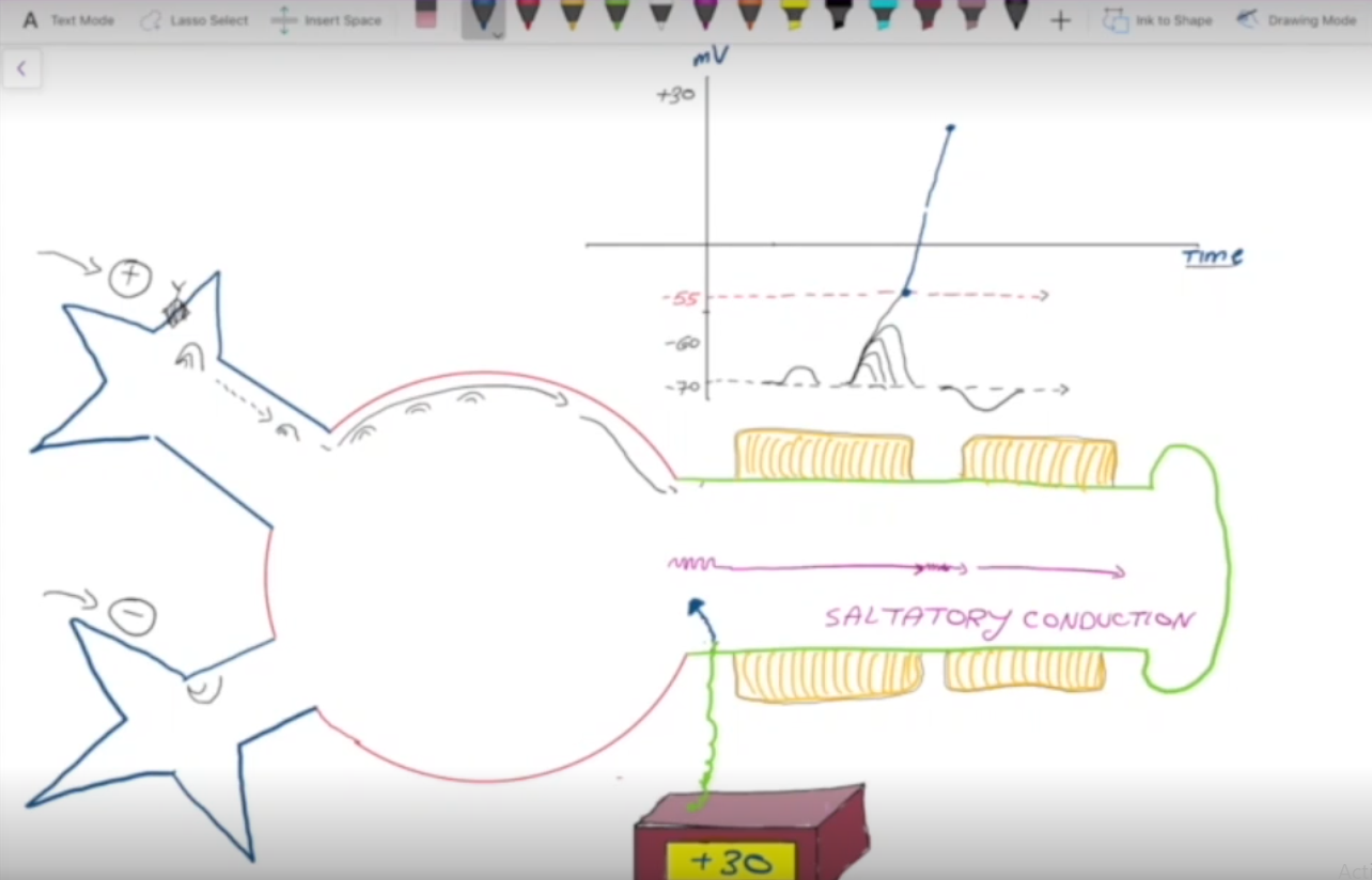
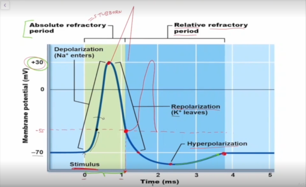
Action Potential Description
As positive ions enter the neuron, they’ll eventually travel towards the axon hillock (trigger zone); if enough make it to the zone then the action potential fires, bring the charge of the cell up from -55 mV to +30 mV. This action potential is expedited by the myelin sheath (saltatory conduction).
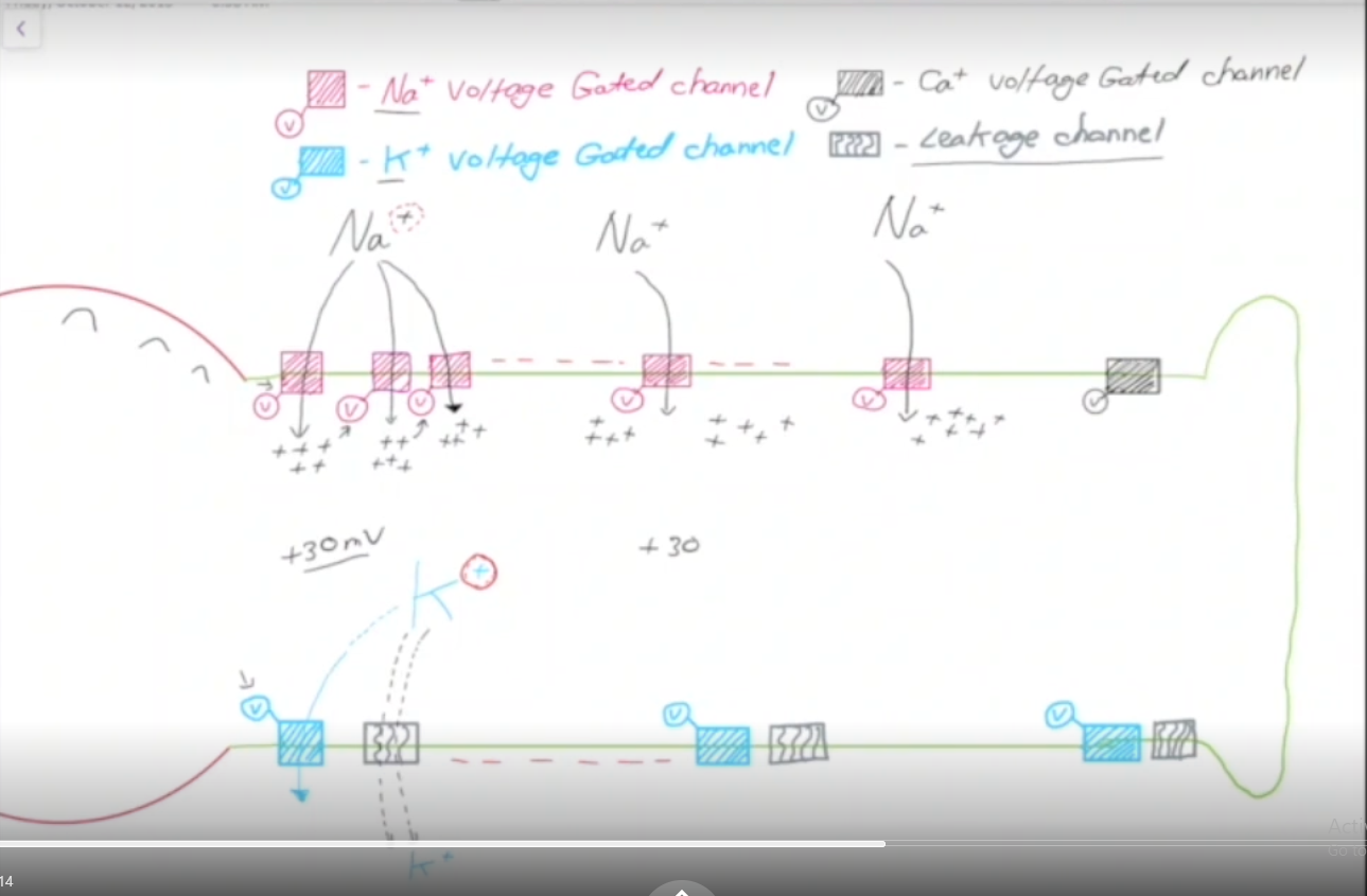
How does an Action Potential mechanism work?
Positively charged Na+ enters the cell via ligand gated channels around the dendrites and the cell body.
After enough Na+ enters, the trigger zone (axon hillock) depolarizes to a charge of -55 mV.
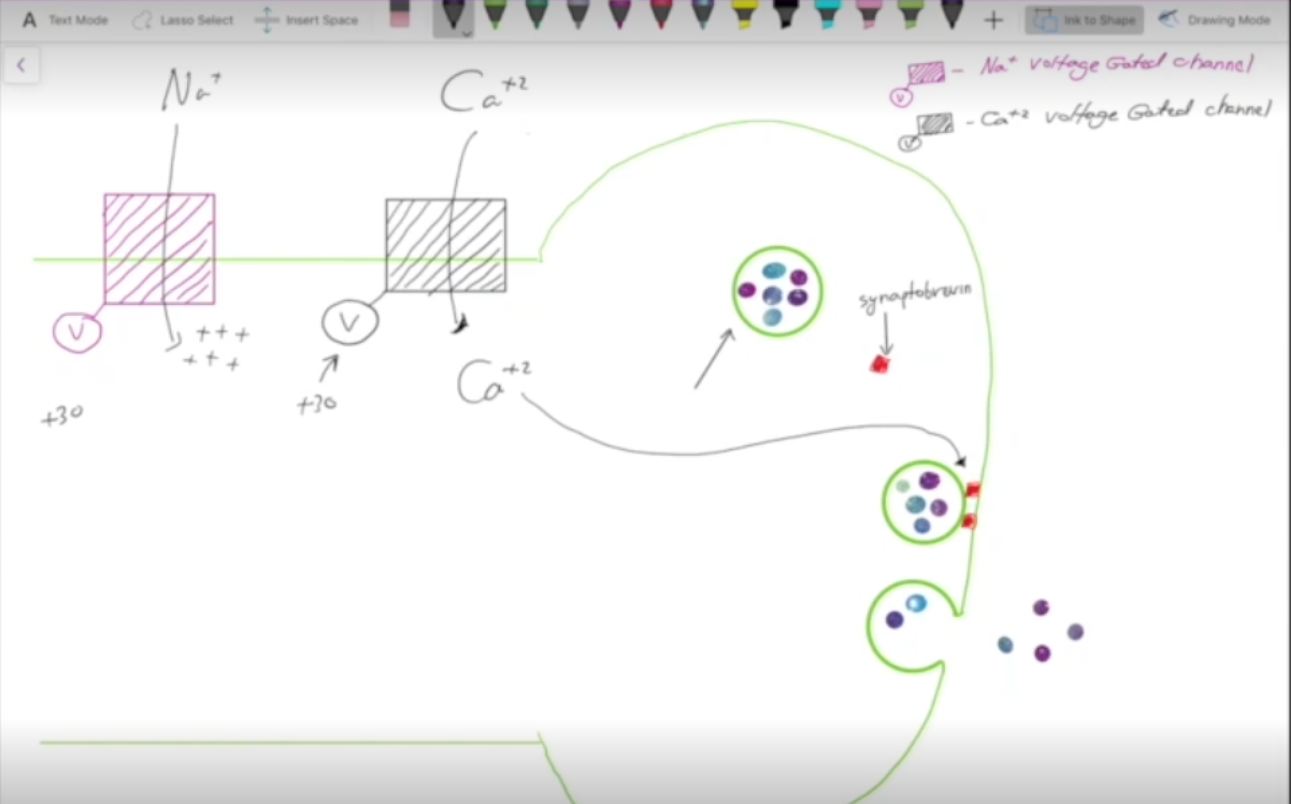
This charge causes Na+ voltage gated channels to open, flooding the cell with positive charge and leads to a cascade effect, depolarizing the cell all the way up to 30 mV.
At 30 mV the Na+ voltage gated channels shut, and K+ voltage gated channels open.
K+ rushes out of the cell via voltage gated channels and leakage channels, repolarizing the cell back down to -70 mV.
K+ voltage gated channels begin to close slowly at -70 mV. Potassium continues to enter for a bit and hyperpolarizes the cell.
The cell will finally reach its resting membrane potential as leak channels for Na+ slowly let sodium back in. -70 mV
Whats the point bruz. Why we even action potential???
You need 30+ mV to activate Ca+2 voltage gated channels.
Ca+2 helps vessicles filled with neurotransmitters attach to synaptobrevin, which allows the neurotransmitters to leave the axon.
Absolute Refractory Period
From -70 mV to -55 mV, at this point the neuron CANNOT fire another action potential.

Relative Refractory Period
From -55mV onwards, can get to another action potential if enough sodium enters.
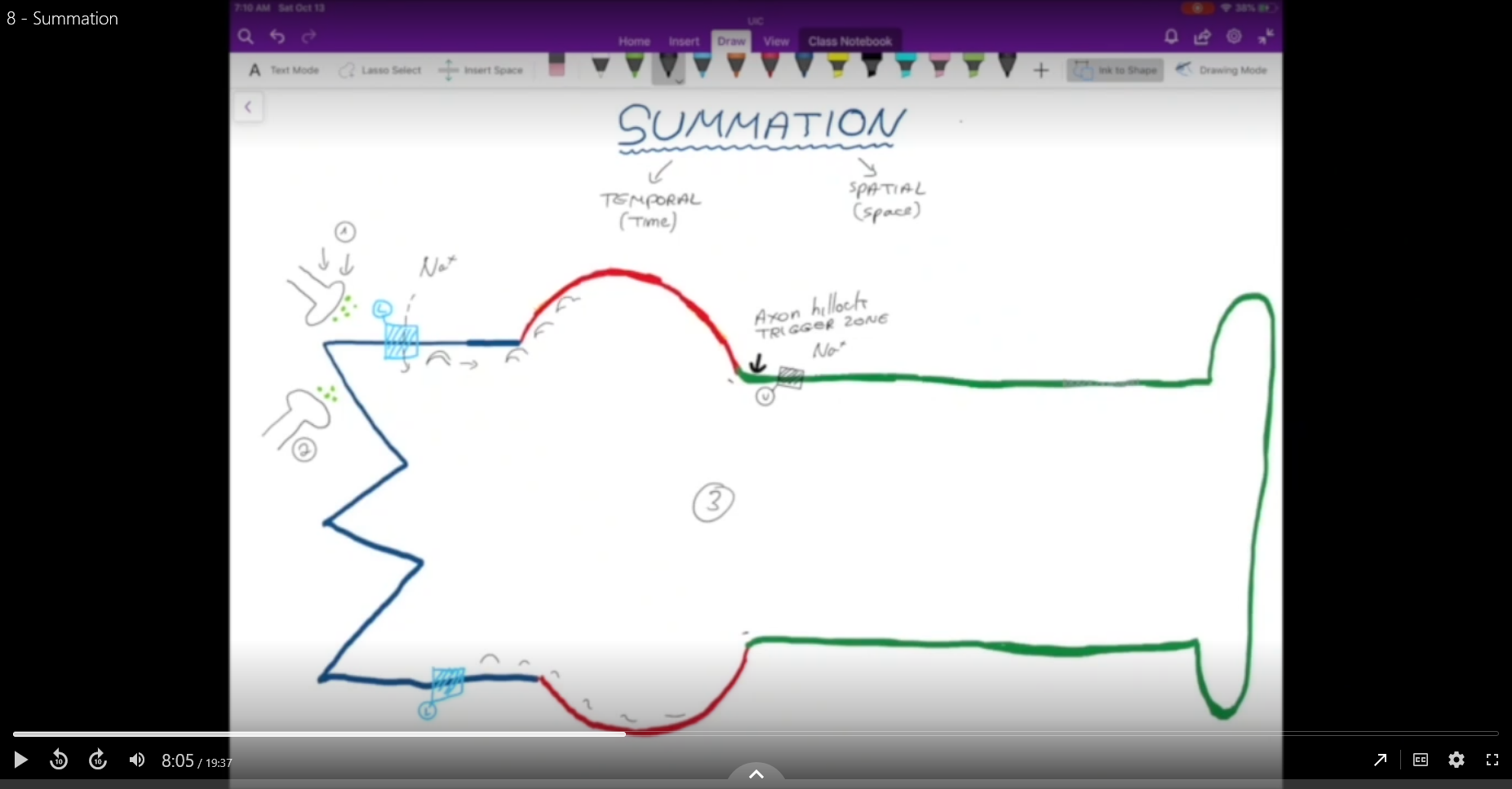
Sumation
Combining gated potentials to reach the trigger zone.
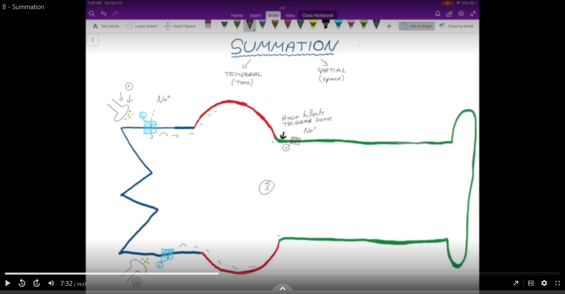
Temporal Sumation
Keep on sending neurotransmitters consistently until -55 mV is reached.

Spatial Sumation
Have multiple axons work together in a single location to combine their neurotransmitters to get a large gated potential for -55 mV.
Excitatory Post Synaptic Potential (EPSP)
Adds positive charge to the inside of the neuron by allowing Na+ in.
Inhibitory Post Synaptic Potential
Decreases charge in the neuron by either allowing Cl- in or taking K+ out.
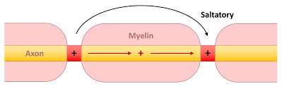
Saltatory Conduction
Where there is no myelin sheathe, the positive charges have to fight through collected negative charges to move, afterall, at resting potential the opposites attract. However, the myeline sheathes put distance between the positive and negative charges at rest; therefore, when a myelin sheathe is present, the positive charge doesnt have to fight through a bunch of negative charge.
Action potential speed in narrow vs wide axons.
Axons have protiens, in narrow channels Na+ gonna bonk into them, not in wide channels

Lidocaine
Local anethstetics, blocks the inner part of sodium, no sodium, no signal, no signal no pain.

Tetrodotoxin
Pufferfish, blocks sodium and kills you

Multiple Sclerosis
Removes myelin sheath, slow signals

Brain
The braon in the head
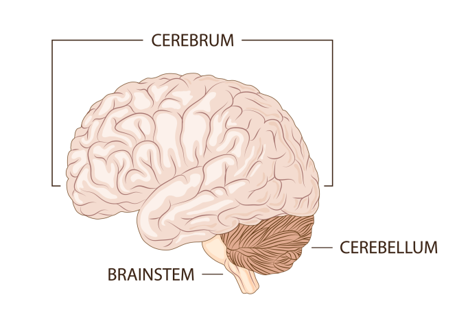
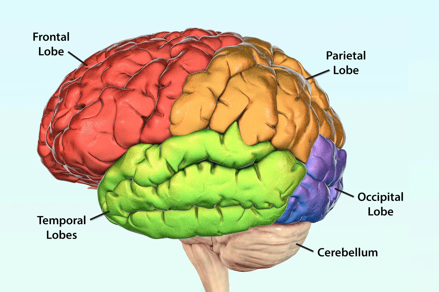
Cerebrum
The big part on the top.
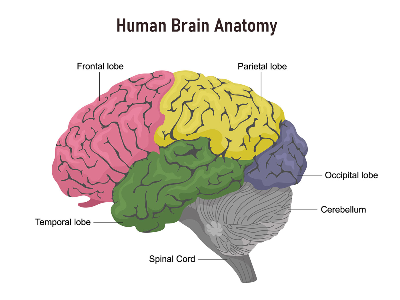
Made of four lobes, makes up 80% of the brain, and is the center for all intelligence, reasoning, memory, and voluntary processes.
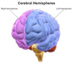
Cerebral Hemisphere
One half of the cerebrum
Each contains four lobes.
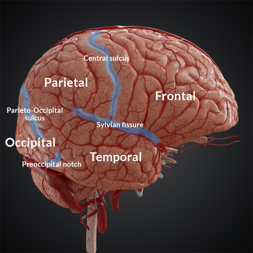
Frontal Lobe
Lobe in da front
Responsible for our personality and decision making.

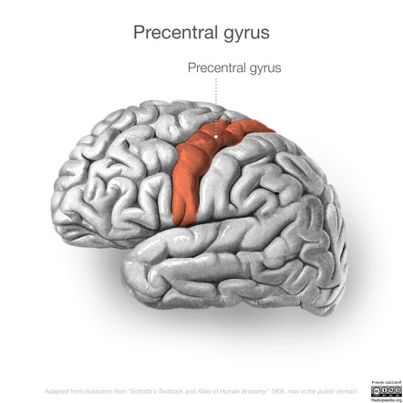
Precentral gyrus (Primary Motor Cortex)
Divides front and back kinda
Responsible for voluntary movement.
Parietal Lobe
Back upper lobe
Responsible for physical sensations like touch from the skin and for joint proprioception.
Contains the post central gyrus
Postcentral gyrus (Primary somatosentory cortex)
Kinda behind the motor cortex
Main sensory receptive area for touch.
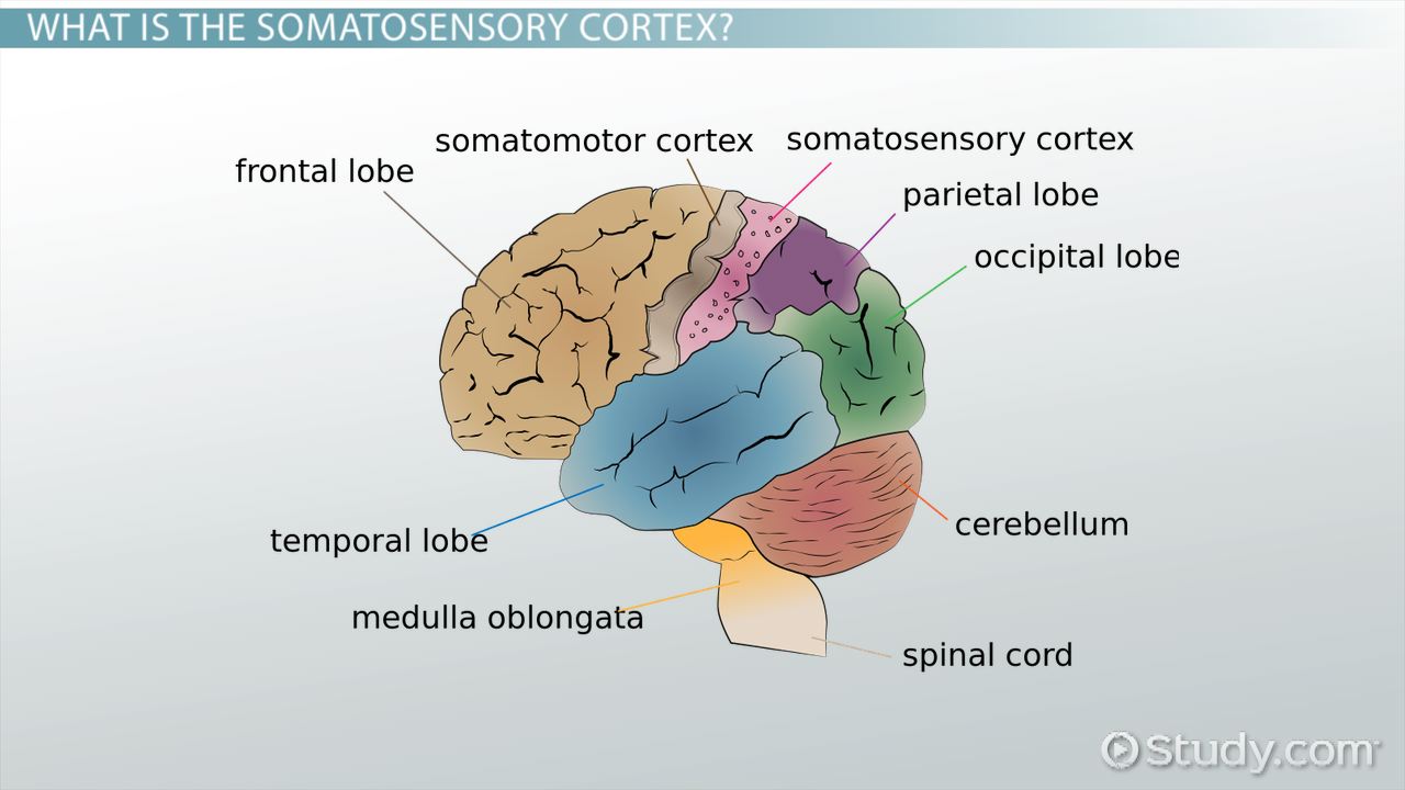
Occipital Lobe
is occipital
Responsible for processing and storing visual information

Temporal Lobe
Da one on the side sliiide to the left
Responsible for our ability to smell and hear.

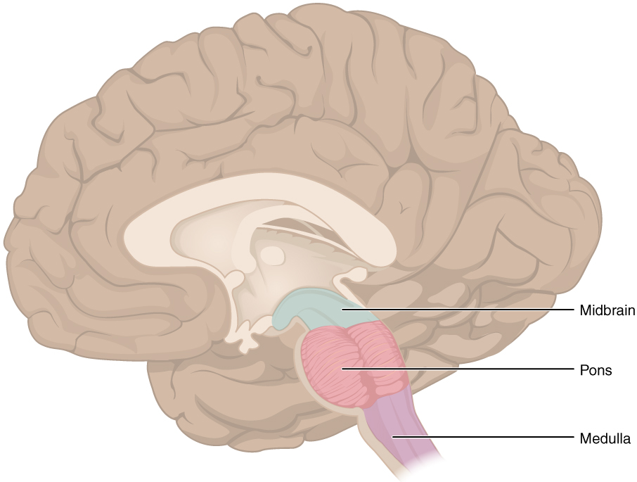
Brainstem
its a stem, that comes out of the brain
Midbrain
Pons
Medulla
Responsible for not dying. Makes us breathe and makes our heart beat.
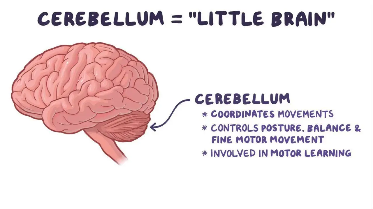
Cerebellum
brain under the brain brain part 2 brain
Responsible for fine motor control, coordination of skeletal muscle, and equilibrium.
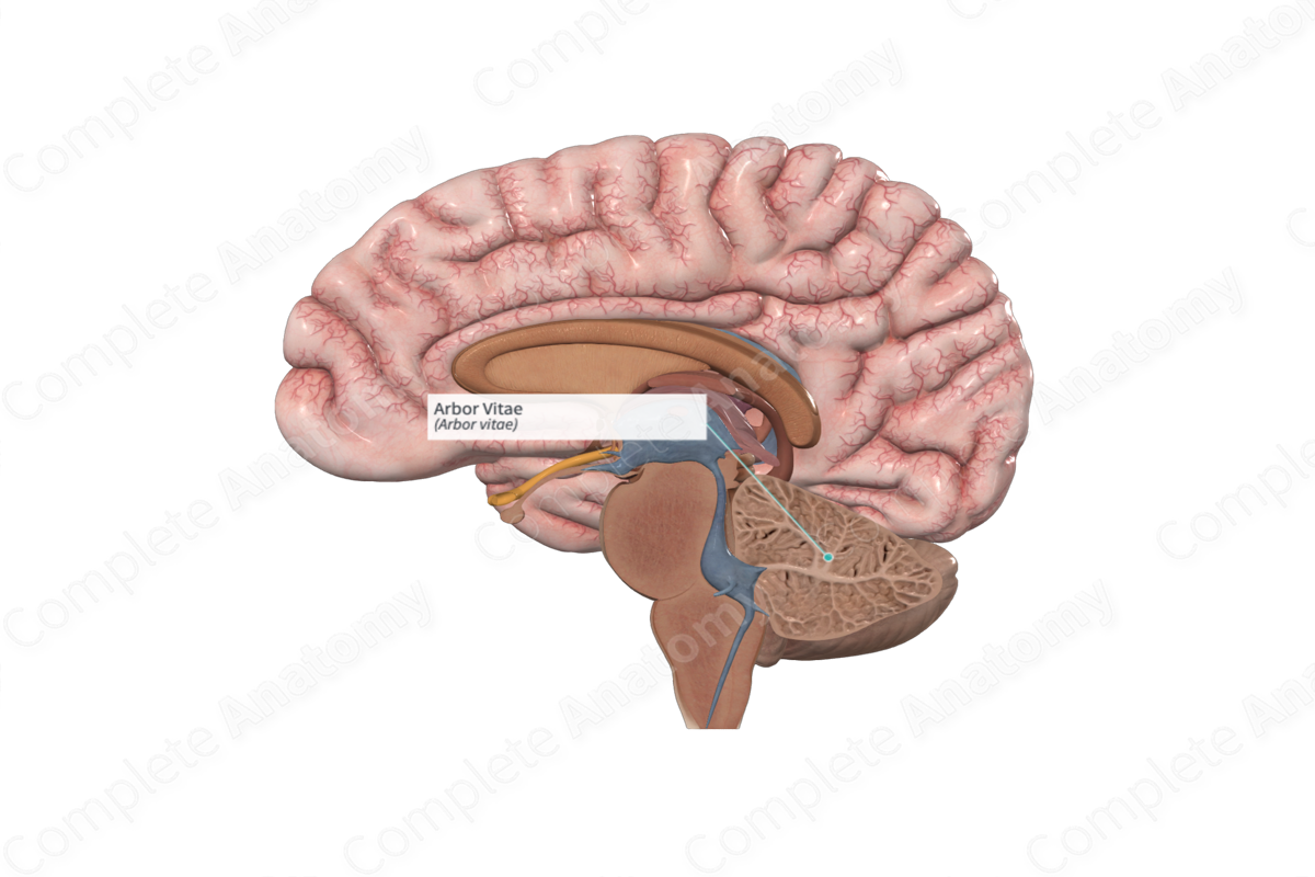
Arbor vitae
Tree thing in the cerebellum
Made of white fat.
Vermis
Down the middle of the cerebellum.
Seperates the 2 halves of the cerebellum.
Means worm.
Fissures / sulci
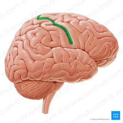
Central sulcus
Lateral sulcus
Longitudinal fissure
Transverse fissure
Parieto-occipital sulcus
Central sulcus
Central
Divides the frontal and parietal lobes.
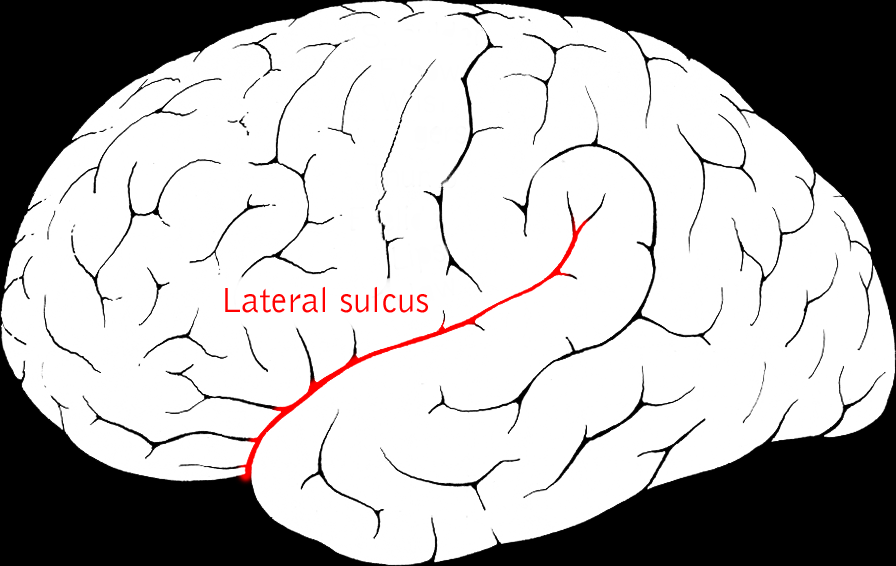
Lateral sulcus
Kinda middle bottom cleft
Seperates the temporal lobe from the frontal and parietal lobes.
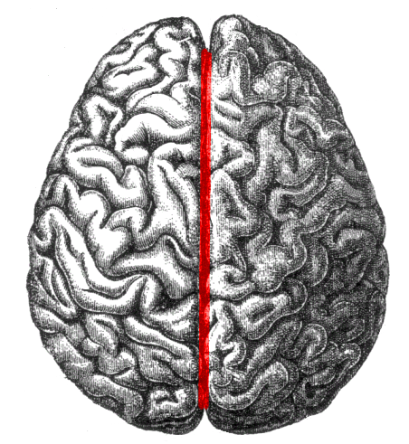
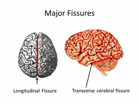
Longitudinal fissure
Right down mid
Seperates left and right cerebrum
Transverse fissure
Between brain and brain pt 2.
Seperates the cerebrum and the cerebellum.
Parieto-occipital sulcus
Between the parietal and occipital lobes
Separates the the parietal and occipital lobes