skeletal, cardiac, smooth
What are the 3 types of muscle tissue?
skeletal
What muscle tissue is voluntary?
cardiac and smooth
What muscle tissues are involuntary?
skeletal and cardiac
What muscle tissues are striated?
How do muscles get bigger ?
myosatellite cells add new myofibrils, where more protein is needed.
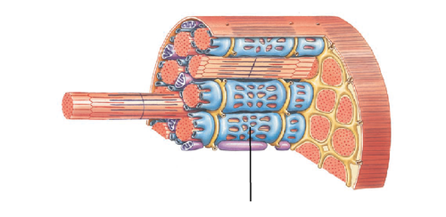
fassicles
bundles of muscle fibers wrapped together by connective tissue
Multinucleated
One muscle fiber that has many nuclei
Sacroplasmic Reticulum (SR)
modified smooth endoplasmic reticulum in muscle cells that surrounds the myofibrils and stores Ca2+
T tubules
tubular infoldings of the sarcolemma which penetrate through the cell and emerge on the other side
motor end plate (neuromuscular junction)
the location where the nerve terminates into the muscle, forms a synapse into which neurotransmitter (ACh) is released
neuromuscular junction (NMJ)
the synapse between a somatic motor neuron and a skeletal muscle fiber
C. Both
The opening of ion channels to the motor end plate is permeable to which ions?
A. Na+ B. K+ C. Both
end plate potential (EPP)
Depolarization of the membrane potential of skeletal muscle fiber, caused by the action of the transmitter acetylcholine at the neuromuscular synapse.
EPSP (excitatory postsynaptic potential)
Is when more Na+ is entering than K+ leaving the membrane or better known as EPP
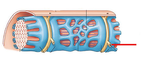
Sarcomere
contractile unit of a muscle fiber
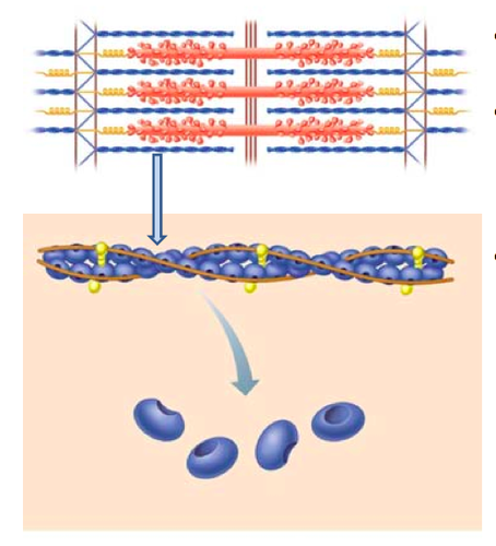
Actin (thin filament)
a protein made up of 2 F stands of actin that form a double helix and (together with myosin) the contractile filaments of muscle cells.
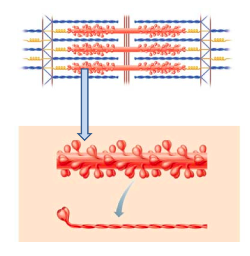
Myosin (thick filament)
a fibrous protein made up of myosin dimers bound together at tails, binding sites on heads (crossbridges) for actin, ATPase site
Sliding filament mechanism
is when the myosin head makes a crossbridge with actin, the head will move back towards the center of the sarcomere, and the filaments will slide on top of each other, making the length of the sarcomere shorter. (muscle contracts)
Tropomyosin
A protein of muscle that forms a complex with Troponin regulating the interaction of actin and myosin in muscular contraction (relaxing)
Troponin
A protein of muscle that together with tropomyosin forms a regulatory protein complex controlling the interaction of actin and myosin and that when combined with Ca2+ to allow muscular contraction
Ca2+ and ATP
What does all muscle contraction require ?
Ca+ levels are low in muscle
troponin keeps tropomyosin on top of the myosin binding site on actin so that crossbridges cannot be formed. (Muscle relaxed)
Ca+ levels are up in muscle
Ca2+ binds to troponin, which moves tropomyosin out of the way so that crossbridges can be formed and muscle contraction can occur.
excitation-contraction coupling
1.Motor Neuron AP 2.End plate potential (Excitation) 3.Increase in muscle cell calcium levels 4.Troponin and Tropomyosin conformational changes 5.Crossbridge cycling>Sliding Filaments (CONTRACTION)
DHP receptor (dihydropyridine)
located on the t-tubule it undergoes shape change in response to action potential, physically attached to RyR
RyR receptor (ryanodine)
are mechanically gated Ca2+ channels on the SR membrane that directly attach to DHP receptors and opens when DHPr's changes shape and release Ca2+ out.
What is the purpose of Ca+ in muscle contraction?
when binding to troponin, it moves tropomyosin out the way to PERMIT contraction
crossbridge cycling
Crossbridge formation
Power stroke
Release of myosin head
Reset myosin head
muscle contraction terminated
-Motor neuron input terminates -EPPs terminate -High myoplasmic Ca2+ concentration shuts SR calcium channels -Active calcium uptake through SERCA pumps on SR -Calcium dissociates from troponin -Tropomyosin covers myosin binding sites on actin
The twitch
is the smallest muscle contraction possible
isotonic twitch contraction
Muscle-generated force CAUSES muscle shortening and lifts a load (load must be less than or equal to muscle tension)--(picking up a book)
isometric twitch contraction
Muscle generates force but does NOT shorten (load/force opposing muscle shortening greater than muscle tension)--(pushing a brick wall)
latent period of muscle twitch
period of time between when the action potential arrives at the muscle and when the muscle is 100% contracted
extraocular muscles
control eye movement (7-8 msec)
gastrocnemius muscle
Calf muscle (40 msec)
Soleus
plantar flexion (90 msec)
slow twitch fibers
contain slow myosin, (hydrolyzes ATP to ADP and P slower, myosin head cocking slower)
fast twitch fibers
contain fast myosin, (hydrolyzes ATP to ADP and P faster, myosin head cocking faster)
Glycolytic (anaerobic)
are muscles that generate more ATP through glycolysis with a high cytosolic concentration of glycolysis enzymes, contain few mitochondria, has a large diameter and is lighter in color. (high intensity exercises)
oxidative fibers
are muscle fibers that generate more ATP through Oxidative Phosphorylation with low concentration of glycolysis enzymes, contain lots of mitochondria, possess myoglobin (oxygen storage molecule), is small diameter (surrounded by capillaries) and is darker in color. (walking, yoga)
low intensity exercise fatigue (aerobic)
is the depletion of energy reserves (glycogen)
high intensity exercise fatigue (anaerobic)
Build up of lactic acid, compression of blood vessels, depletion of acetylcholine (neuromuscular)
smooth muscle
Involuntary muscle found inside many internal organs of the body that do not contain sarcomeres
smooth muscle contraction
will contract when fibers are supplied with an external supply of Ca2+
Smooth Muscle Excitation-Contraction Coupling
Free Ca2+ in cytoplasm triggers contraction by binding with Ca- calmodulin which then in the sarcoplasm activates myosin light chain kinase (MLCK) to form the crossbridge. (just like troponin and triptomyothin)
Shutting off smooth muscle contraction
Inactivation of myosin by phosphatases, which remove phosphate group from myosin light chain and causes the muscle to relax.
cardiovascular system
The transport system of the body responsible for carrying oxygen and nutrients to the body and carrying away carbon dioxide and other wastes; composed of the heart, blood vessels, and blood.
Erythrocytes
are red blood cells that carry O2 bound to hemoglobin (color of rust)
Leukocytes
are white blood cells (clear) that mediate immune responses
Platelets
are cell fragments or megakaryocytes that allow blood to clot
Plasma
is the liquid portion of blood
Arteries
are very large blood vessels that carry blood away from the heart to arterioles
Arterioles
are small blood vessels that carry blood away from the heart from arteries to the capillaries
Capillaries
Microscopic blood vessel that are in charge of Exchange (O2 in blood) take place between the blood and cells of the body and blood then flows to venules. (leaky, blood moves slow)
Venules
are small blood vessels that transport blood to the heart from capillaries to veins
Veins
are large blood vessels that transport blood back to the heart from venules.
septum
Divides the right and left chambers of the heart
Left Heart (left atrium and left ventricle)
Supplies blood to systemic circuit (body)
Right Heart (right atrium and right ventricle)
Supplies blood to pulmonary circuit (lungs)
deoxygenated blood (burgandy)
blood that is low on O2 and high in CO2 where its returning from your tissues to the lungs and is located on the right side of the heart
oxygenated blood (cherry)
blood that is low on CO2 and high in O2 where it is distributed throughout the rest of the body from the lungs
left ventricle pump blood throughout entire body and right only has to go to lungs.
Why is the left ventricle thicker than the right?
Aorta
The large arterial trunk that carries blood from the heart to be distributed by branch arteries through the body.
PDA (patent ductus arteriosus)
connects pulmonary artery to the aorta, bypasses the lungs. will find this in pre-born infants.
pulmonary circuit
carries blood to the lungs for gas exchange and returns it to the heart
systemic circuit
transports blood to and from the rest of the body
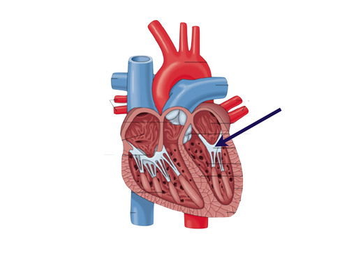
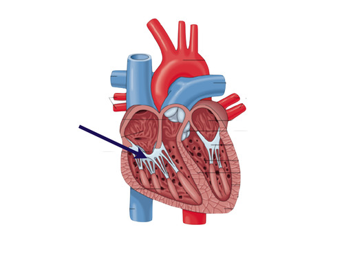
Artioventricular valves
aka AV valves that allow blood to go from atria to ventricles with out back flow
Mitral valve (left AV valve, bicuspid valve)
made up of two cusps and lies between left atria and ventricle
tricuspid valve (right AV valve)
made up of three cusps and lies between right atria and ventricle
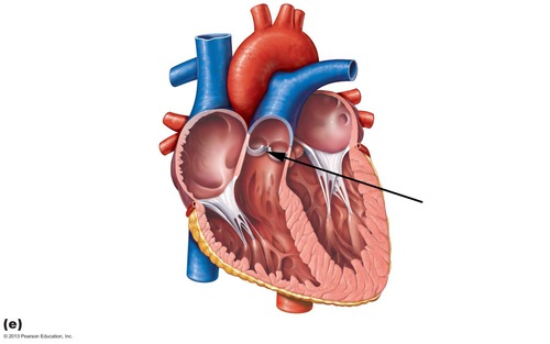
semilunar valves
aka SL valves that separate ventricles and arteries.
Aortic Smilunar Valve (left SL)
separates the left ventricle from the aorta
pulmonary semilunar valve (right SL)
heart valve opening from the right ventricle to the pulmonary artery
purpose of heart valves
prevent back flow of blood and to allow flow of blood from high to low pressure.
chordinae tendinae
tethers that hold the atrioventricular valves in place while the heart pumps blood
papilary muscles
muscles located in the ventricles of the heart
arteries
Where is blood pressure the highest?
veins
Where is blood pressure the lowest?
tissue cells in systemic capillaries.
Where is CO2 pressure high?
cardiac cycle
A complete heartbeat consisting of contraction and relaxation of both atria and both ventricles
Myogenic
Describes muscle tissue (heart muscle) that generates its own contractions.
Rhythm (heart)
depicts how quickly the heart beats due to its electrical activity
Force (heart)
depicts how hard the heart is beating duse to the activity of contractile cells.
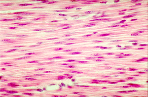
cardiac muscle
Involuntary muscle tissue found only in the heart that contains sarcomeres, gap junctions, and have action potentials longer than skeletal muscle cells.
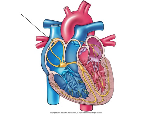
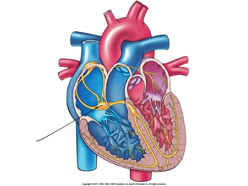
cardiac pacemaker cells
is the set rhythm of the heartbeat which contains nodes that provide action potentials during contractions using SA and AV nodes.
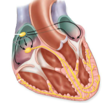
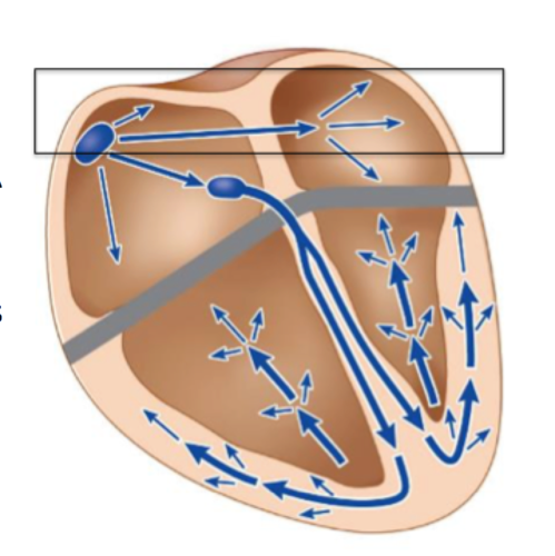
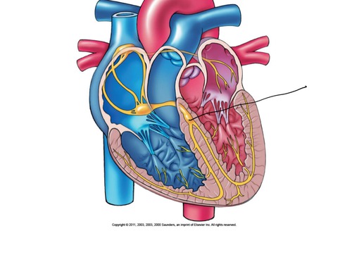
Sinoatrial Node (SA Node)
is in the the top right of the atrium and produces an action potential that makes both atria contract
Atriaventricular Node (AV Node)
is at the top of the ventricles that produces a 2nd action potential that makes both ventricles contract.
cardiac conduction fibers
is the electrical wiring of the heart that allow the action potentials (transmit rhythm) that are produced from the nodes to propagate throughout the heart. ( Don't respond or produce action potentials)
internodal pathway
conduction pathway from the SA node to the AV node
interatrial pathway
a pathway of specialized, cardiac cells that conducts pacemaker activity from the right atrium to the left atrium
Purjunkie fibers
specialized fibers that stimulate ventricular syncytium
Contractile cells
are cells that generate the contractile force of the heart.
Bundle of His (AV bundle)
located next to the AV node; provides the transfer of the electrical impulse from the atria to the ventricles
AV nodal delay
The delay in impulse transmission between the atria and ventricles at the AV node, which allows enough time for the atria to become completely filled with blood and contract, emptying their contents into the ventricles, before ventricular depolarization and contraction occur
bottom up
What Is the direction of ventricular contraction ?
Pacemaker cells
a group of cells located in the right atrium that sends out signals that make the heart muscle contract and that regulates heartbeat rate
pacemaker rapid depolarization
occurs after threshold, where Na+ and L-type voltage-gated channels opens where Na+ and Ca2+ causes a steep rise.
pacemaker potential
A self-initiating action potential that triggers the START action potentials in the heart.
Pacemaker action potential
occurs in SA and AV node lacks phases 1 and 2 of cardiac action potential
pacemaker repolarization
is where L-type Ca2+ channels close, Na+ channels to inactivate and cause K+ channels to open where K+ leaves the cell leading to hyperpolarization to the next action potential.
pacemaker threshold
minimal voltage needed for capture (-40mV)