BIO211 ~ Anatomy Kinesiology
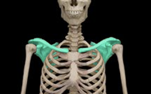
Shoulder Girdle
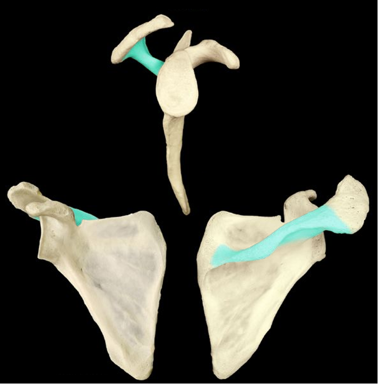
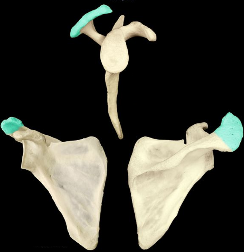
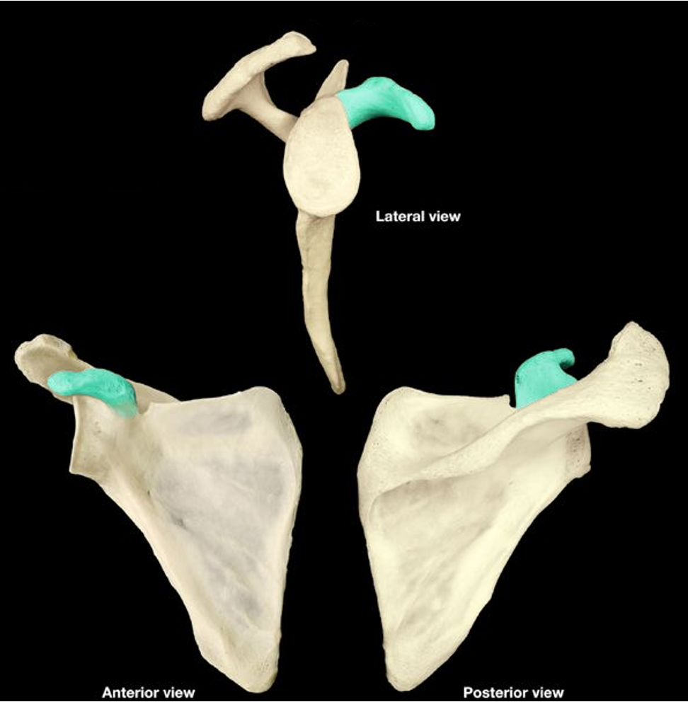
Clavicle & Scapula
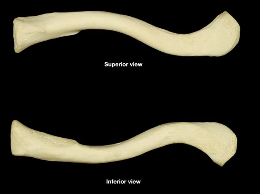
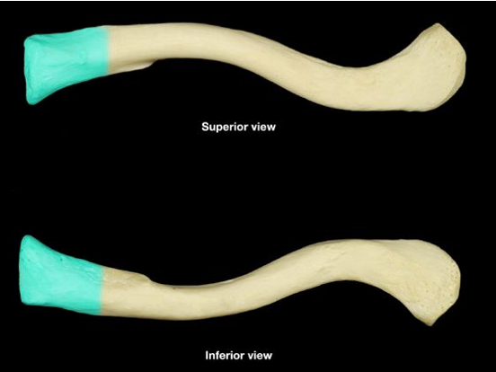
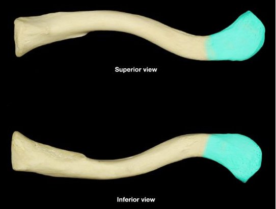
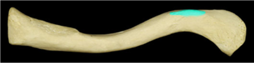
Clavicle
collar bone
Medial (sternal) end of clavicle
the end of the clavicle that articulates with the sternum
Lateral (acromial) end of clavicle
the end of the clavicle that articulates with the scapula
Conoid Tubercle
the bump on the end of the clavicle (posterior, inferiror & lateral)
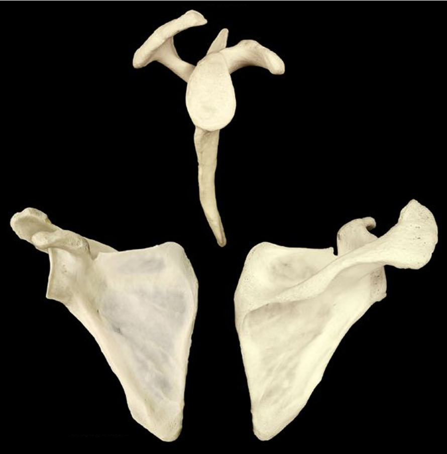
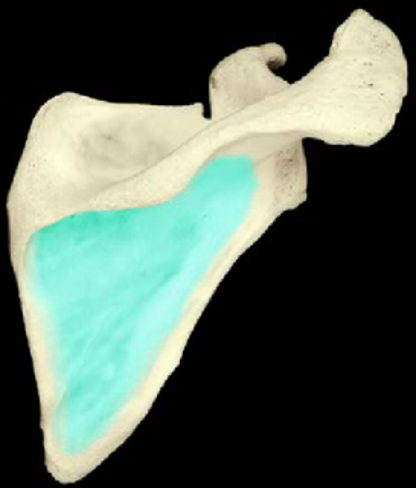
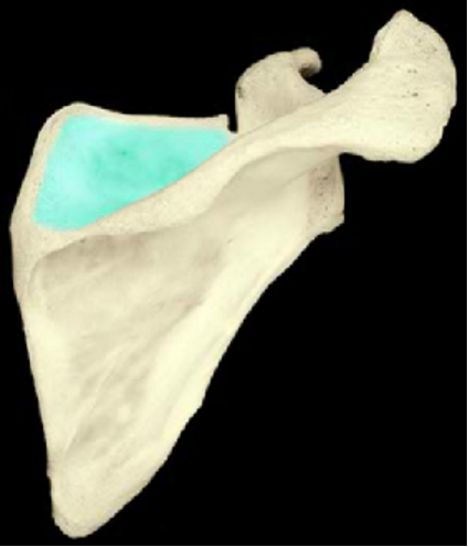
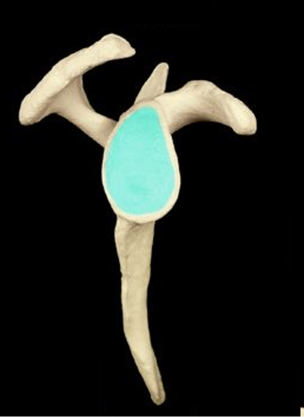
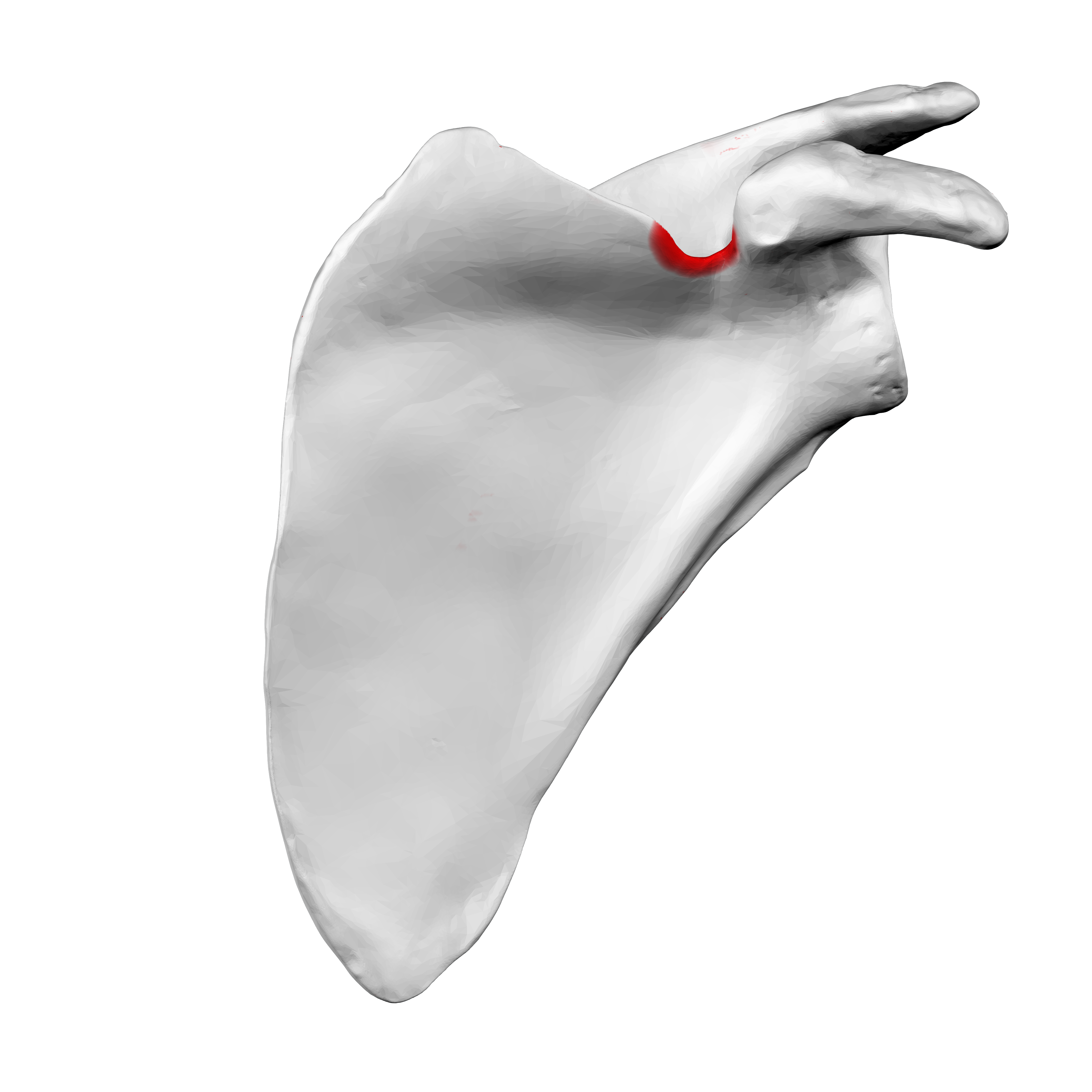
Scapula
shoulder blade
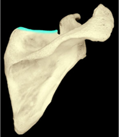
Superior border of scapula
The border of the scapula that stretches across the top
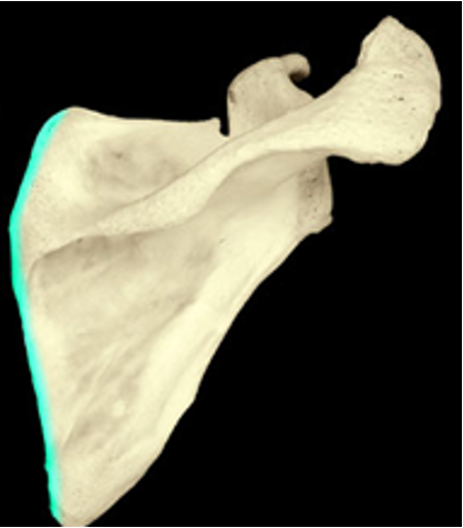
Middle border of scapula
The border of the scapula that is parallel to the vertebral column
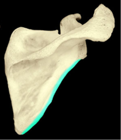
Lateral border of scapula
the border of the scapula that points at the shoulder
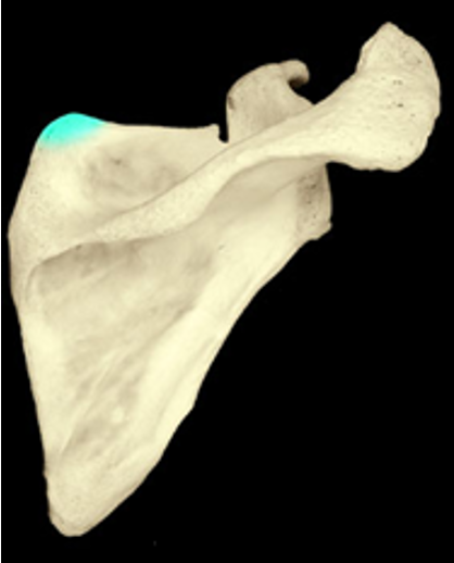
Superior angle of scapula
the angle of the scapula where the medial and superior borders meet
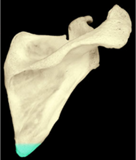
Inferior angle of scapula
the pointy inferior edge of the scapula
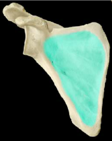
Subscapular fossa
the flat area on the anterior surface of the scapula
Scapular spine
The raised ridge on the posterior side of the scapula
Acromion
the lateral end/tip of the scapular spine that articulates with the scapula
Infraspinous fossa
the flat area inferior to the scapular spine
Supraspinous fossa
the flat area superior to the scapular spine
Coracoid process
the projection on the anterior side of the scapula
Glenoid cavity
the lateral indentation the the scapula that forms the socket of the shoulder joint
Suprascapular notch
the small notch at the top of the scapula
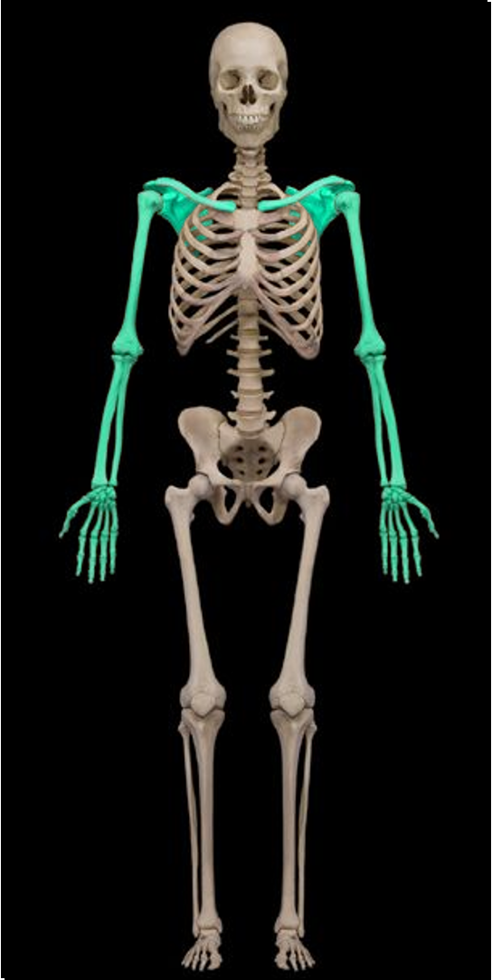
Upper limb
Arm, forearm, and hand
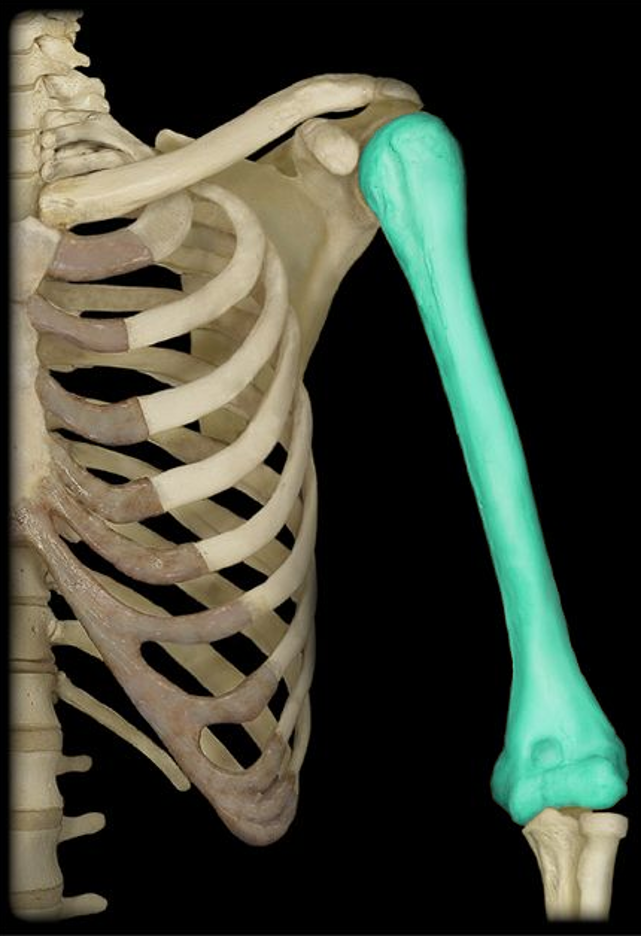
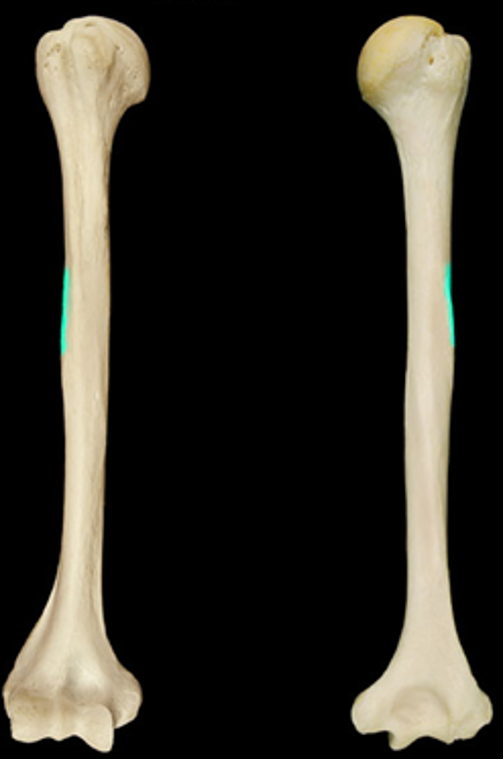
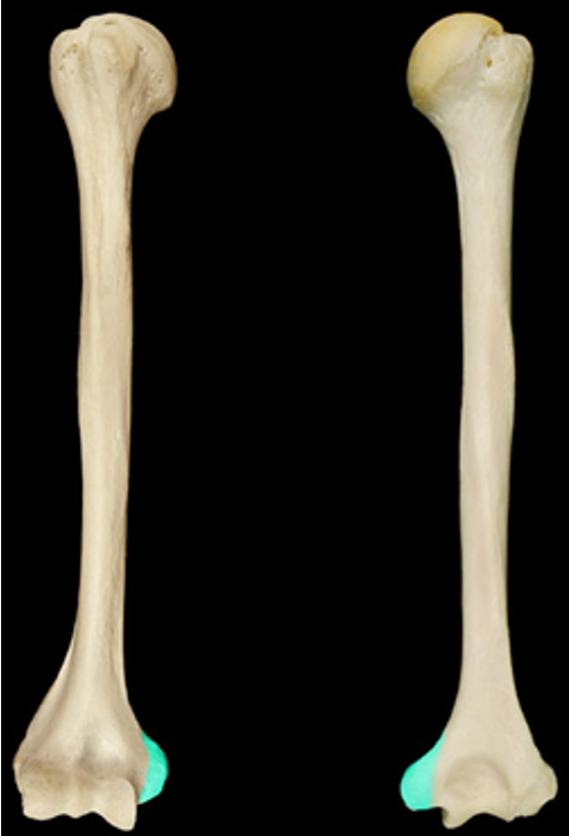
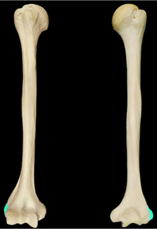
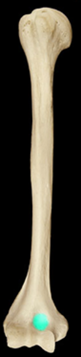
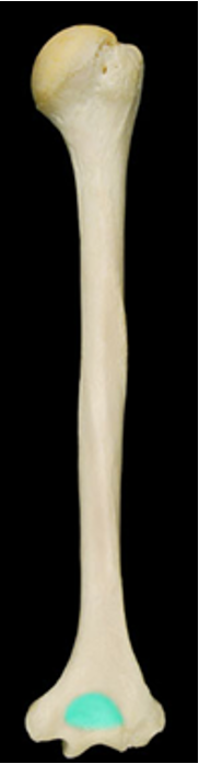
Humerus
Large upper bone of the upper limb
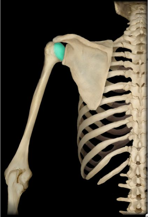
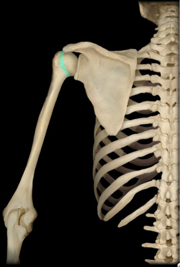
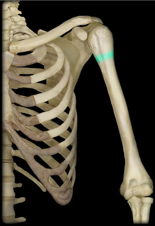
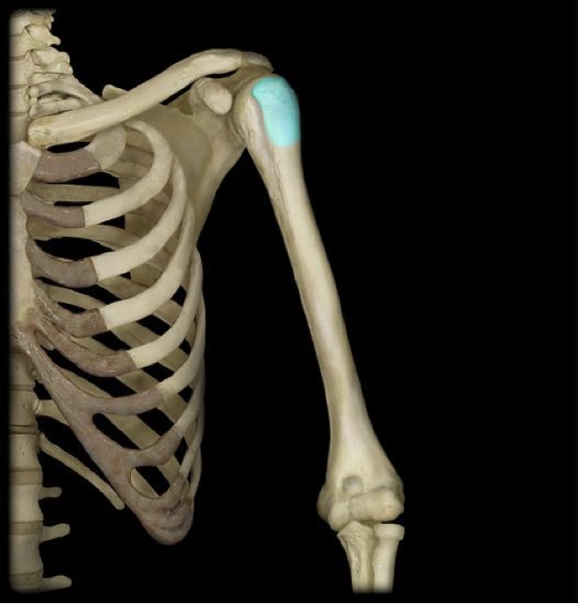
Humerus head
the proximal rounded end of the humerus; the ball of the shoulder joint
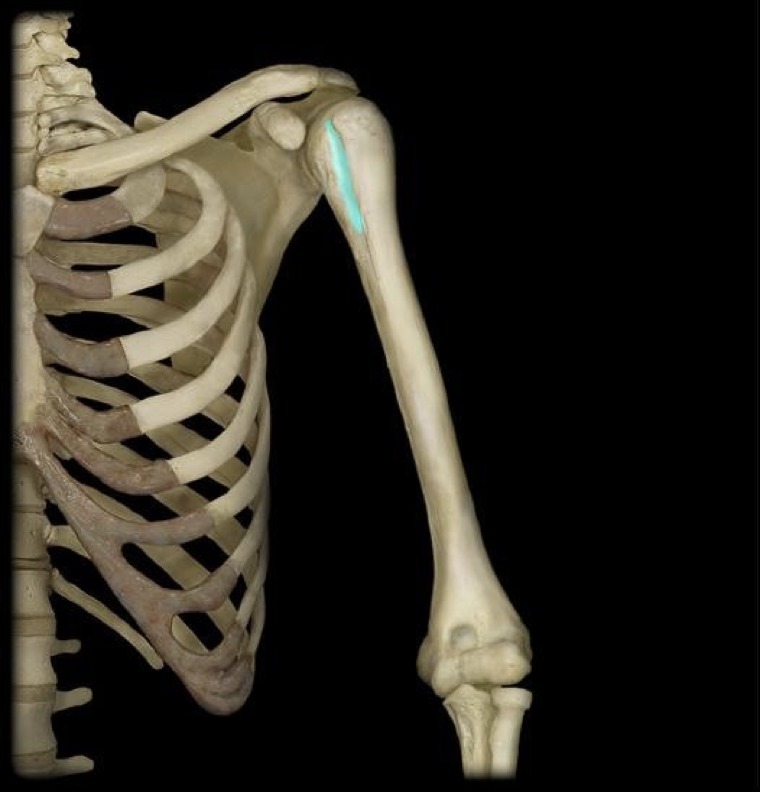
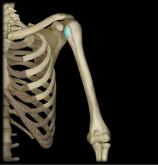
Intertubercular groove
groove between the tubercles of the humerus
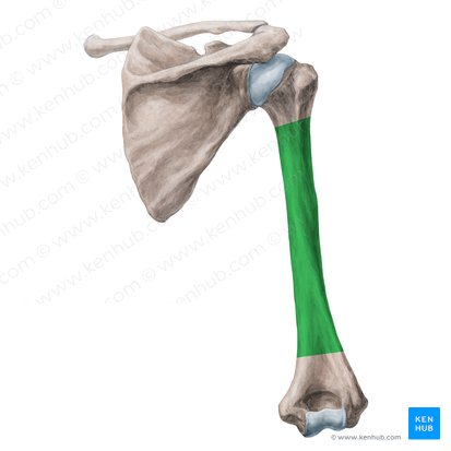
Humerus body/shaft
main portion of the humerus
Anatomical neck of humerus
the narrow region just below the head of the humerus
Surgical neck of humerus
the narrow region beneath all the superior structures of the humerus
Greater tubercle
raised area just opposite/lateral to the head of the humerus
Lesser tubercle
small raised area between the head the the greater tubercle of the humerus
Deltoid tuberosity
rough area distal and lateral, midway down from the greater tubercle
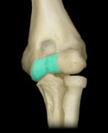
Trochlea
pulley shaped area on the medial side of the distal humerus
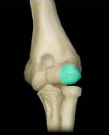
Capitulum
rounded condyle on the lateral side of the distal humerus
Medial epicondyle (humerus)
the large, pronounced, medial bump on the distal end of the humerus
Lateral epicondyle (humerus)
the less pronounced bump on the lateral side of the distal humerus
Coronoid fossa
An indentation just above the trochlea on the anterior surface of distal humerus
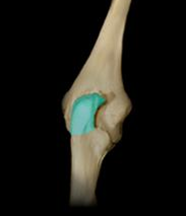
Olecranon fossa
a large indentation on the posterior surface of the distal humerus
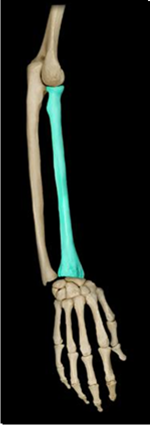
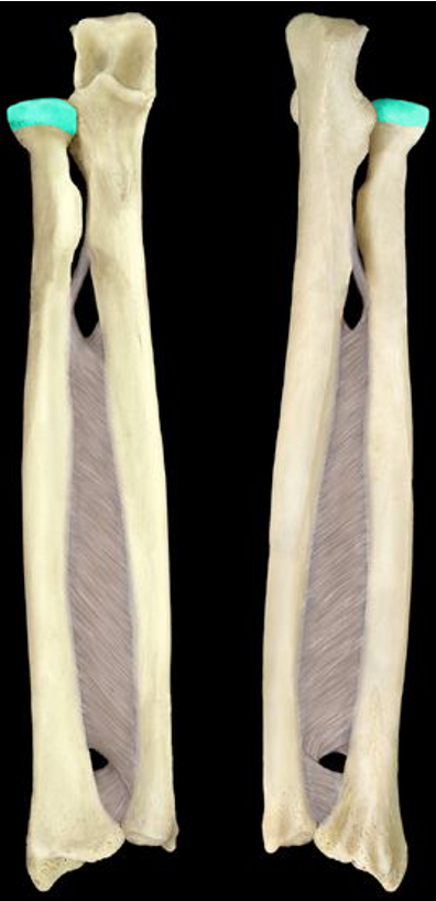
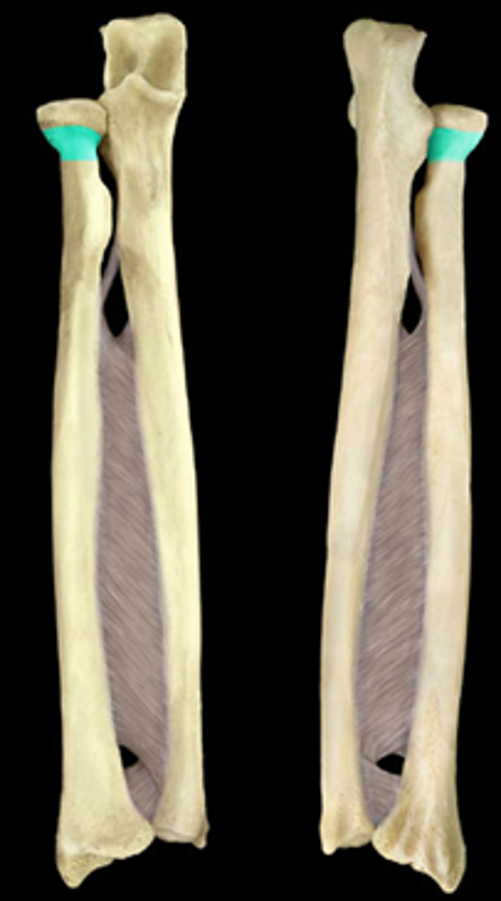
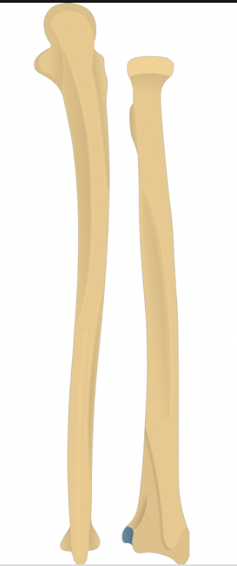
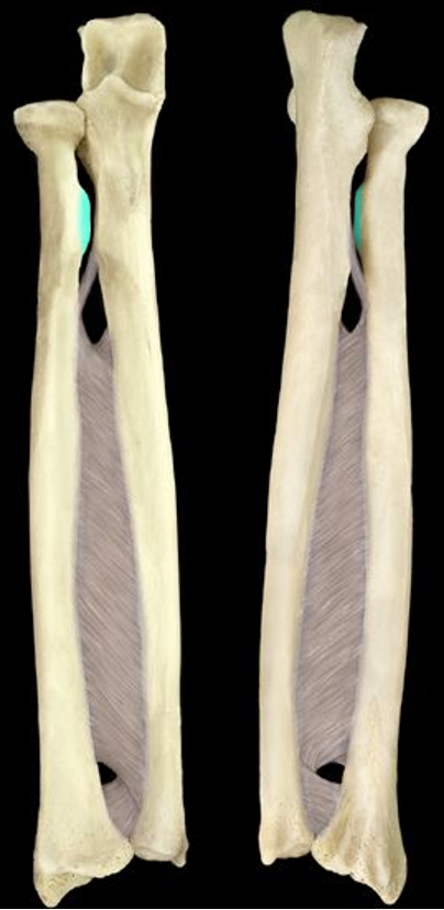
Radius
lower arm bone of the medial/thumb side
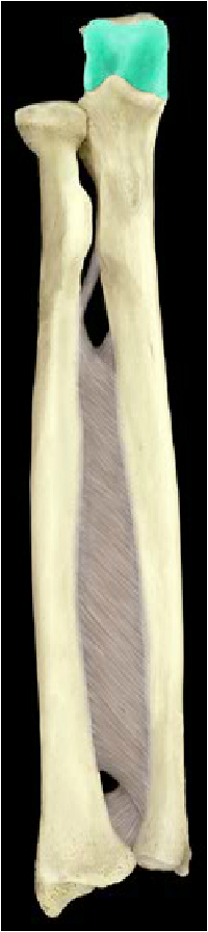
Head of radius
the proximal/superior end of the radius; disc-like
Neck of radius
narrow area beneath the head of the radius
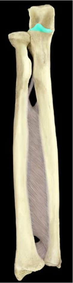
Ulnar notch
an indentation next to the styloid process on the distal end of the radius
Radial tuberosity
a raised area on the proximal radius that faces the ulna
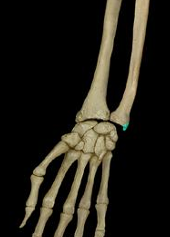
Styloid process of radius
a pointy projection at the distal end of the radius; points to the thumb
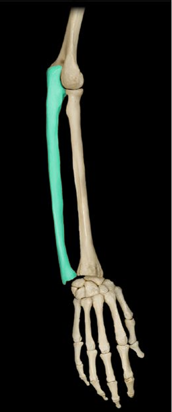
Ulna
lower arm bone of the lateral/pinky side
Head of ulna
distal end of the ulna
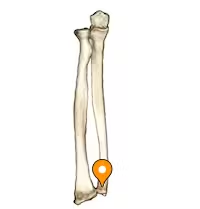
Olecranon
the top of the C shape on the proximal ulna that inserts in the olecranon fossa
Coronoid process
the bottom of the C shape on the proximal ulna that interacts the the coronoid fossa
Trochlear notch
a notch between the olecranon and coronoid process
Radial notch
the contact point between the ulna and radius on the proximal ulna
Styloid process of ulna
a pointy projection at the distal end of the ulna; points to the pinky
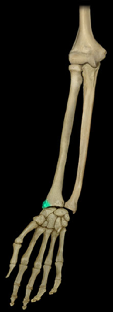
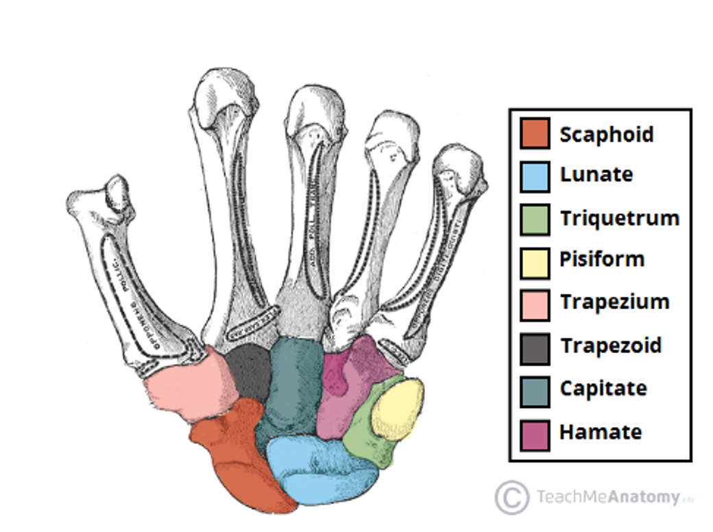
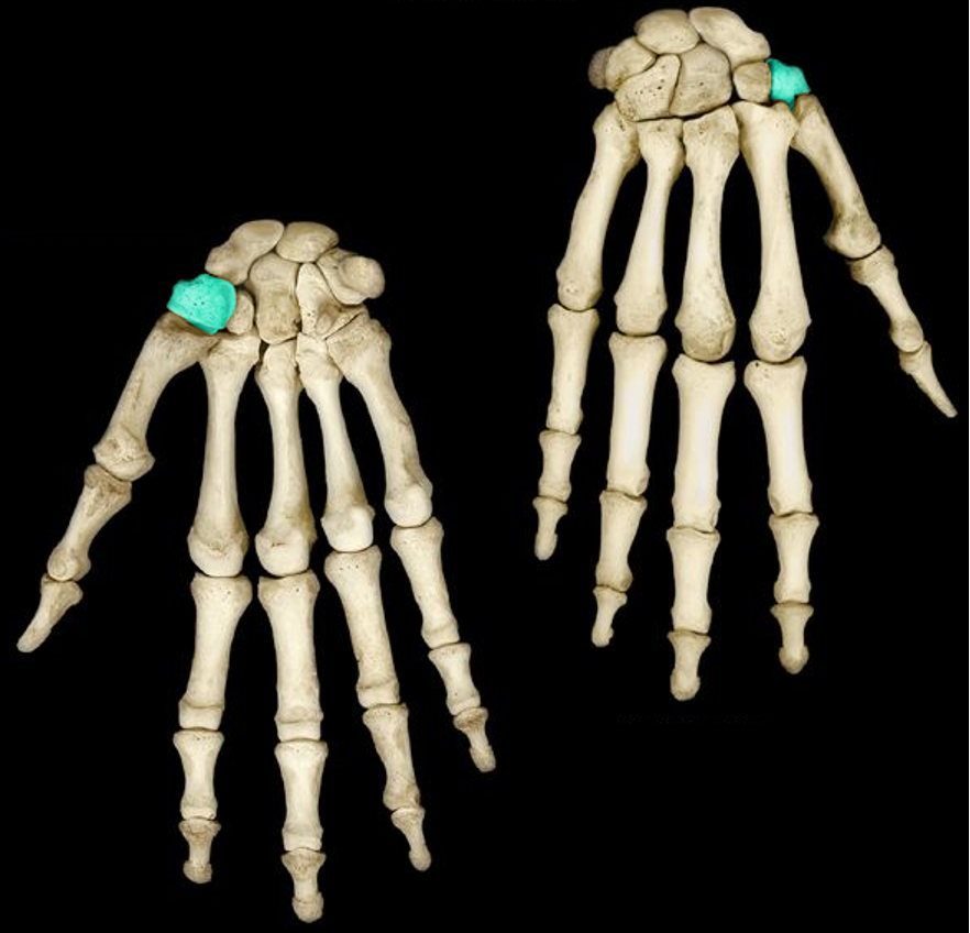
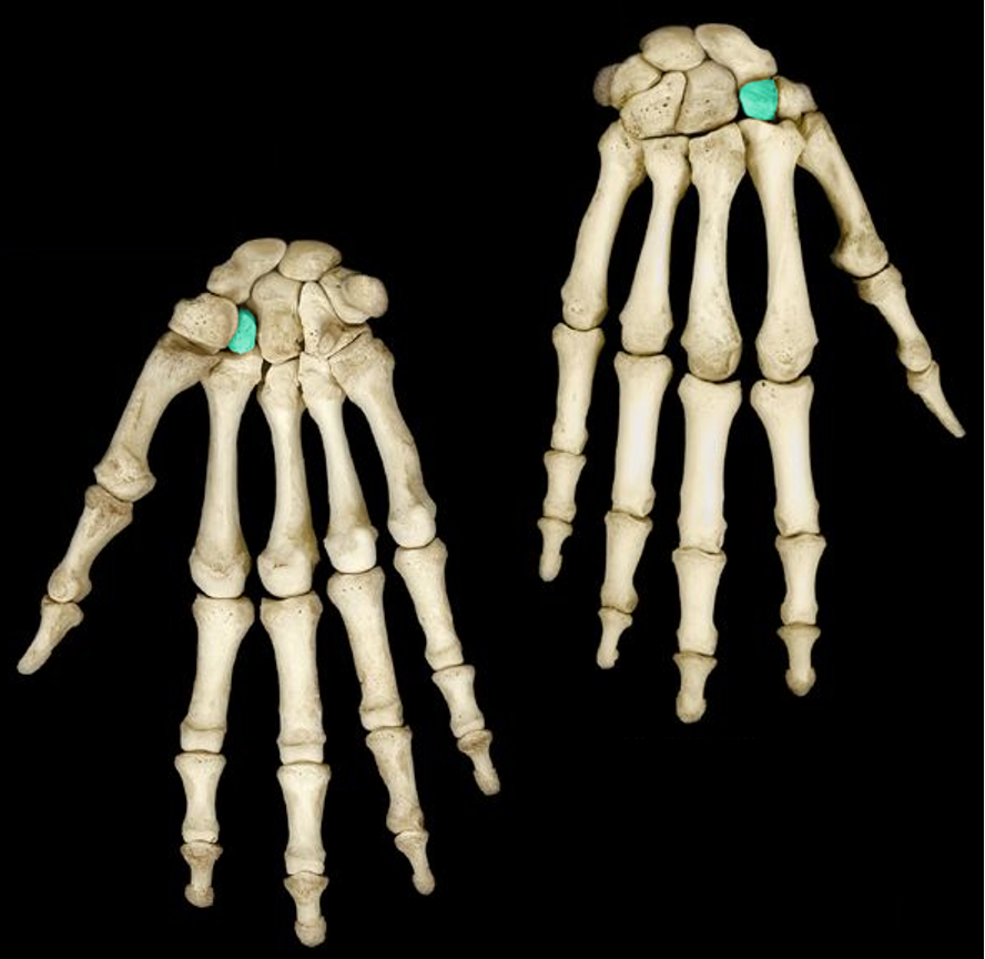
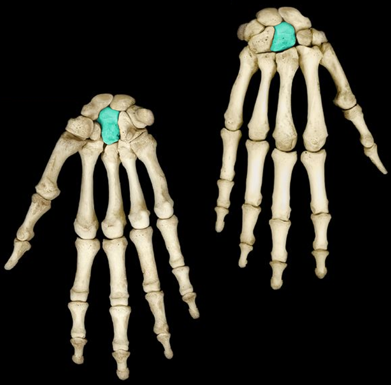
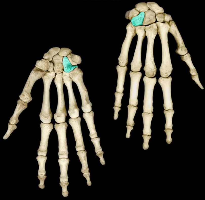
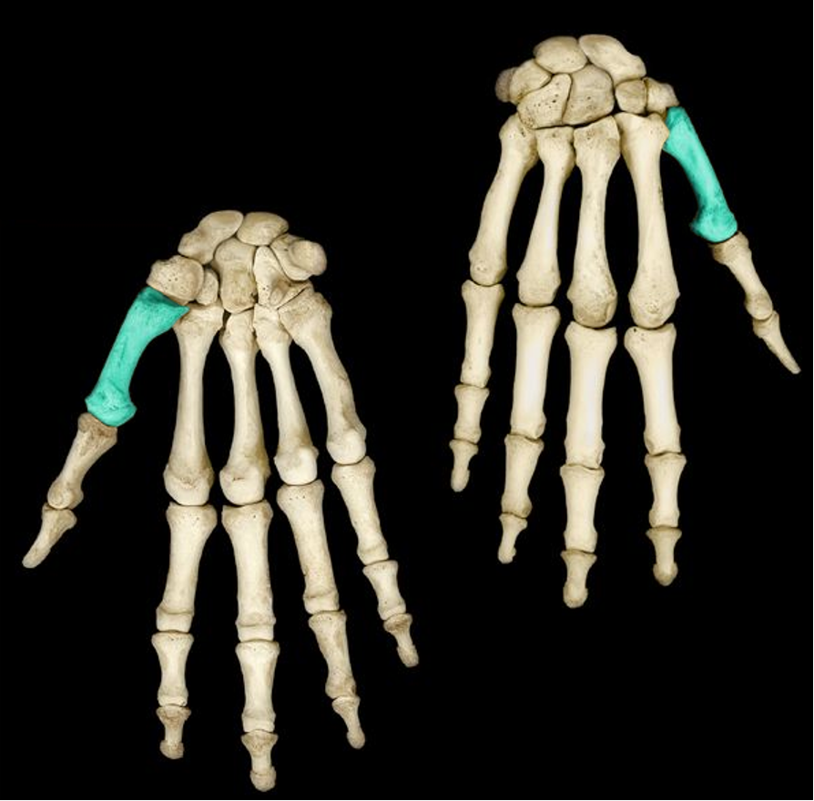
Carpals
8 small bones in the proximal hand
Pneumonic device for carpals (thumb to pinky)
She Looks Too Pretty = Scaphoid Lunate Triquetrum Pisiform
Try To Catch Her = Trapezium Trapezoid Capitate Hamate
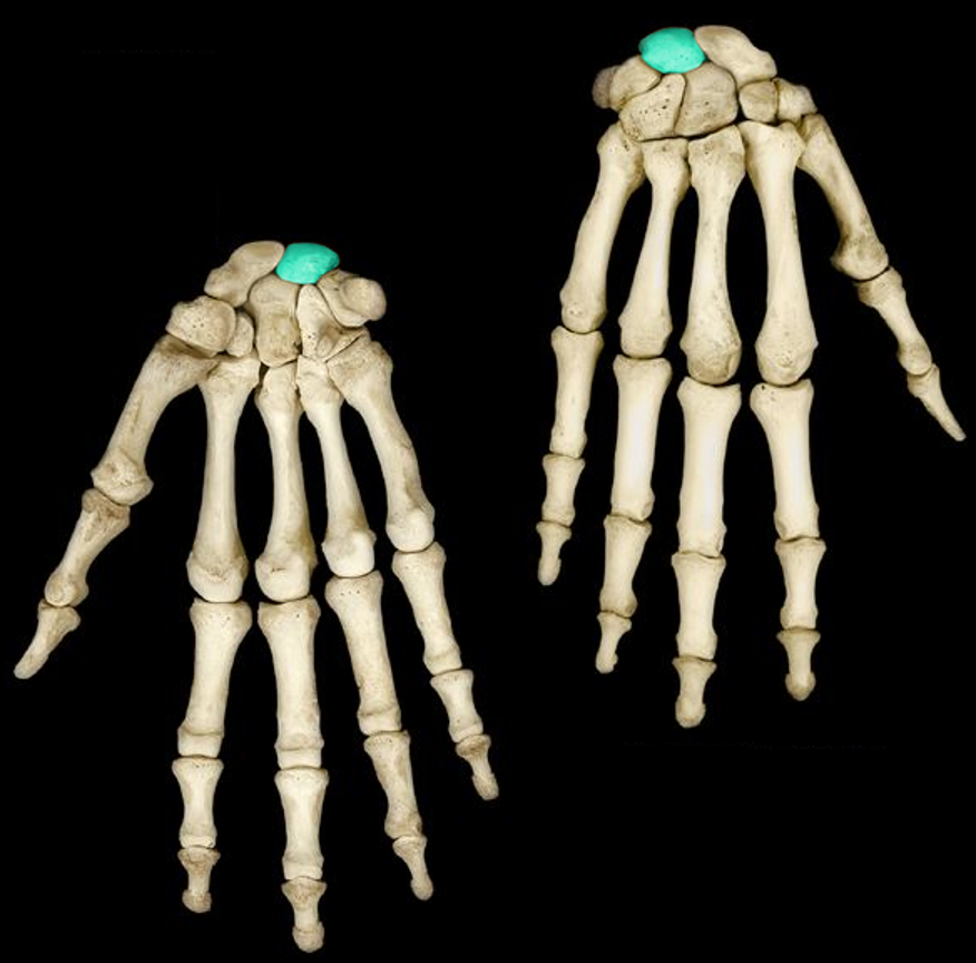
Scaphoid
“She”
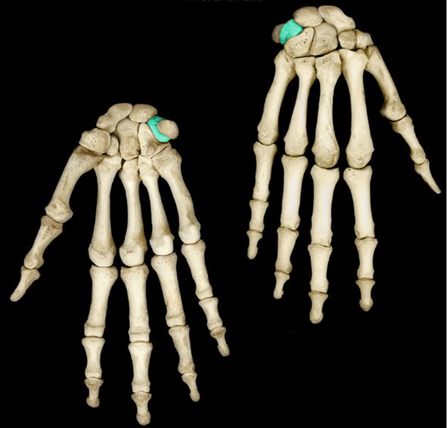
Lunate
“Looks”
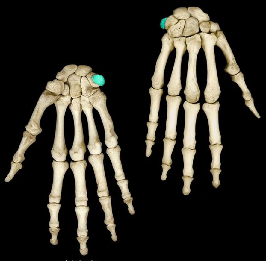
Triquetrum
“Too”
Pisiform
“Pretty”
Trapezium
“Try”
Trapezoid
“To”
Capitate
“Catch” - big: captivates
Hamate
“Her” - hood shaped
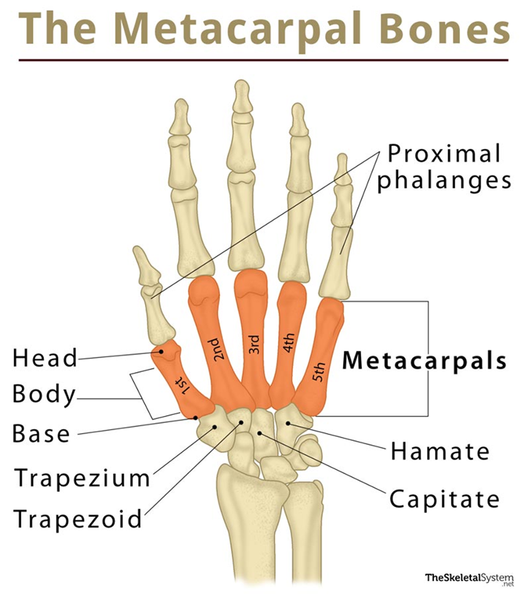
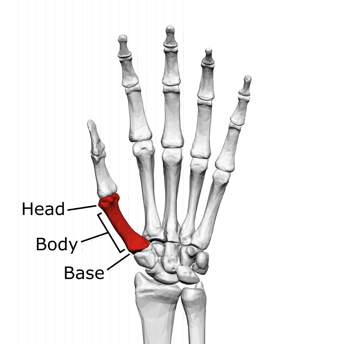
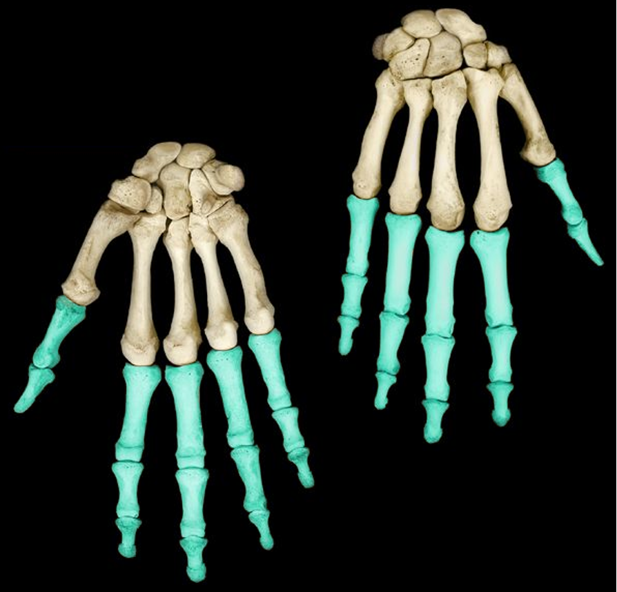
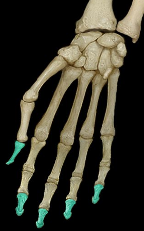
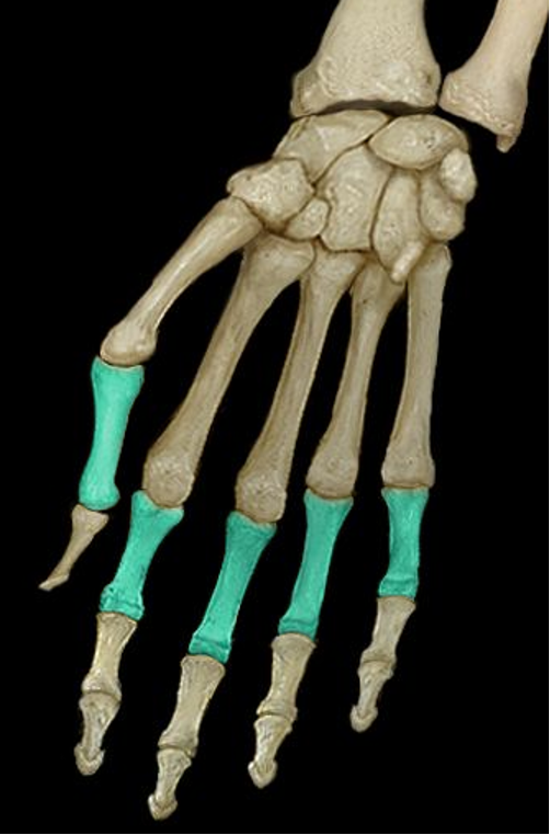
Metacarpals
number I II III IIV V from thumb side to pinky side
Proximal base (metacarpals)
bottom of the metacarpals
Intermediate shaft (body) (metacarpals)
middle of the metacarpals
Distal head (metacarpals)
top of the metacarpals
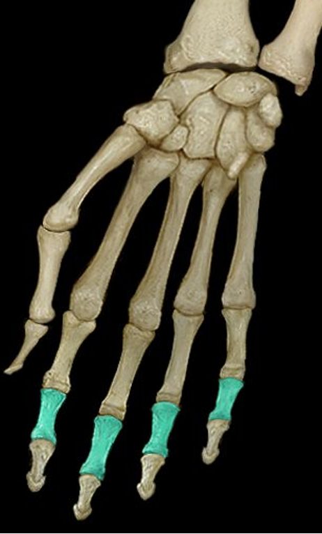
Phalanges (phalanx)
Numbered I II III I V from thumb side to pinky side
Distal phalanx (hand)
tip of the finger
Middle phalanx (hand)
Middle of the finger; not found on the thumb
Proximal phalanx (hand)
Articulates with the head of the metacarpal
Pollex
thumb; has no middle phalanx
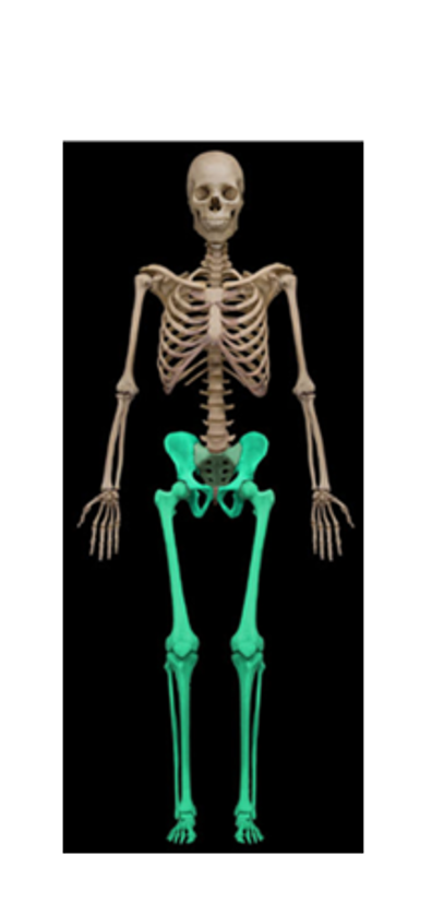
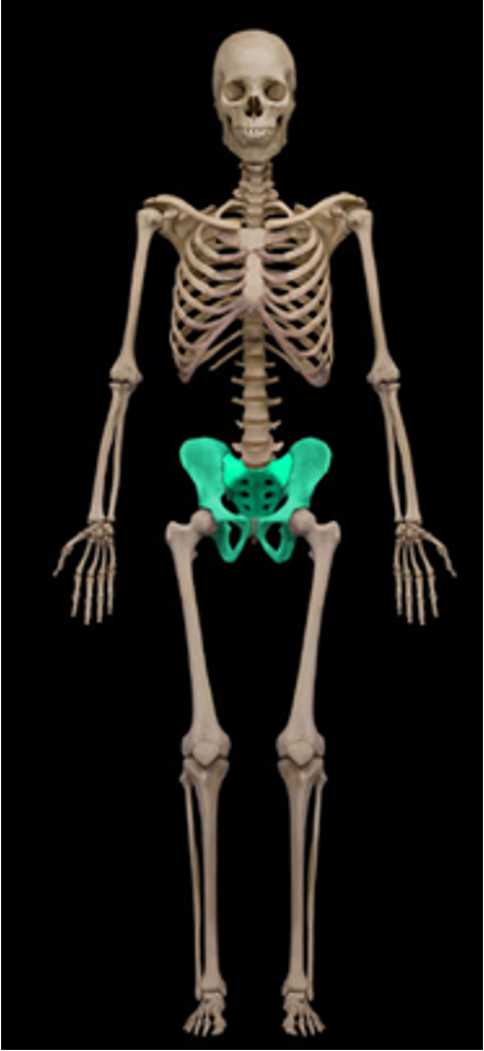
Lower limbs
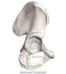
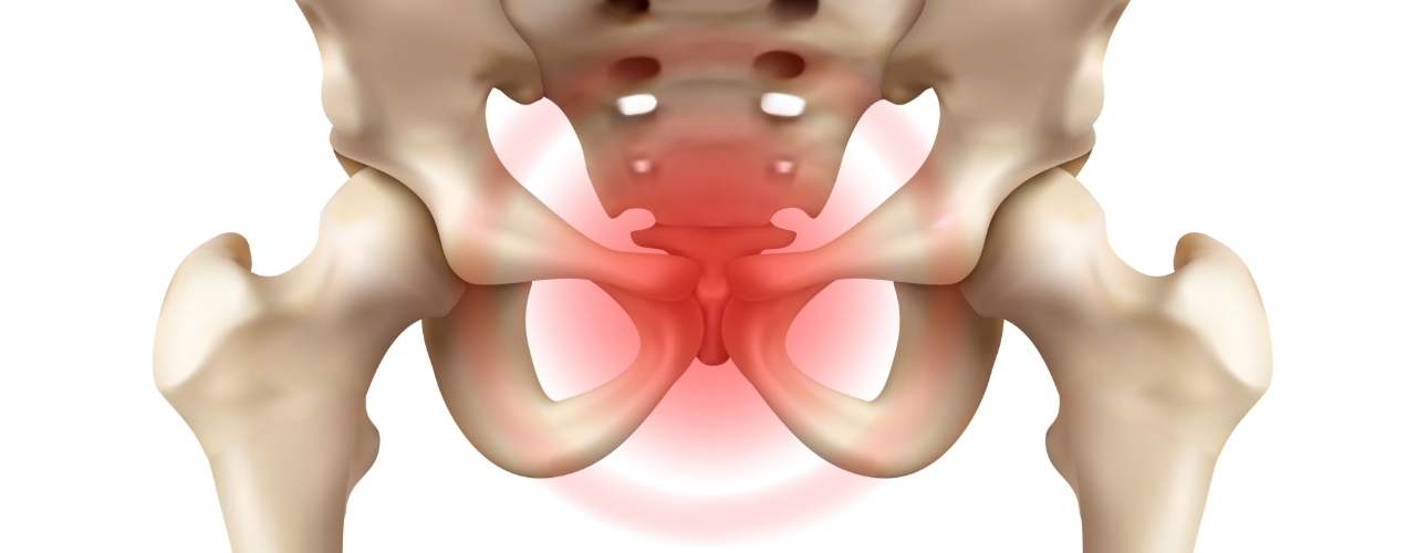
Pelvic girdle/coxal bone
pelvis
os coxa/ossa coxae
forms pelvic inlet with the sacrum and coccyx
pubic symphysis
joint at the base where ossa coxae fuse; amphiarthrodial
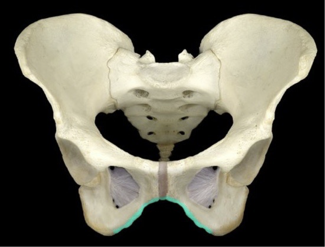
pubic arch (subpubic angle)
inferior angle of the pelvis
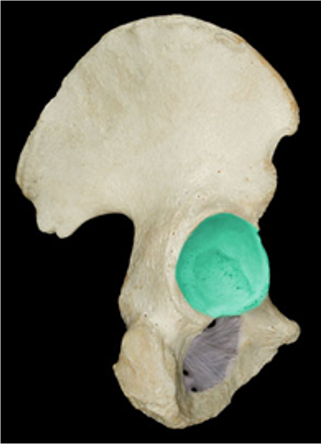
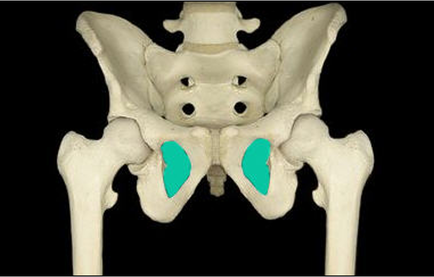
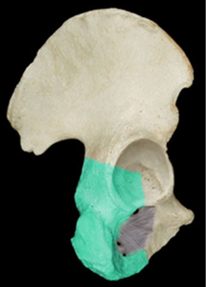
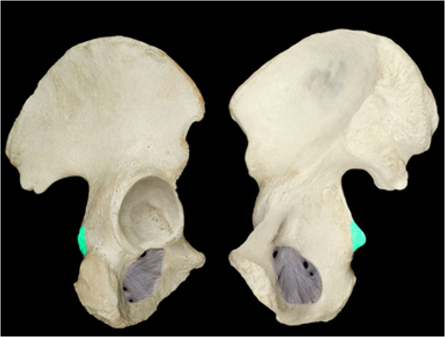
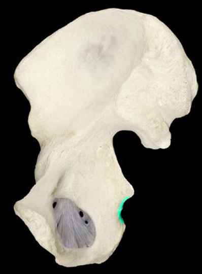
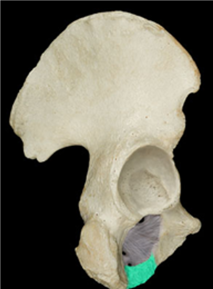
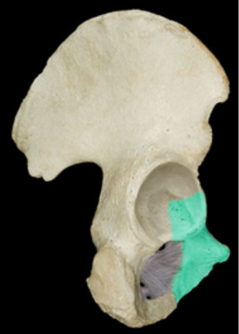
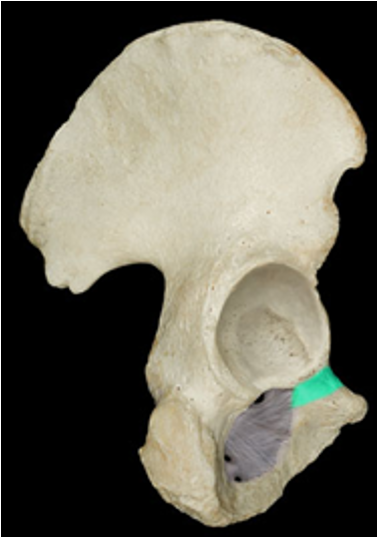
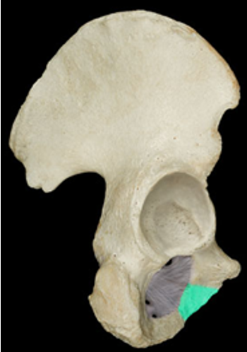
Acetabulum
deep socket of the hip joint
Obturator foramen
large inferior holes for tendons/muscles to pass through
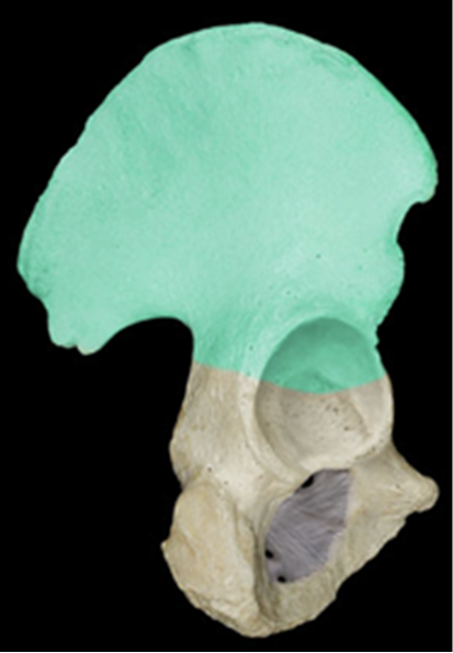
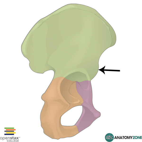
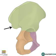
ilium
large upper region of the os coxa; broad and superior
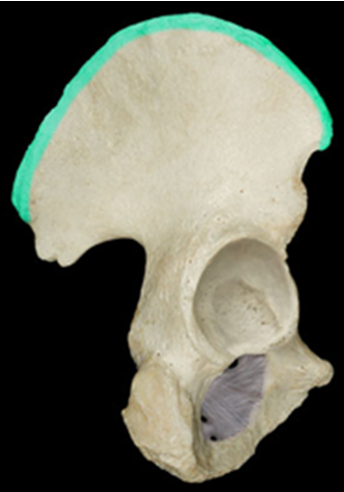
Iliac crest
broad upper boarder of the os coxa
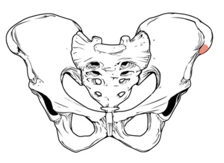
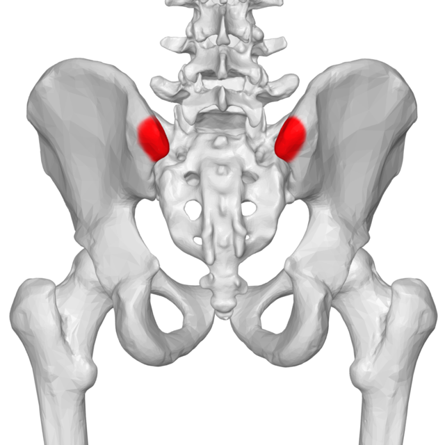
Anterior superior Iliac spine
cliff-like edge on the anterior side of iliac crest
Posterior superior iliac spine
cliff-like edge on the posterior side of the iliac crest
Anterior inferior iliac spine
inferior to the anterior superior iliac spine, below a the indentation
Posterior inferior iliac spine
inferior to the posterior superior iliac spine, below the small bump
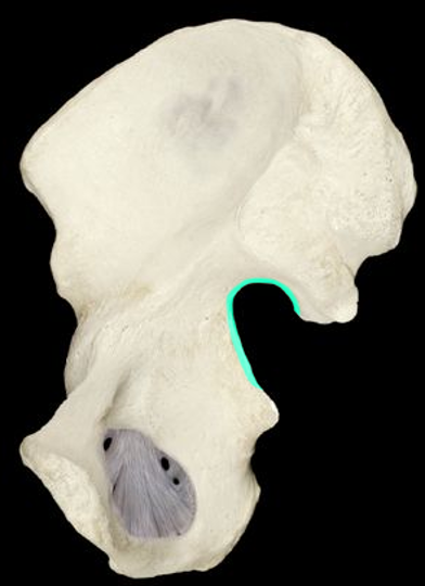
Greater sciatic notch
large notch inferior to the posterior spines
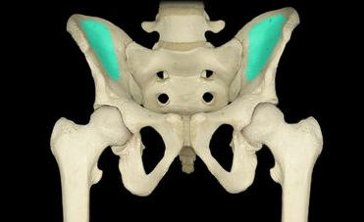
Iliac fossa
large, smooth, concave surface ob the internal surface of the ilium
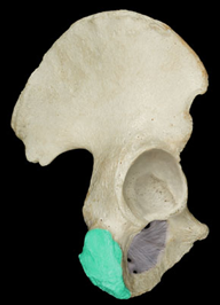
Ischium
inferior, posterior section that contacts a surface when sitting
Ischial tuberosity
rough, chunky bump of the ischium - “sit bump”
Ischial spine
projection between the sciatic notches
Lesser sciatic notch
small notch inferior to the greater sciatic notch
Ischial ramus
inferior/bottom edge of the obturator foramen
Pubis
inferior, anterior section; pubic region
Superior ramus
continuation of the inferior ramus of the obturator foramen
Inferior ramus
continuation of the ischial ramus around the obturator foramen
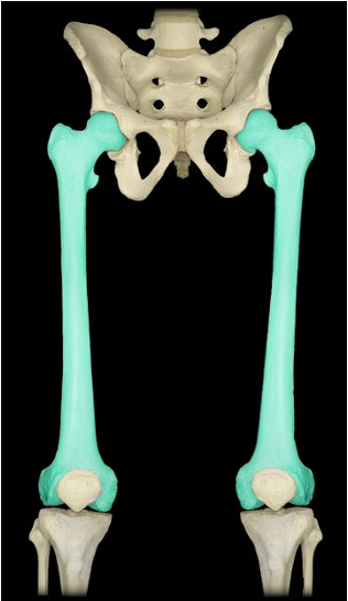
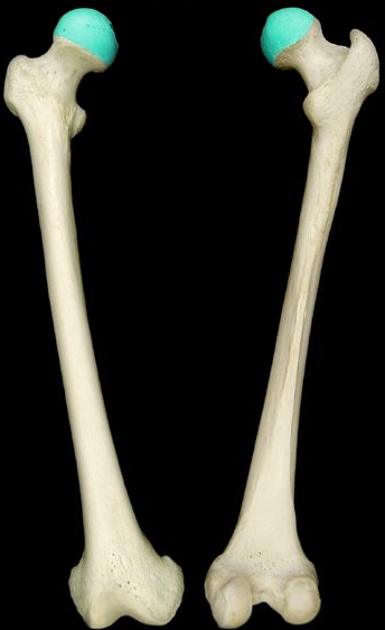
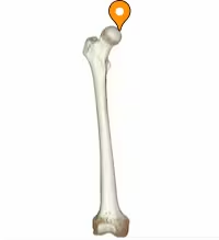
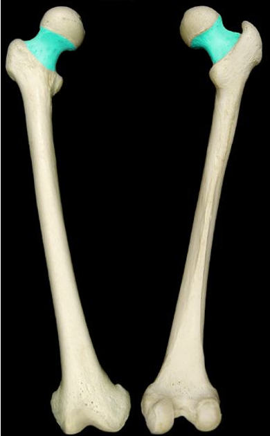
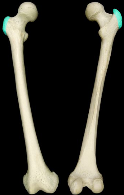
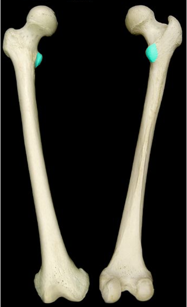
Femur
largest/heaviest bone; upper leg bone
Femur head
proximal end of the femur; ball of the hip joint
Fovea capitis
small indentation in femur head
Femur Neck
narrow structure just inferior to the head of the femur
Greater trochanter
small projection opposite the femur head (lateral)
Lesser trochanter
small projection on the medial/head side of the femur
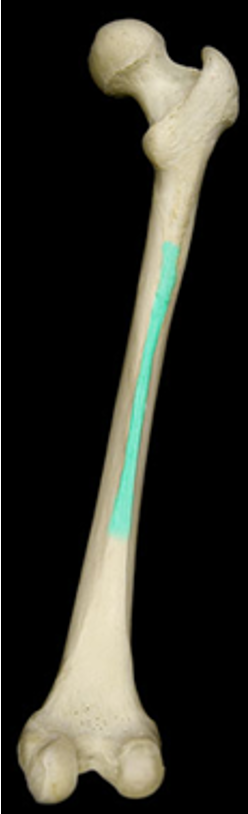
Gluteal tuberosity
rough area just inferior of the greater trochanter
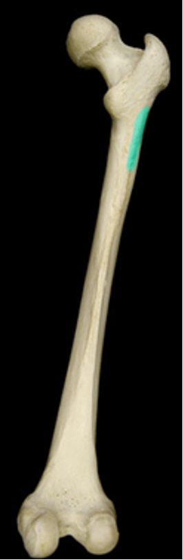
Linea aspera
long raised ridge along the shaft of the femur
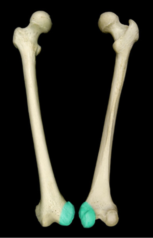
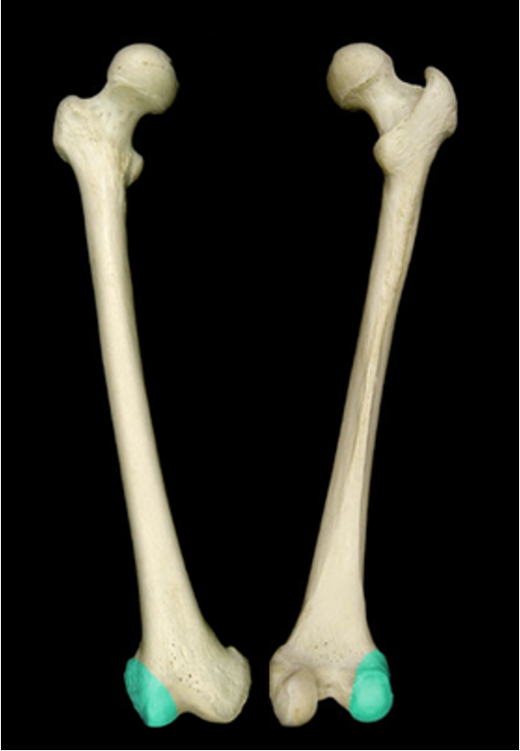
Medial condyle (femur)
smooth knob-like surface on the medial femur
Lateral condyle (femur)
smooth knob-like on the lateral femur