Looks like no one added any tags here yet for you.
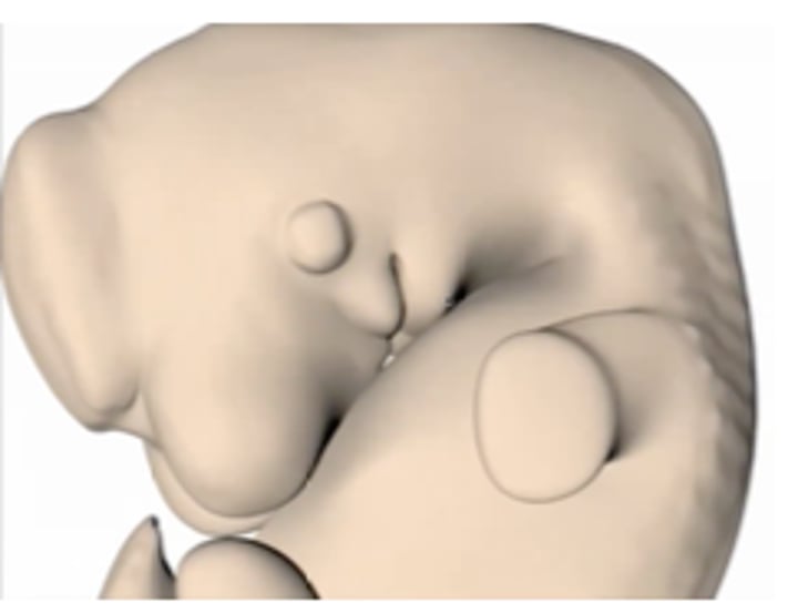
at what week of gestation does tooth formation begin?
week 6
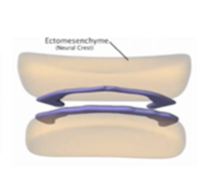
mesenchymal cell differentiation in developing jaws of embryo
upr/lwr jaw develop from max. and mand. processes of 1st pharyngeal arches respectively
stages of early tooth development
bud, cap, bell
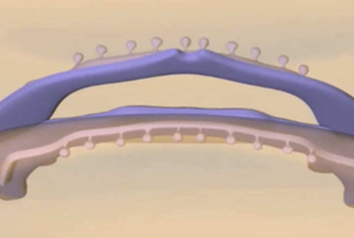
dental lamina
growth from the oral epithelium that gives rise to the tooth buds
Thickened band of oral epithelium that follows the curve of each developing arch
beginning of tooth development
forms max and mand arch (at same time)
where are buds first seen?
along dental lamina; buds will eventually become teeth
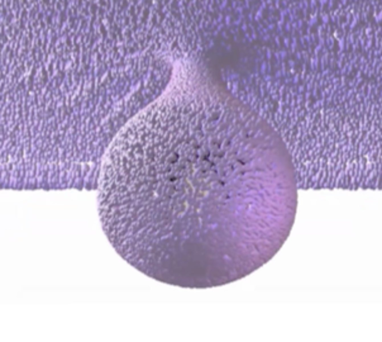
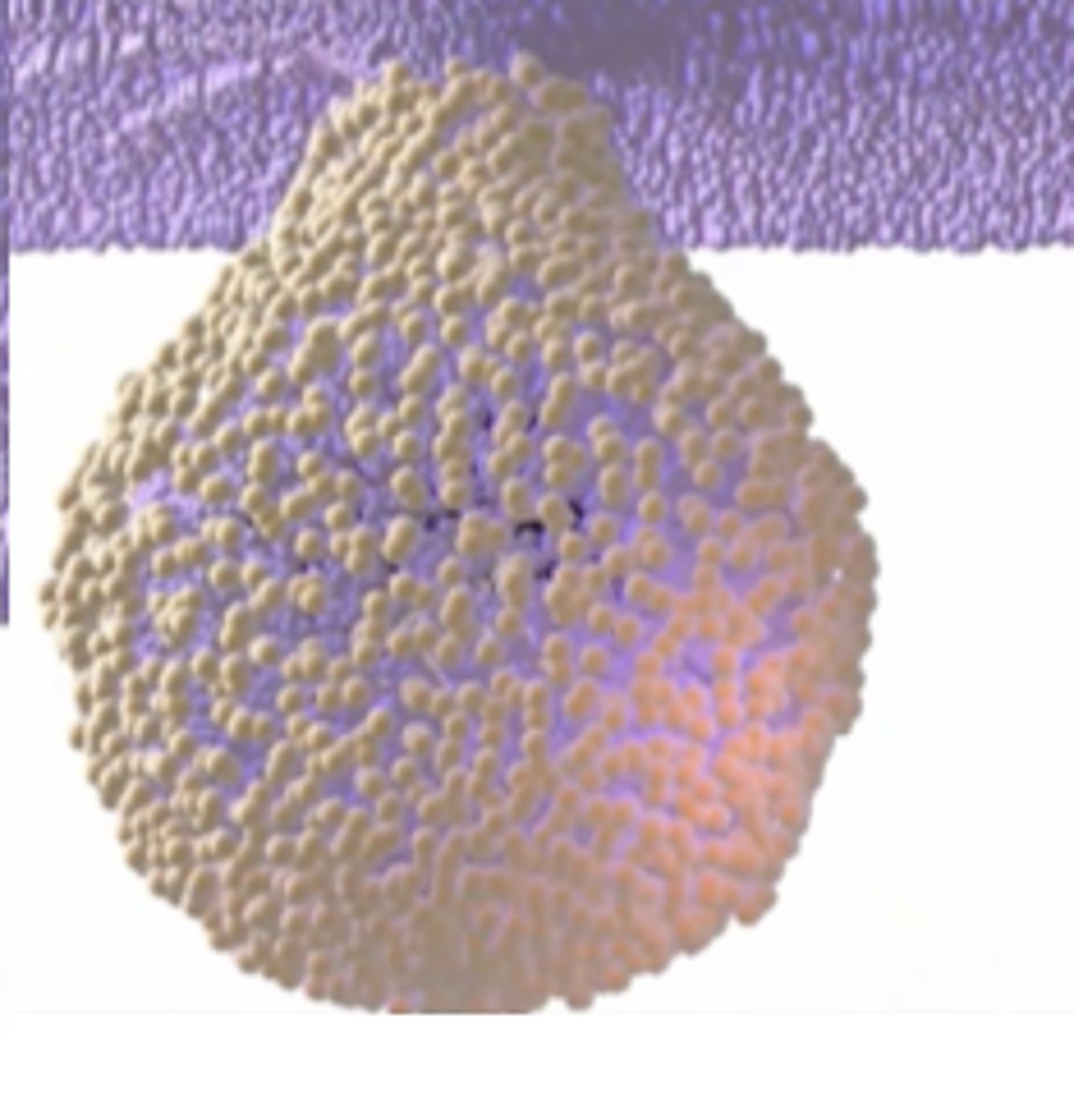
bud stage
first stage to create each single tooth
the first stage of odontogenesis, initiation of the tooth begins
second stage of tooth development marked by growth of dental lamina into buds
bud
mesenchymal cells move towards bud
bud folds inward (leading to cap stage)
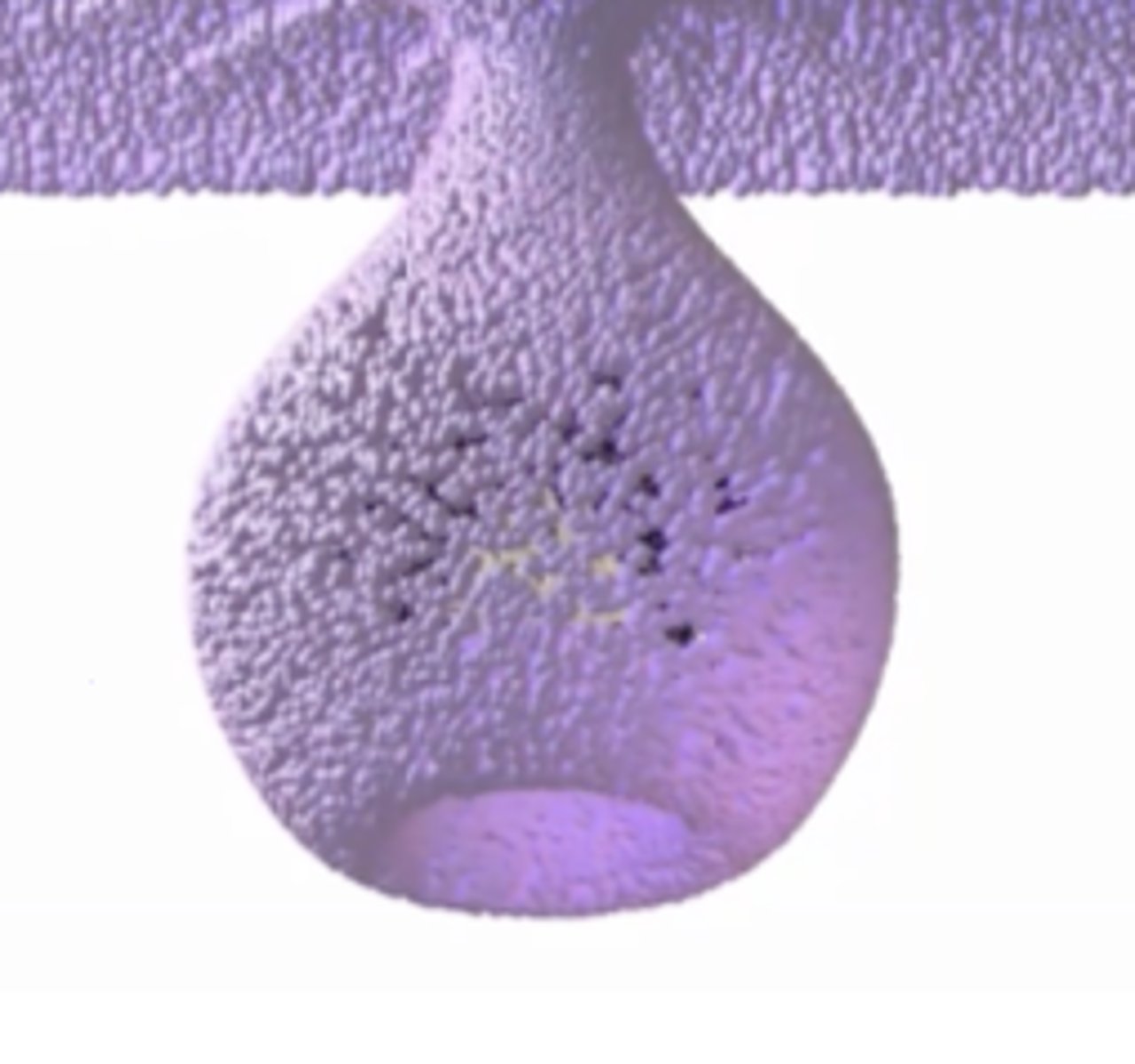
what is the beginning of the cap stage?
when the ingrowth is seen, proliferation of cells and the bud folds inward; beginning of dental structure formation
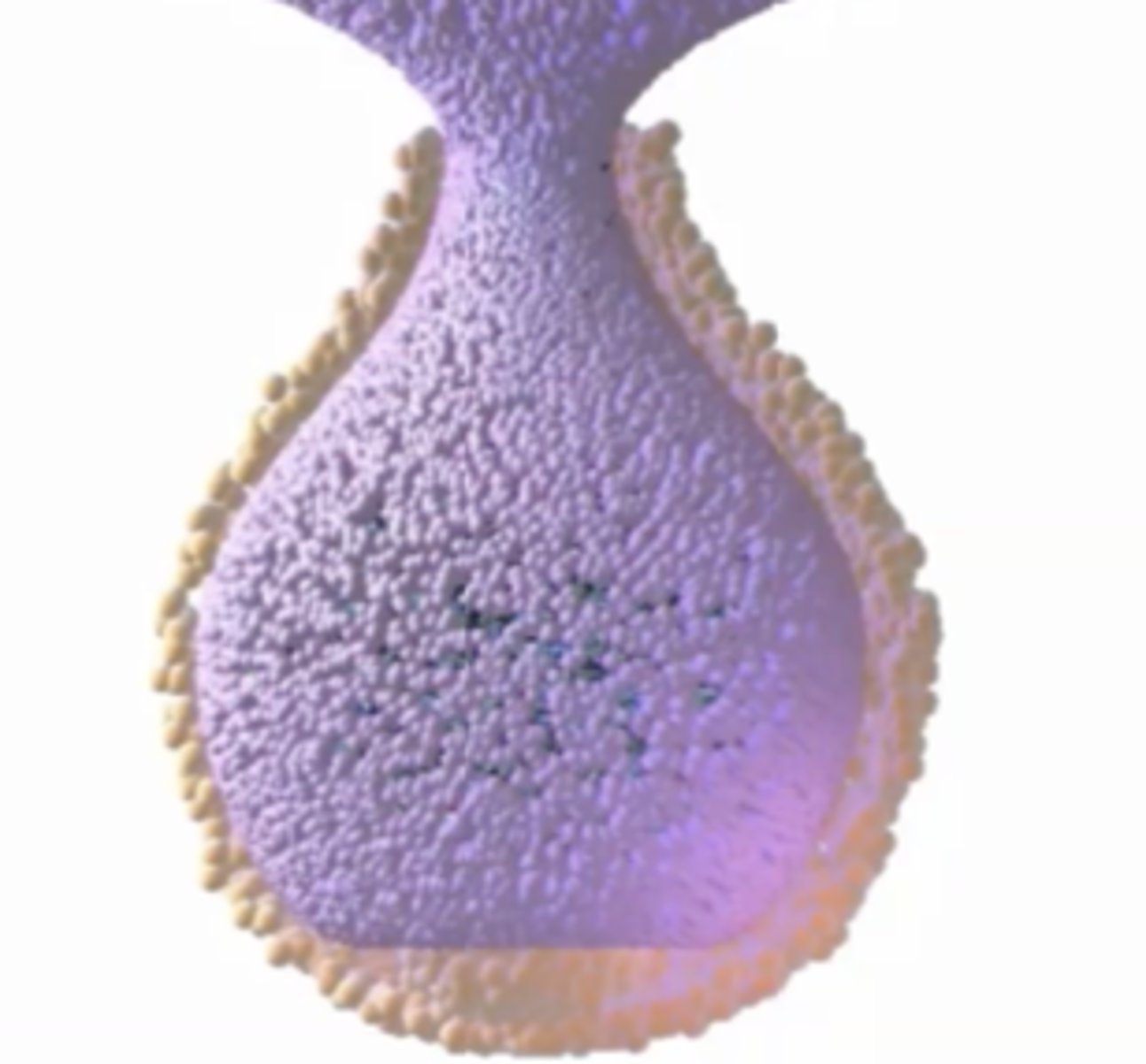
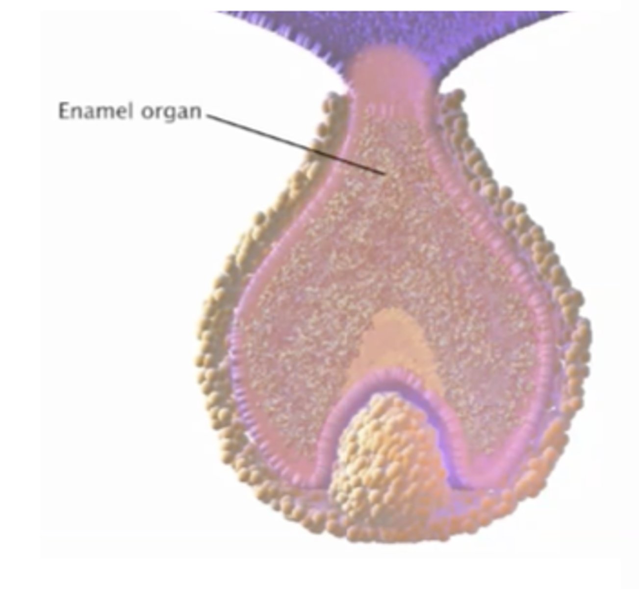
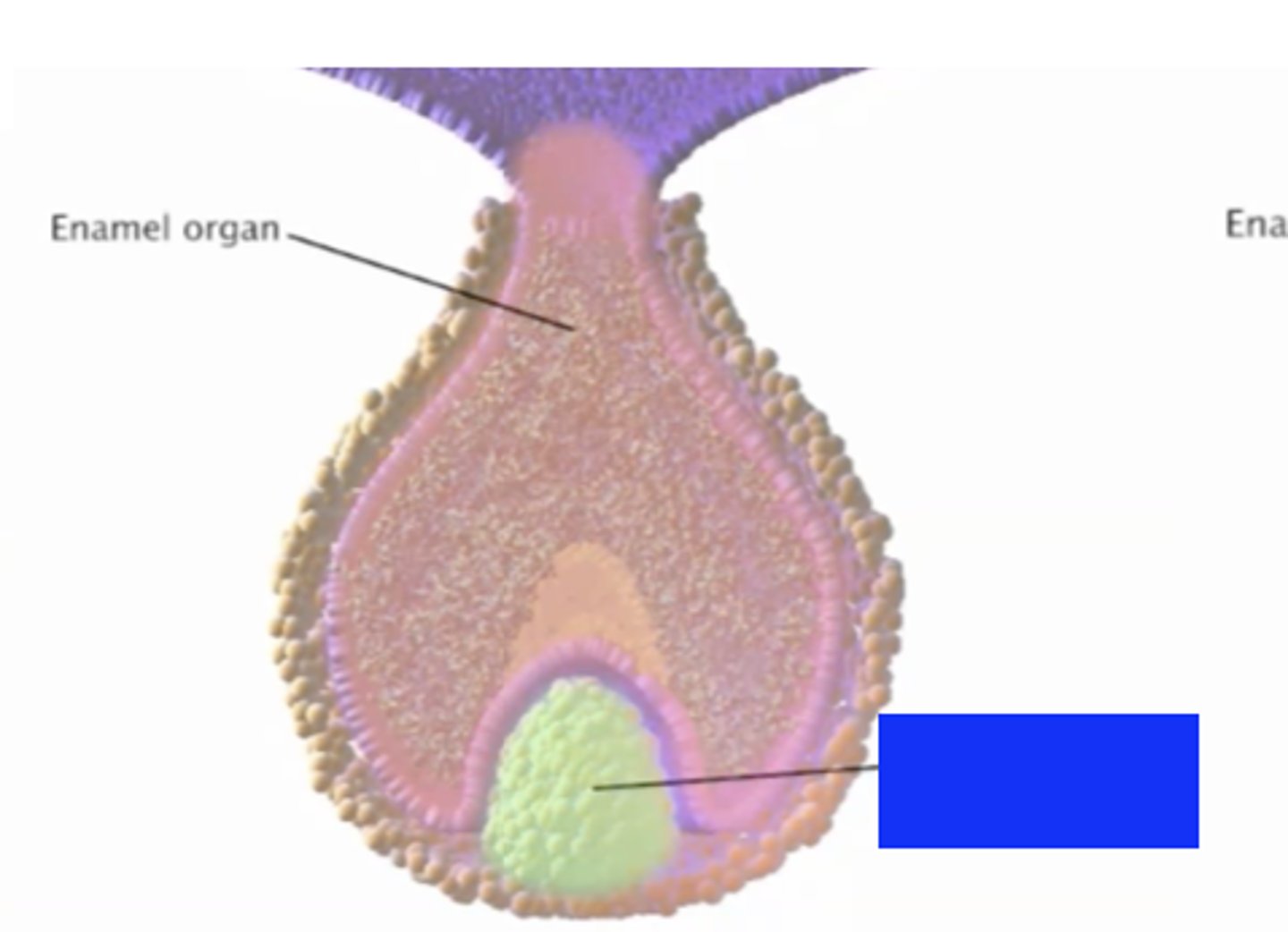
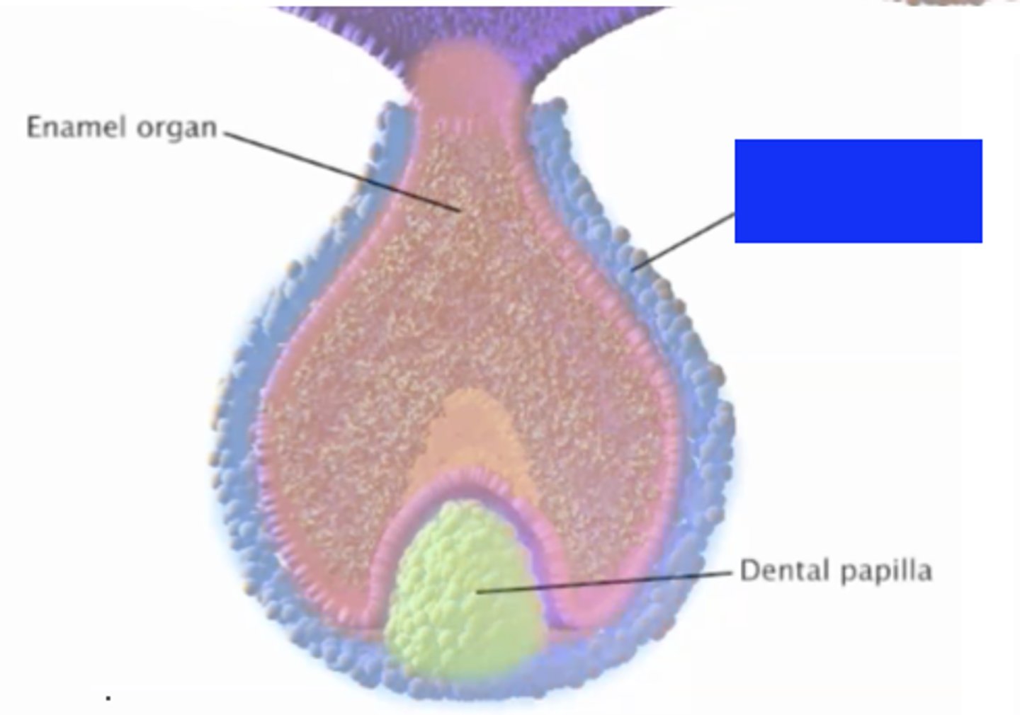
enamel organ
cap or bell-shaped part of tooth germ that produces enamel
part of a developing tooth destined to produce enamel
forms when the bud bends inwards
shifts/modifies shape for each tooth at bud stage
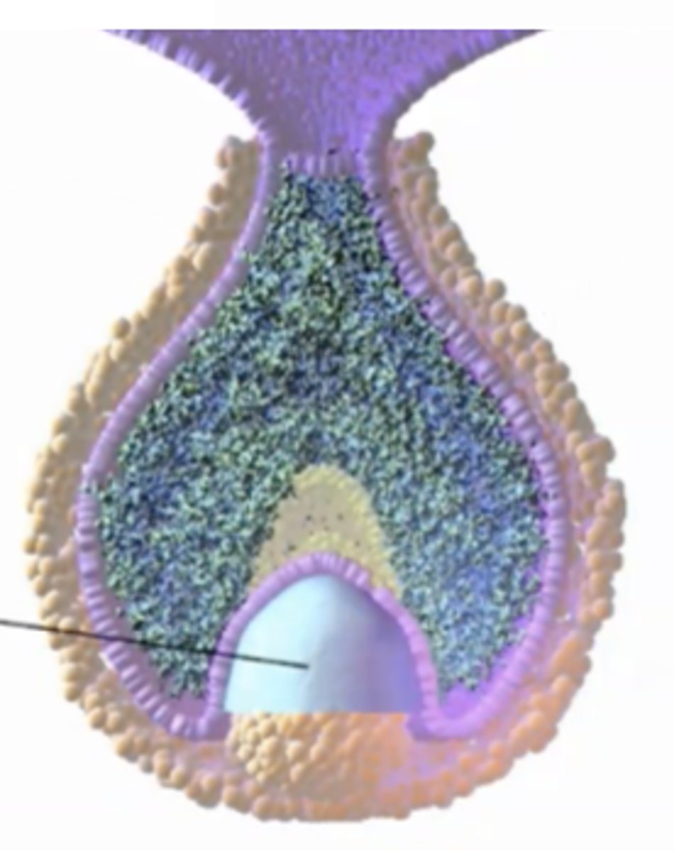
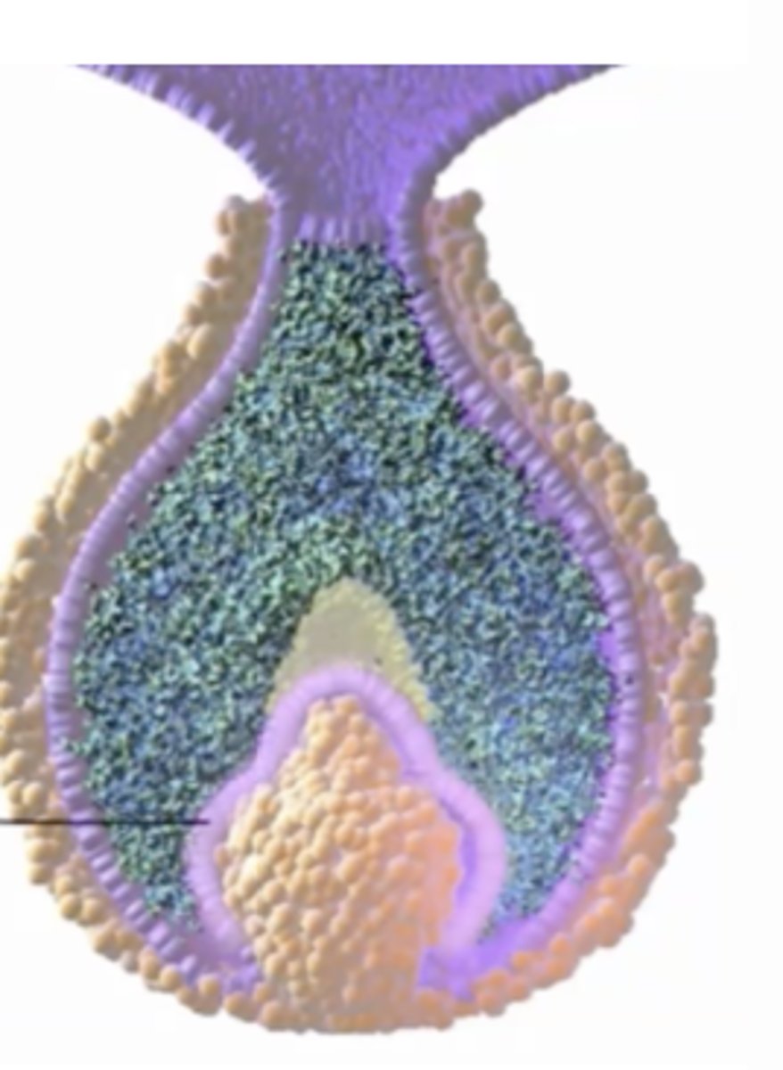
dental papilla
dental follicle
outer protection until tooth formation
basement membrane
separates papilla from enamel organ
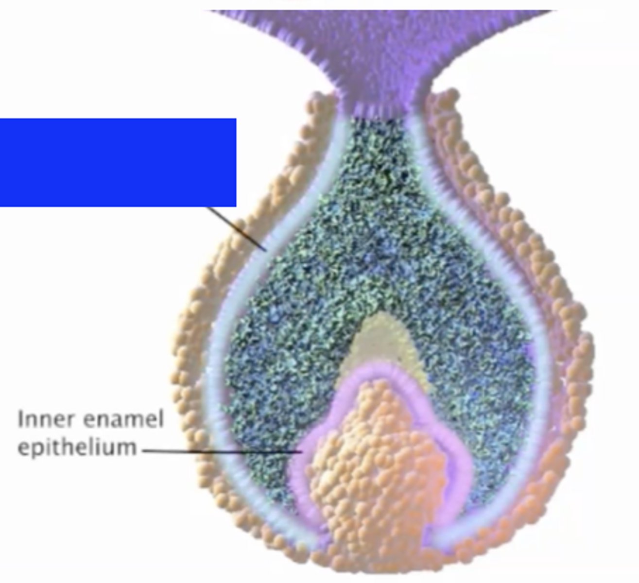
inner enamel epithelium
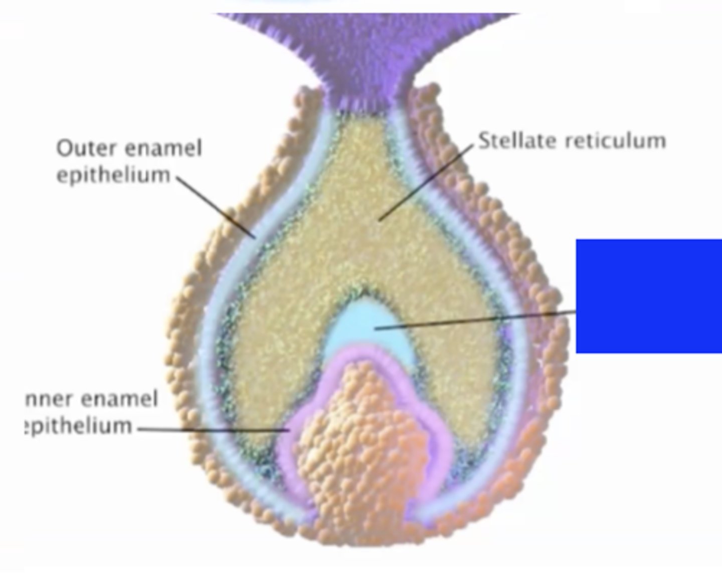
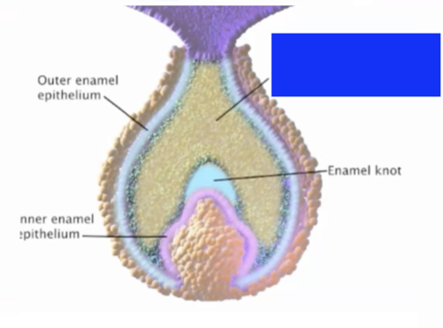
outer enamel epithelium
enamel knot
stellate reticulum
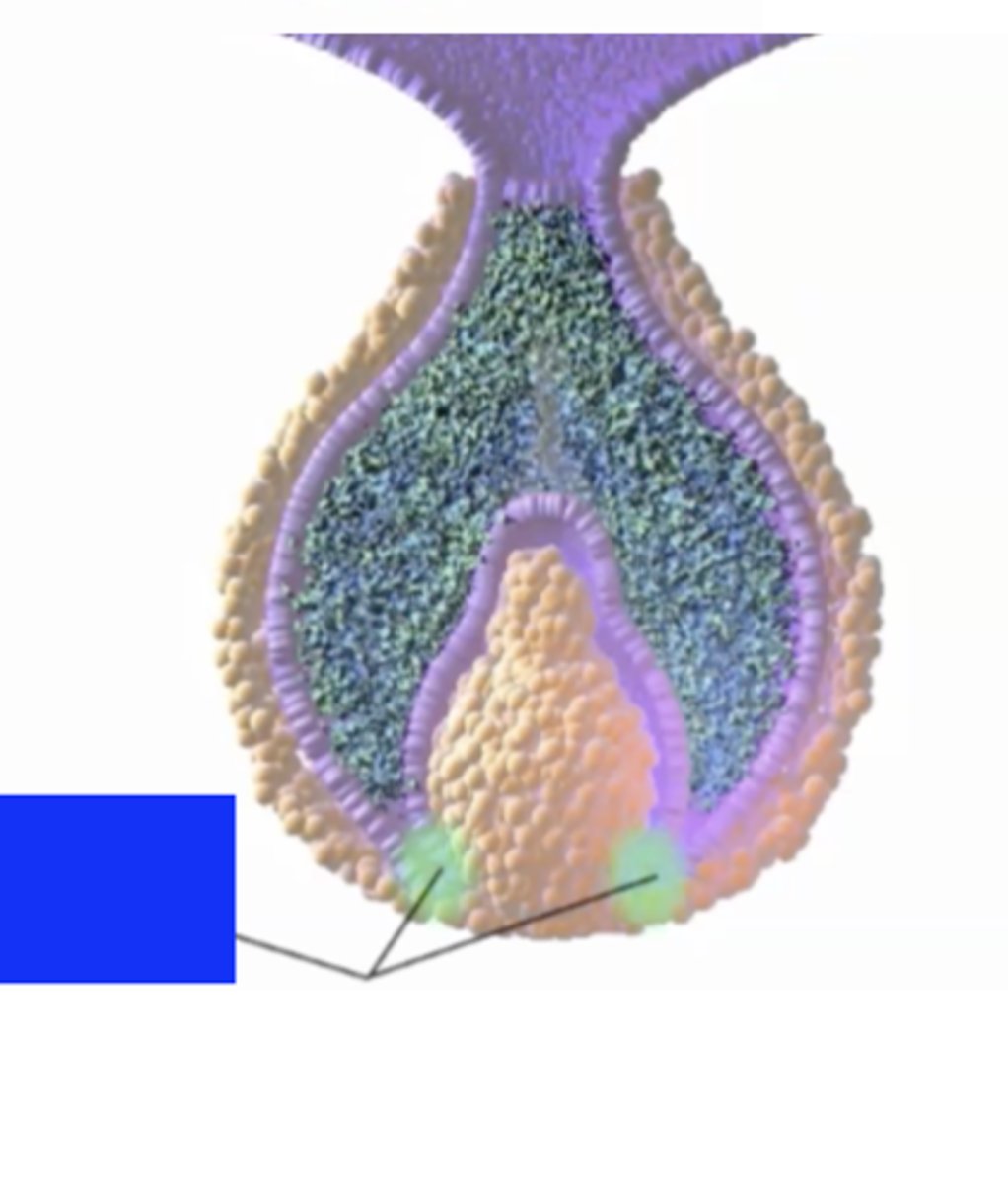
cervical loupes
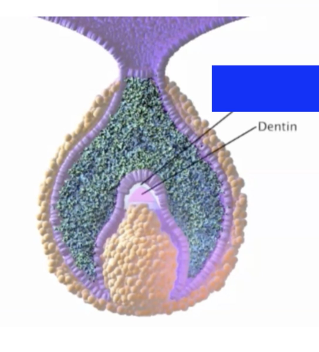
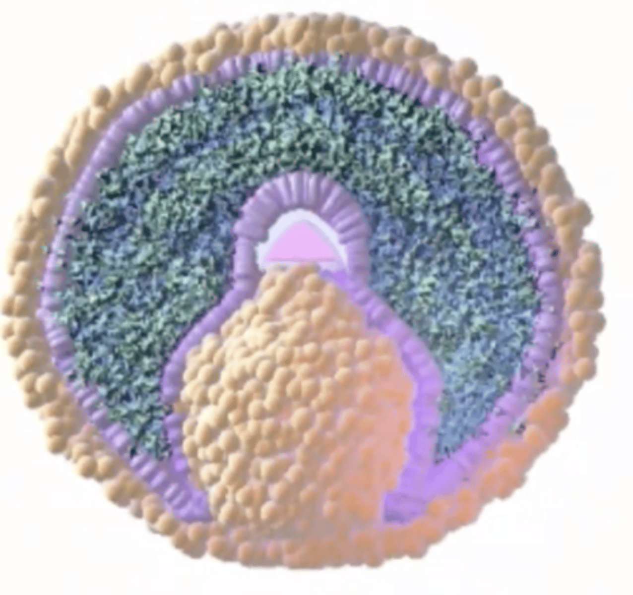
what 4 structures are all present in the bell stage?
outer enamel epithelium
stellate reticulum
enamel knot
inner enamel epithelium
cervical loupes
begin to form shape of tooth's crown as like a mold
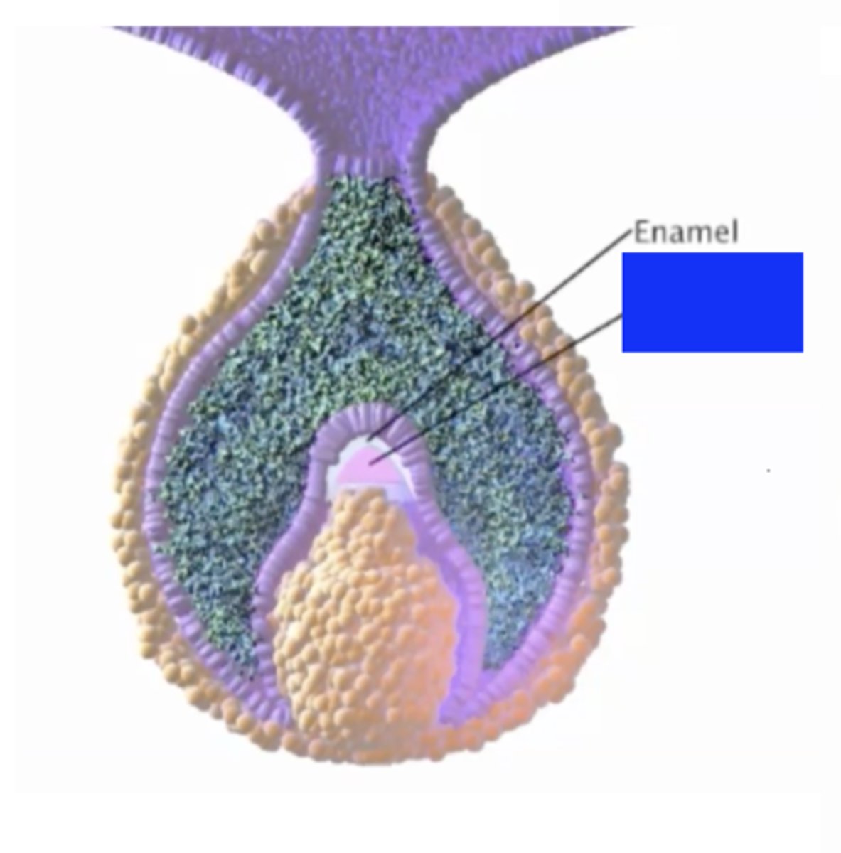
enamel
dentin
at what stage is enamel and dentin formed?
late bell stage
mold forms --> then see enamel and dentin
what signifies the end of bell stage?
shape of each tooth is created; develops differently
shrinks enough to break away from dental lamina --> becomes own tooth
enamel
outermost layer of crown; protects entire tooth
cementum
along the root between teh CEJ and apex
amelogenesis
formation of enamel by ameloblasts
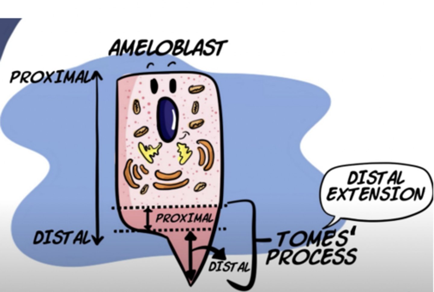
tome's process function
secrete enamel
what do ameloblast cells secrete?
hydroxyapatite- main mineral to form enamel and make layers
Tome's process
45º at cell edges
90º at cell center
creates rod/inter-rod formation
enamel formation looks like a honeycomb; daily growth
maturation stage
enamel fully calcified
secretes minerals and absorbs proteins
enamel hardens and is fully formed
during enamel formation, after teh cells elongate, what do the cells of the inner enamel epithelium differentiate into?
ameloblasts --> cells that form enamel
secretion/formation of enamel after the nucleus shifts to the upper part of cells
Tome's process
distal end of the ameloblast cells bend at 45º inclination at distal end of cell
secretes enamel
what is the function of the Tome's process?
secrete enamel
hydroxyapatite
main crystal to form enamel after calcification; secreted by ameloblasts
describe the structure of the Tome's process layers of enamel
creates layers of enamel
40º at cell edges
90º at cell center
angulation during hydroxyapatite crystals
forms rod/inter-rod enamel formation
enamel formation creates what greater shape?
honeycomb due to different-angulations of hydroyapatite
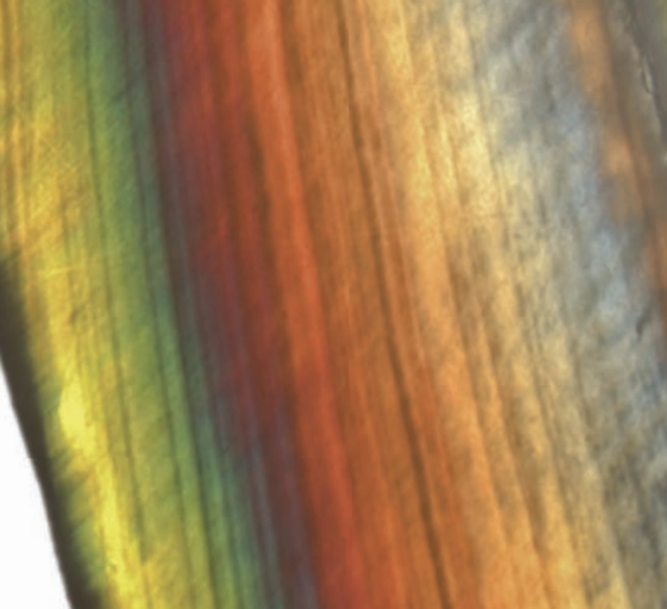
Stria of Retzius
Incremental lines of growth formed by ameloblasts
- Represent weekly changes in enamel formation
- Stria may also represent growth of the enamel organ
May be caused by incremental pattern of enamel secretion during tooth development
composition of enamel
inorganic 88%
organic 2%
water 10%
describe the arrangement of enamel rods
arranged parallel in a direction perpendicular to the dentin-enamel junction from dentin to the outer enamel surface
microstructure of enamel: inorganic
Enamel consists of hydroxyapatite crystallites 25 m thick, 100 m wide and 500-1000 nm long
Crystallites are arranged into 5 m diameter rods that are encapsulated by 1 um thick protein rich sheaths.
Enamel rods are arranged parallel in a direction perpendicular to the dentin-enamel junction from dentin to the outer enamel surface.
Within the rod units, the directional arrangement of the apatite crystallites varies. Crystallites in the central part of the rod are parallel to the rod axis while those near the edge of the rod usually have an angle of near 15°-45° to the longitudinal axis of the rods.
describe the arrangement of crystallites in the central part of the rod
parallel to the rod axis while those near the edge of the rod usually have an angle of near 15°-45° to the longitudinal axis of the rods.
what is the significant of rod edges beign 15-45º inclined to the longitudinal axis?
lessens enamel stress when eating
enamel rods
hold proteins and water
inter rod
enamel rods at 45º/15º perpendicular to others
microstructure of enamel: organic
.Enamel organic components consists of proteins and enzymes
• Proteins: Amelogenin, Ameloblastin, Enamelin, and Tuftlelin
• Enzymes: Mettaloproteinases (MMP), Proteinase, Phosphatase
proteins in organic matrix of enamel
Amelogenin, Ameloblastin, Enamelin, and Tuftlelin
enzymes in protein matrix of enamel
Mettaloproteinases (MMP), Proteinase, Phosphatase
what component of enamel regulates enamel growth?
organic proteins and enzymes
enamel thickness along the tooth
incisal 1/3 > middle 1/3 > cervical 1/3
Striae of Retzius
tooth growth in lines
incremental growth lines or bands seen in tooth enamel. They represent the incremental pattern of enamel, the successive apposition of different layers of enamel during crown formation
• identifies enamel growth
• enamel grows in layers/increments during crown formation
perikymata
external manifestation of the striae of Retzius showing as wavy transverse lines 30-100 microns apart
wavy transversal lines on external surface
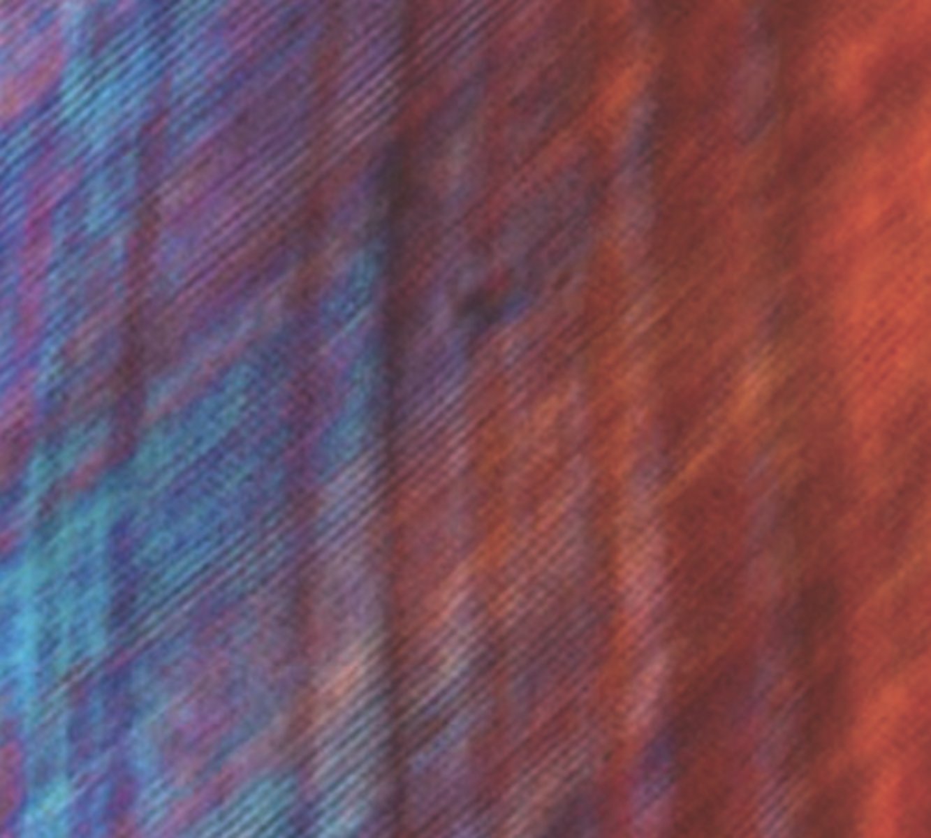
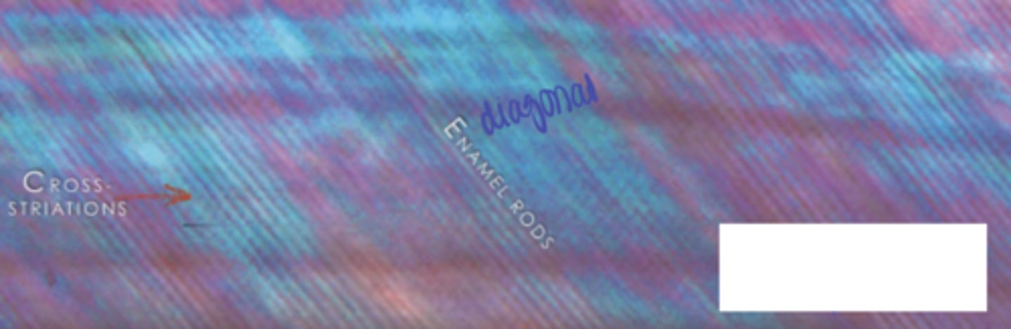
enamel cross-striations
demarcate the amount of enamel deposited by ameloblasts in a single day. The average rate is approximately 4 um/day in humans.
The average distances between cross-striations in human teeth are about 2.5 um at the DEJ and 6.5 um at the enamel surface.
can calculate average growth rate for an individual
When tooth forms from DEJ to outer tooth surface --> closer together at DEJ, growth increases as move towards outer enamel surface
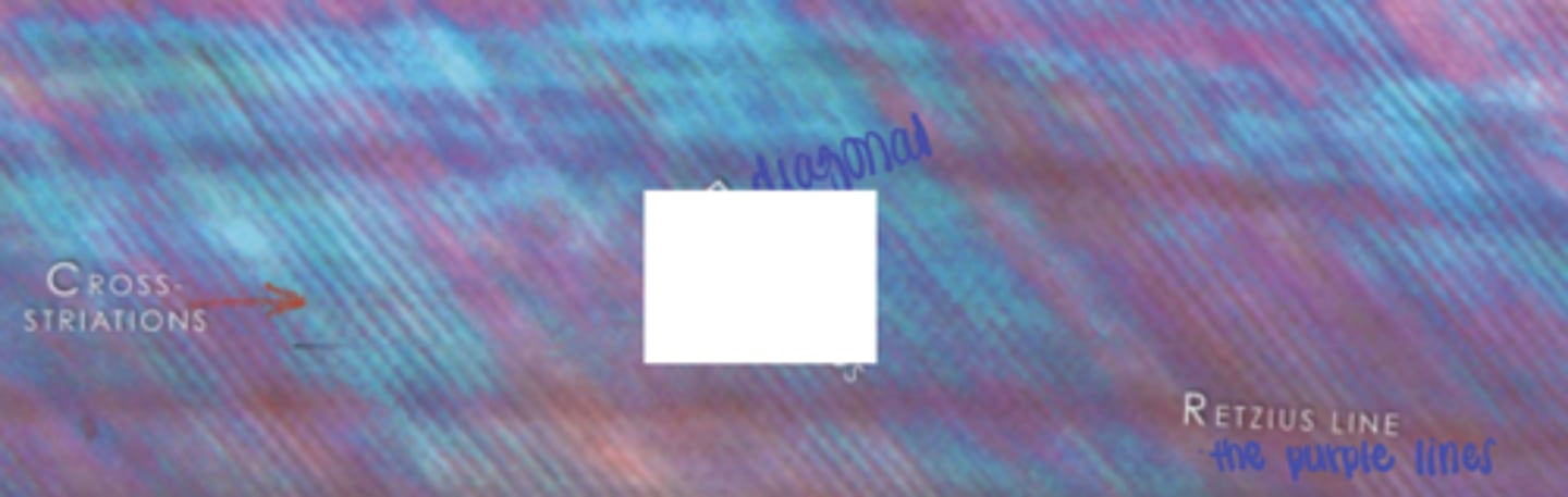
cross-striations
enamel rods
retzius line (purple lines)
outer enamel surface
enamel rods are // to each other when move from outer to inner surface enamel rods change direction into "S" or
wave shape --> decussation
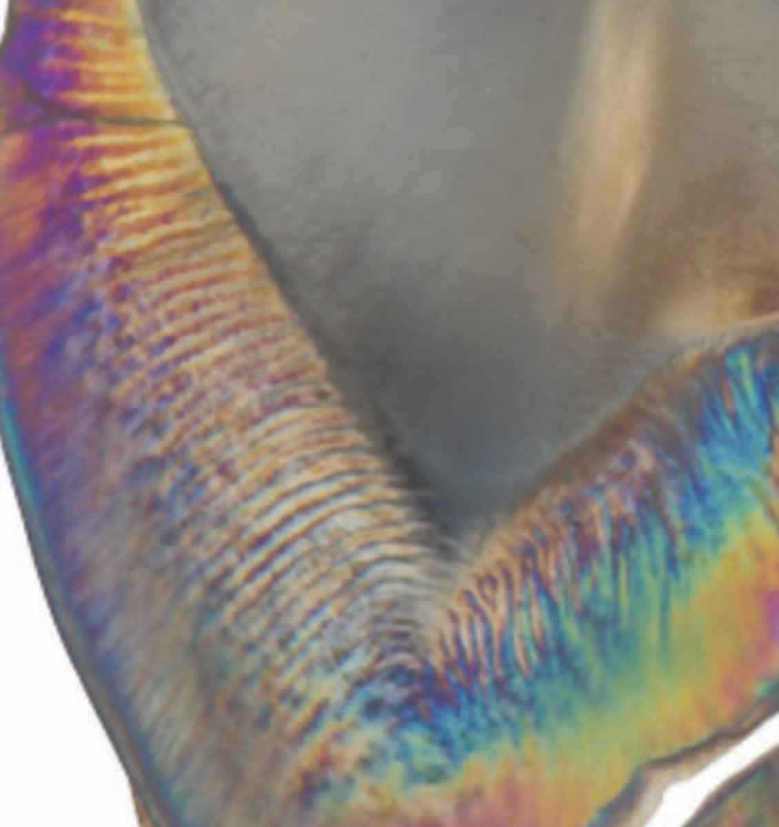
decussation
wave shape of enamel these bands are seen in places of increased functional demand
Hunter-Schreger bands (HSB)
composes set of 10 or more layers of enamel rods
is related to the synchronous decussation of enamel rods in the horizontal plane and is probably caused by reflection of light by inter-prismatic material
are most concentrated in regions exposed to the greatest functional demand, such as the occlusal surfaces of posterior teeth for chewing and the surfaces of maxillary and mandibular canines for guiding mandibular movement
where are HSBs most commonly found?
most concentrated in regions exposed to the greatest functional demand, such as the occlusal surfaces of posterior teeth for chewing and the surfaces of maxillary and mandibular canines for guiding mandibular movement
area with more stress/occlusal contact has more HSB to protect the tooth
difference in inclination on areas with more stress
what is the name of the bands with different inclinations of enamle?
HSB
• difference in inclinations of enamel rods especially on areas with high stress
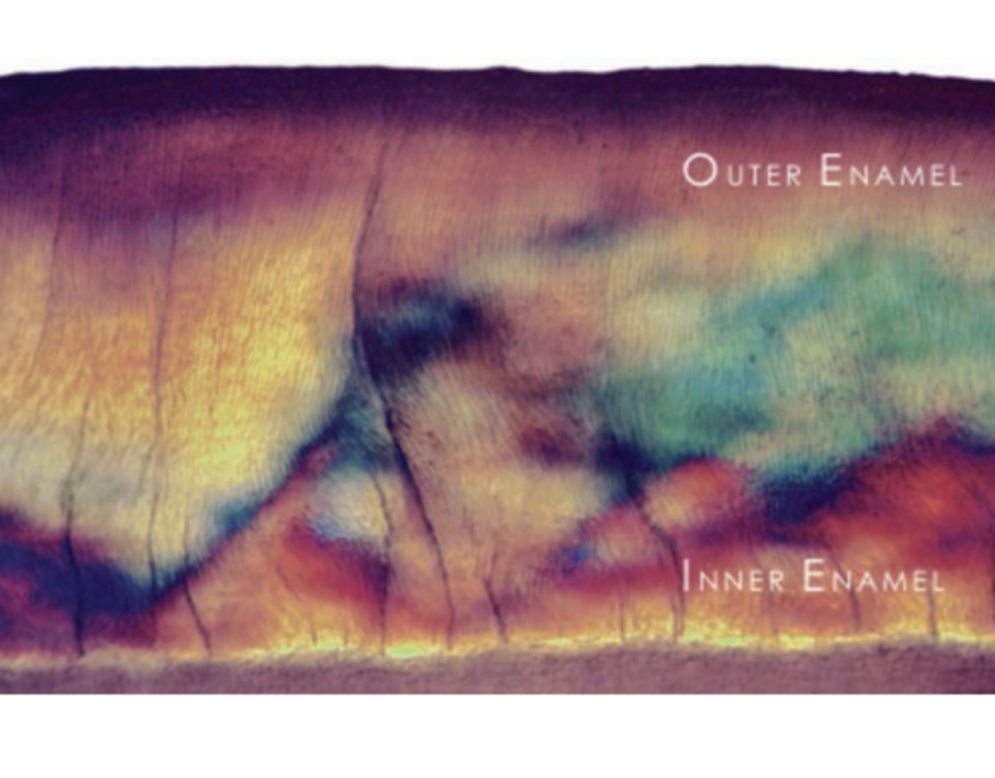
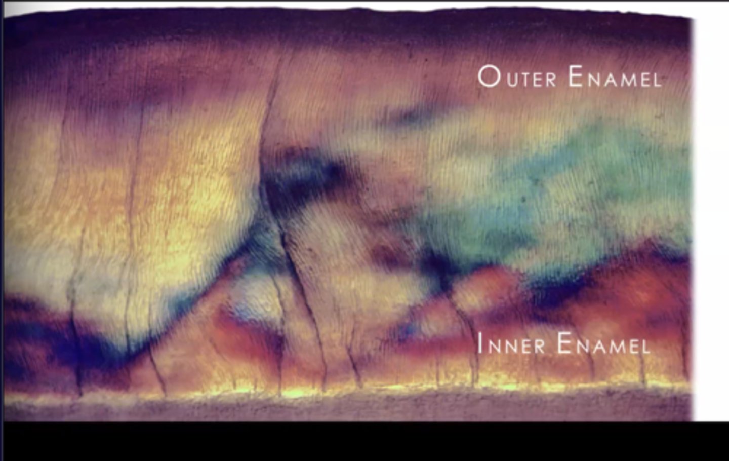
describe the change of rod arrangement as you move from outer to inner enamel
outer enamel has parallel enamel rods
enamel rods deviate from original parallel path as you move downward
inner enamel has decussation
outer enamel
enamel rods are mostly oriented parallel to each other and perpendicular to the DEJ
where is aprismatic enamel found?
in outer enamel
what area of enamel has the highest modulus of elasticity?
outer enamel
• very rigid, strong (tears food) bc of inclination of // enamel rods
• handles lots of occlusion
aprismatic enamel
outer enamel
ameloblasts secrete hydroxyapatite and in the last layer pack lots of hydroxyapaite and make the area dense
when ameloblasts secrete hydroxyapatite, area with densely packed hydroxyapatite but dont' have rod/inter-rod formation
inner enamel
Closer to the DEJ, the enamel rods decussate (wave pattern) in layers (or bands)
Area of enamel with lower modulus of elasticity compared to that of the outer enamel
• still rigid, out not as much as much as outer surface
Increased organic content
Contains enamel tufts
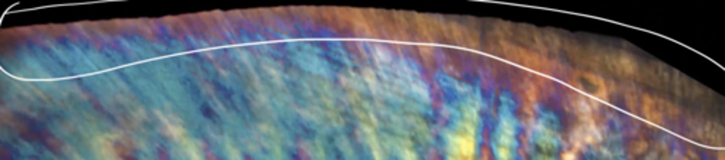
describe how enamel decussation prevents crack propagation
hard for crack to propagate if at 90º with enamel decussation
crack initiates here, enamel turns 90º so crack can't propagate
fracture/crack initiates between the // enamel, so the crack would fracture in half
enamel decussation
•Decussation: constitute a structural reinforcement of teeth
• Decussation is caused by crossing enamel rods bundle within alternating bands that follows a sinusoidal path
• Prevent crack propagation --> hard for crack to propagate if at 90º with enamel decussation; crack initiates here, enamel turns 90º so crack can't propagate
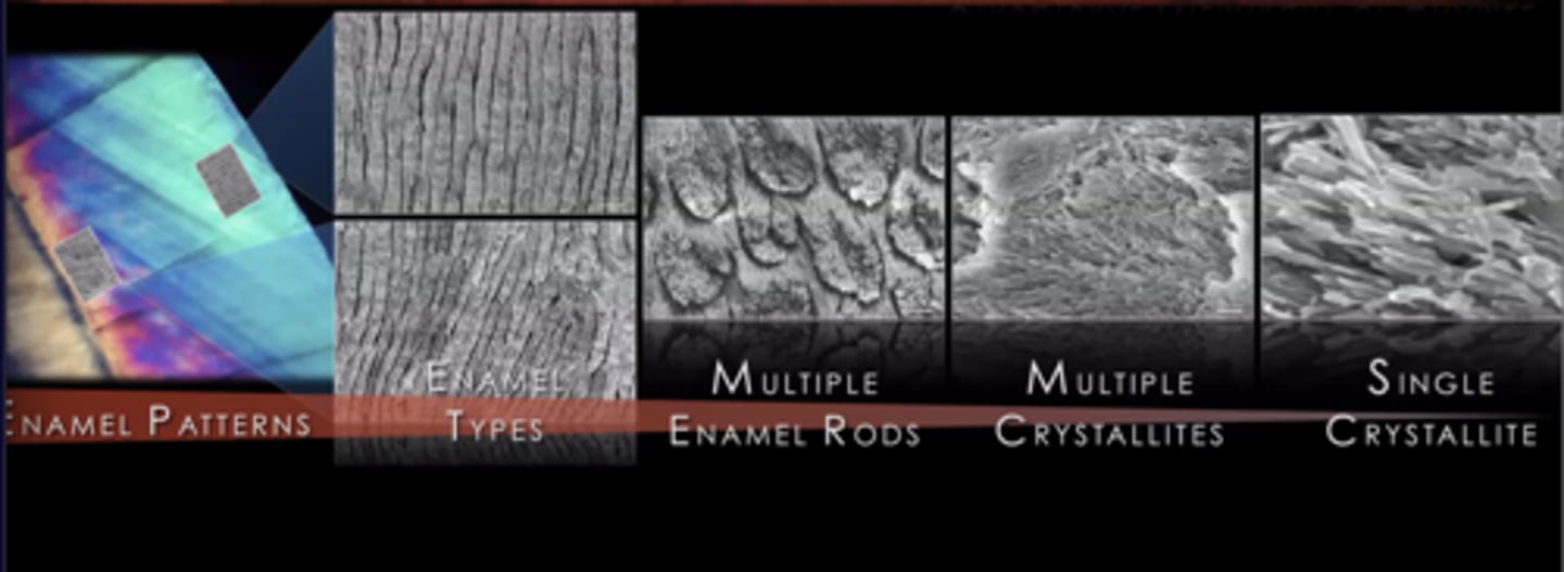
structural hierarchy of enamel
(levels of enamel protection)
enamel patterns
enamel types
• outer enamel // to each other
• inner enamel- decussation (strength)
multiple prisms
• enamel rods, rod/iner-rod formation
multiple crystallites
single crystallites
bridging
if crack is too strong and progresses to a different area enamel tries to prevent this
in addition to decussation what helps prevent a crack/fracture from propagating?
rod/inter-rod
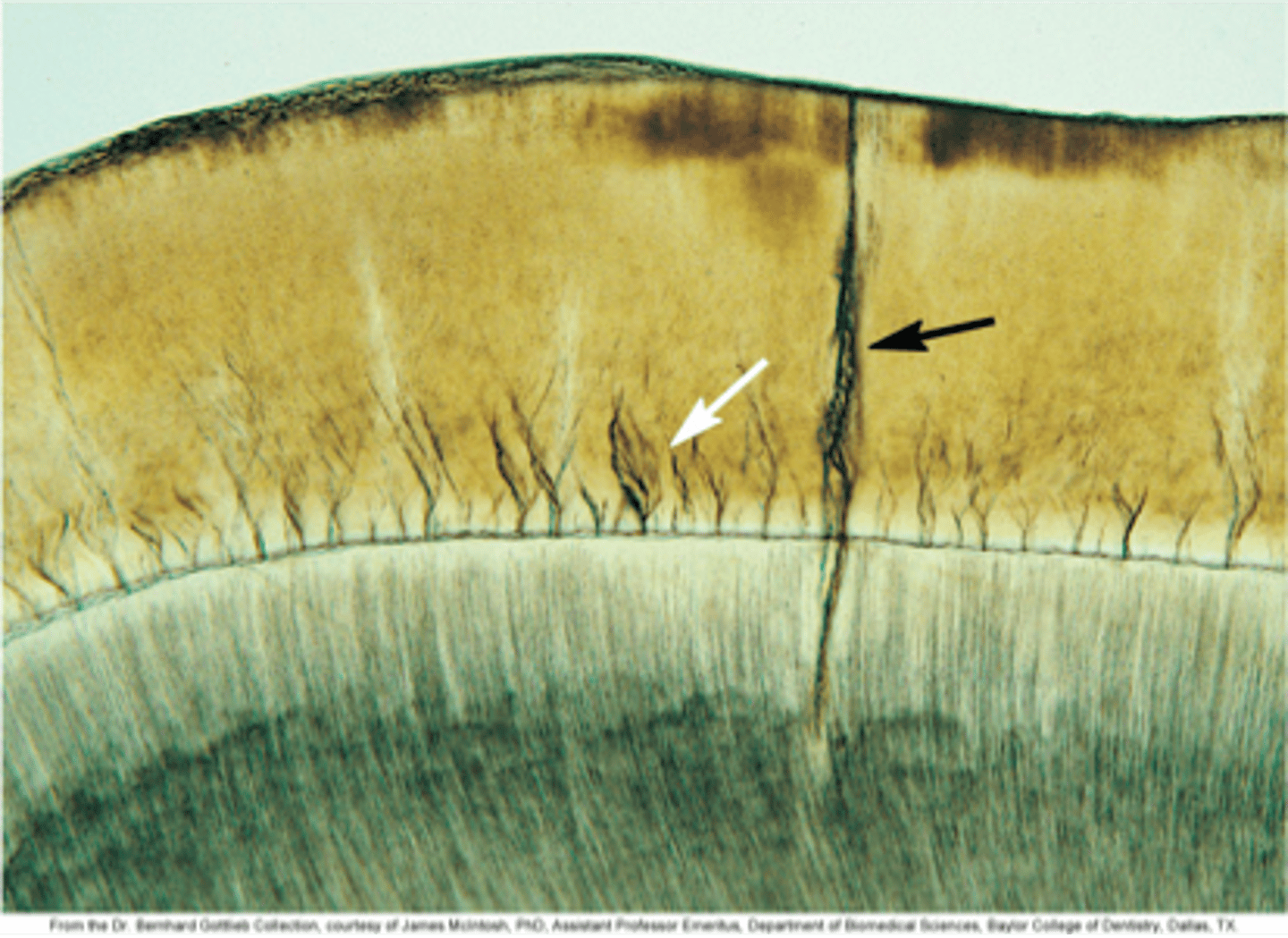
where are enamel tufts located?
on inner enamel, extend outward from the DEJ
enamel tufts
Brush-like structures extending outward from the dentin enamel junction (DEJ).
They are hypomineralized regions containing increased residual enamel matrix proteins, thought to be due to changes in direction of adjacent enamel rods originating from different areas of the scalloped dentin enamel junction.
May assist in the resilience of enamel bc of amount of proteins
inner enamel has enamel decussation and protein-rich
or hypo-mineralized structures => enamel tufts
can be a problem if too long and reach enamel surface
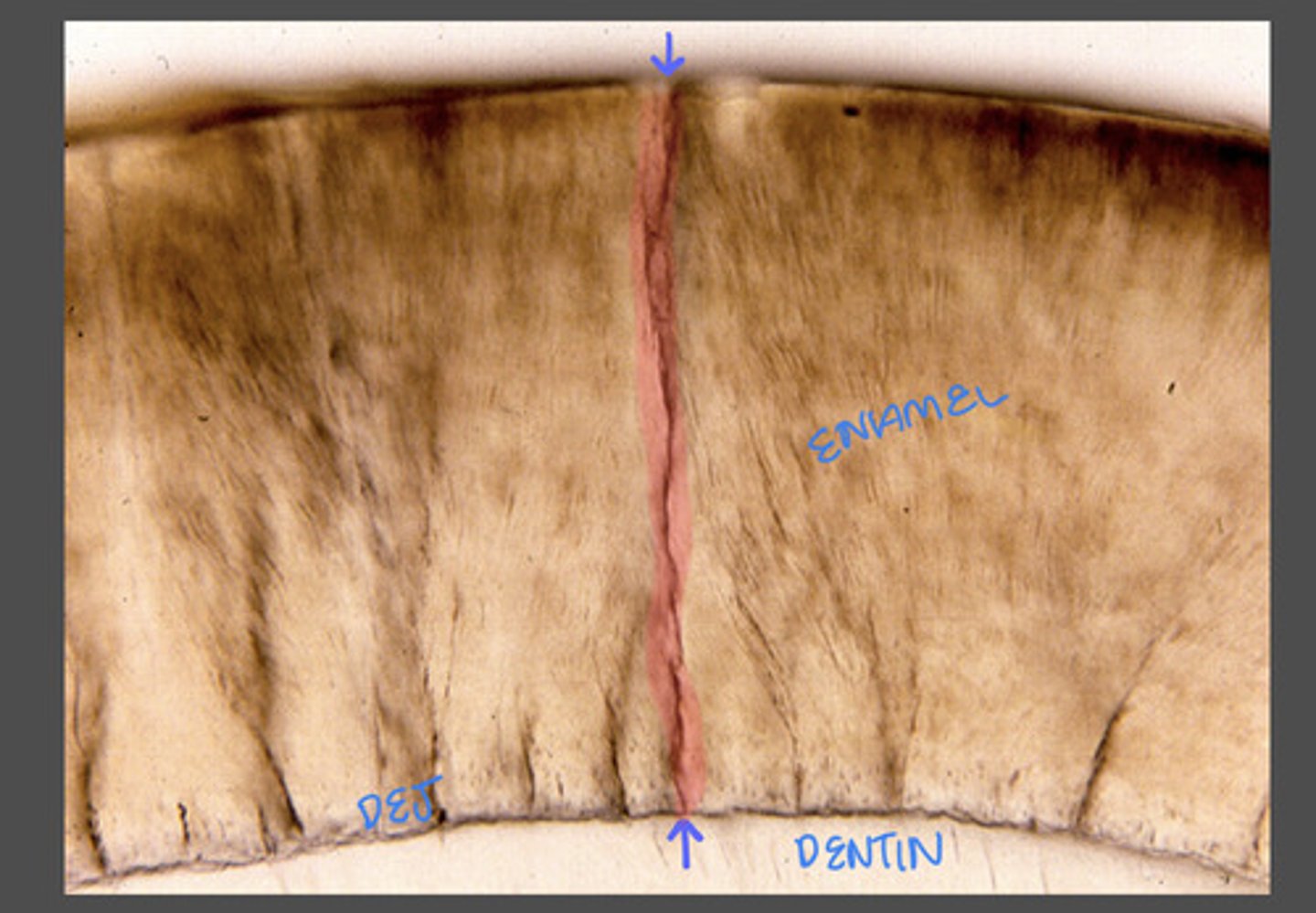
enamel lamellae
Fissure-like linear enamel defects containing proteins proteoglycans, and lipids.
Enamel lamellae extends along the longitudinal axis of the tooth perpendicular to the dentin enamel junction.
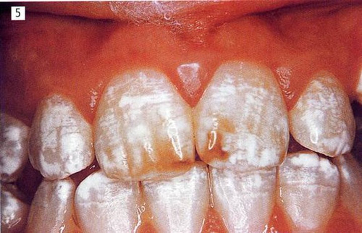
fluorosis
too much F- exposure
competes with calcium from hydroxyapatite and form fluoroapatite which weakens enamel } all during tooth formation
dental fluorosis
Increased enamel porosity along the striae of Retzius.
Hypomineralized lesions that extend throughout the enamel.
Pits, bands, attrition, abrasion and loss of extensive areas occur post eruptively.
too much exposure to F- during tooth development
results in staining
possible to remove stains, minimally invasive
dentin > ? > enamel tufts > ?
(outer to inner)
dentin > DEJ > enamel tufts > inner enamel
amelogenesis imperfecta
Caused by mutations in the AMELX, ENAM, MMP20, and FAM83H genes.
can be treated minimally invasively
Types: Hypomaturation (defect in the final growth and development of the tooth enamel),
Hypocalcification (defect in the initial stage of enamel or tooth formation followed by defective tooth growth), and
Hypoplastic (defects in the amount of enamel)
types of amelogenesis imperfecta
Hypomaturation (defect in the final growth and development of the tooth enamel)
Hypocalcification (defect in the initial stage of enamel formation followed by defective tooth growth)
Hypoplastic (defects in the amount of enamel)
Hypomaturation
defect in the final growth and development of the tooth enamel
Hypocalcification
defect in the initial stage of enamel formation followed by defective tooth growth
Hypoplastic
defects in the amount of enamel
what is the majority of the tooth made of?
dentin
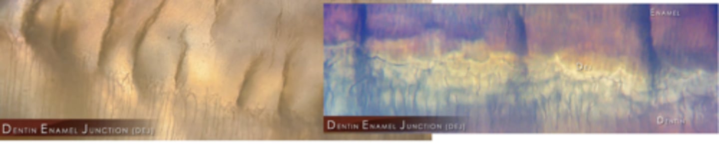
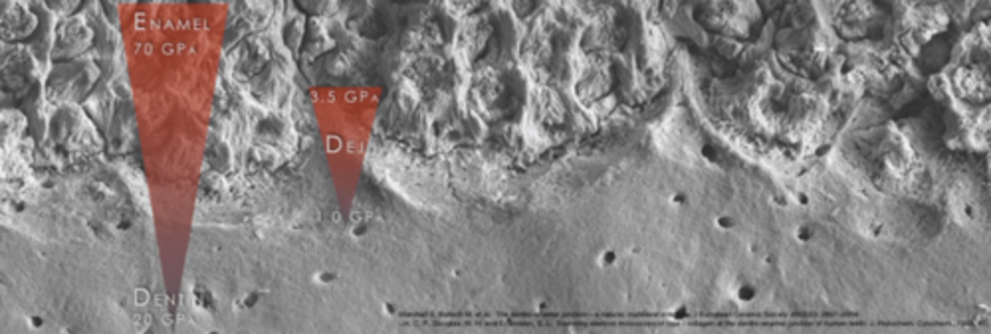
microstructure of DEJ
• The dentin-enamel junction (DEJ) unites two dissimilar calcified tissues: enamel and dentin
(even though they're made of the same thing)
• Three-dimensional scalloped appearance has concavity towards dentin with its convexities directed toward the dentin and concavities
directed toward the enamel.
• Scallop size: 30 um on incisors and 43 um on molars
• Prevent enamel cracks from propagating across the interface, thus preventing catastrophic tooth fractures
• absorbs stress, disipates load
what is the significance of the scalloped pattern of the DEJ?
helps the DEJ (mix of collagen, hydroxyapatite) absorb stressful loads
enamel has lower elasticity than dentin; more rigid, organized
dentin- dentinal tubules have more organic components
scalloping- small area of wave patterns that differentiates enamel and dentin
makes the dentin side of DEJ more elastic
composition of dentin
30% organic (collagen)
20% water
50% inorganic (hydroxyapatite)
microstructure of dentin: organic
Type I Collagen (90%; majority is Type I) (small amounts of Type IlI and Type V collagen are present - 1-3%)
• Non-collagenous components (10%): phosphorylated and non-phosphorylated proteins, proteoglycans, lipids, growth factors
• Proteins: Amelogenin, Osteonectin, Osteocalcin
• Enzymes: to control dentin growth; Matrix Mettaloproteinases (MMP-1, -2, -3, and -9), tissue inhibitors of metalloproteinases (TIMPS), Acid & alkaline phosphates
what is most of dentin?
30% is collagen (type I at 90%, type III and V are also present)
50% hydroxyapatite
microstructure of dentin: inorganic
Crystallites: 2-5 nm in thickness and 60m in length and randomly fill interfibrillar spaces
Intertubular crystallites have a needle like appearance. They are located either at the surface the collagen fibrils, and parallel with the collagen fibril axis.
what goes between collagen fibers?
hydroxyapatite
what is dentin a mix of?
collagen and hydroxyapatite
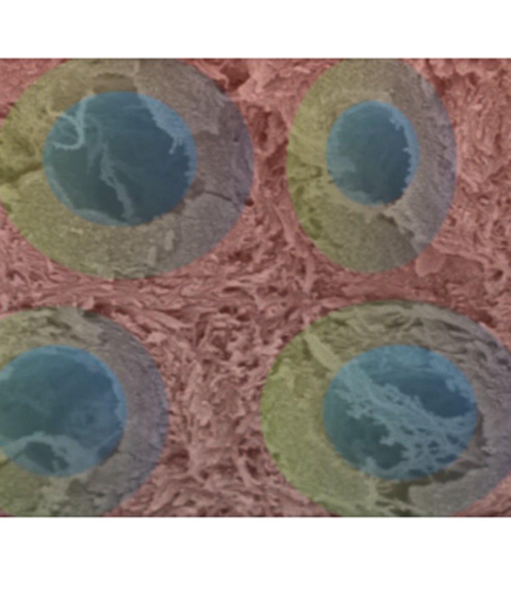
dentin consists of:
dentinal tubules
peritubular dentin
intertubular dentin
dentinal tubules
odontoblast cells --> living part of the tooth
(blue)
peritubular dentin
calcified, heavily densed dentin
protects dentinal tubule
(green)
intertubular dentin
less calcified dentin in between the peritubular dentin
(pink)
intertubular dentin
Collagen-rich dentin associate with proteins along and between the collagen fibrils (organic content 12%)
Hydroxyapatite crystallites of 2-5 pm and 60 nm long (inorganic content: 88%)
Hardness: near DEJ - 0.49 to 0.52 GPa and near pulp 0.12 to 0.18 GPa (becomes softer)
• less inorganic content closer to pulp
• more inorganic content closer to DEJ
is there more or less inorganic content closer to pulp?
less inorganic content closer to pulp
is there more or less inorganic content closer to DEJ?
more inorganic content closer to DEJ