CH 3: Embryonic Development and Cell Cycle
1/61
There's no tags or description
Looks like no tags are added yet.
Name | Mastery | Learn | Test | Matching | Spaced |
|---|
No study sessions yet.
62 Terms
gestation period
-40 weeks total
- Takes 8 weeks for a human embryo to develop into a foetus
-Takes 32 weeks after it develops into a functional child
stages of embryonic development
1: conception- zygote- 1 cell
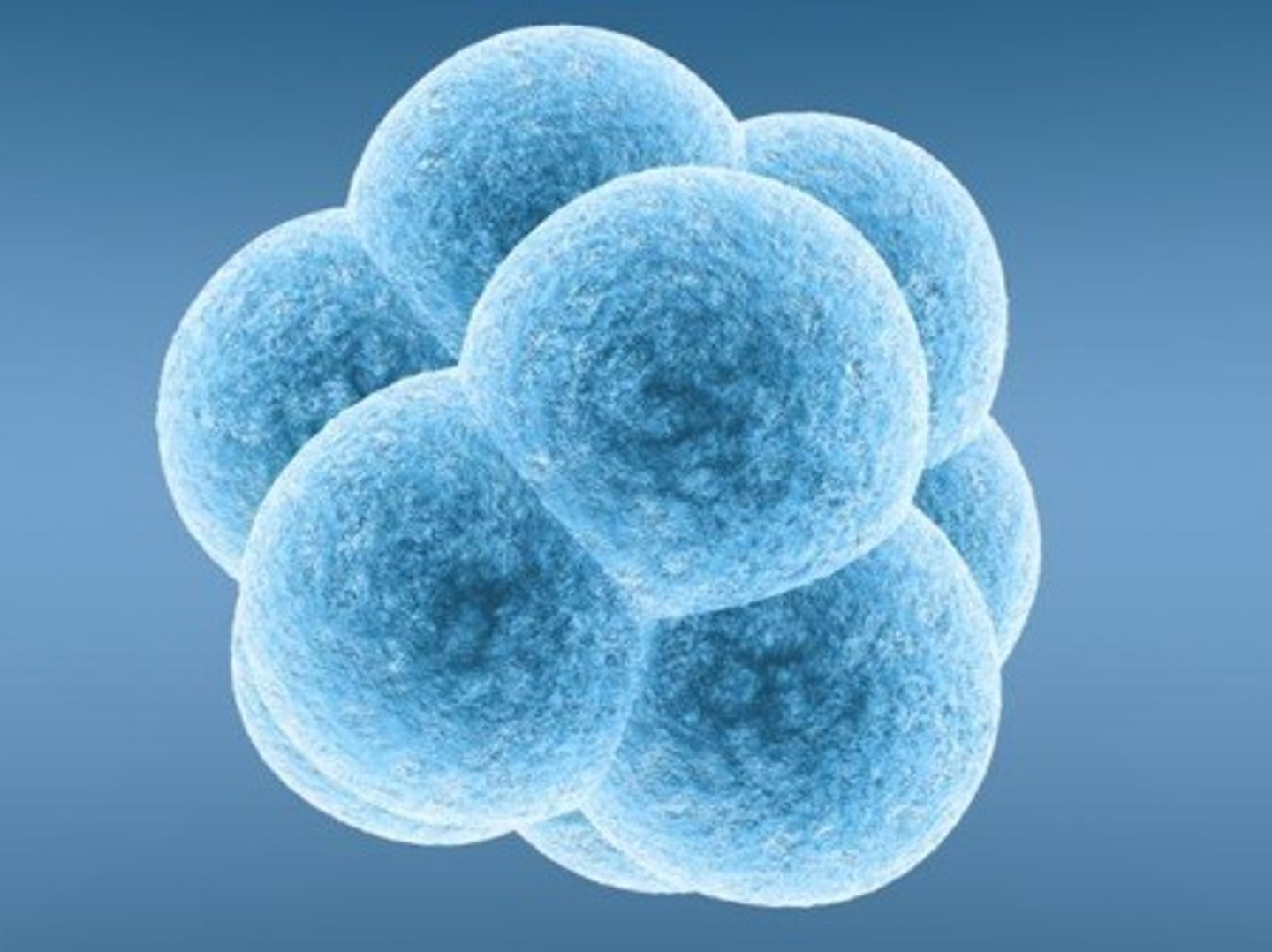
2: 3 days- Morula- 16 cells
3: 4 days early blastocyst- 58 cells
4: day 5- late blastocyst- 107 cells
5: 5+ days- gastrula- formation of the primary germ layers
6: beginning of week 3- embryo- formation of critical structures
7: beginning of week 9- foetus- growth and development of critical structures
Zygote
Fertilised egg that undergoes mitosis.
morula
a solid ball of cells resulting from division of a fertilised ovum,
blastocyst
a cluster of dividing cells made by a fertilised eg
gastrula
Process forming germ layers from blastocyst.
embryo
Early developmental stage before 9 weeks.
foetus
Stage after 9 weeks with formed organs.
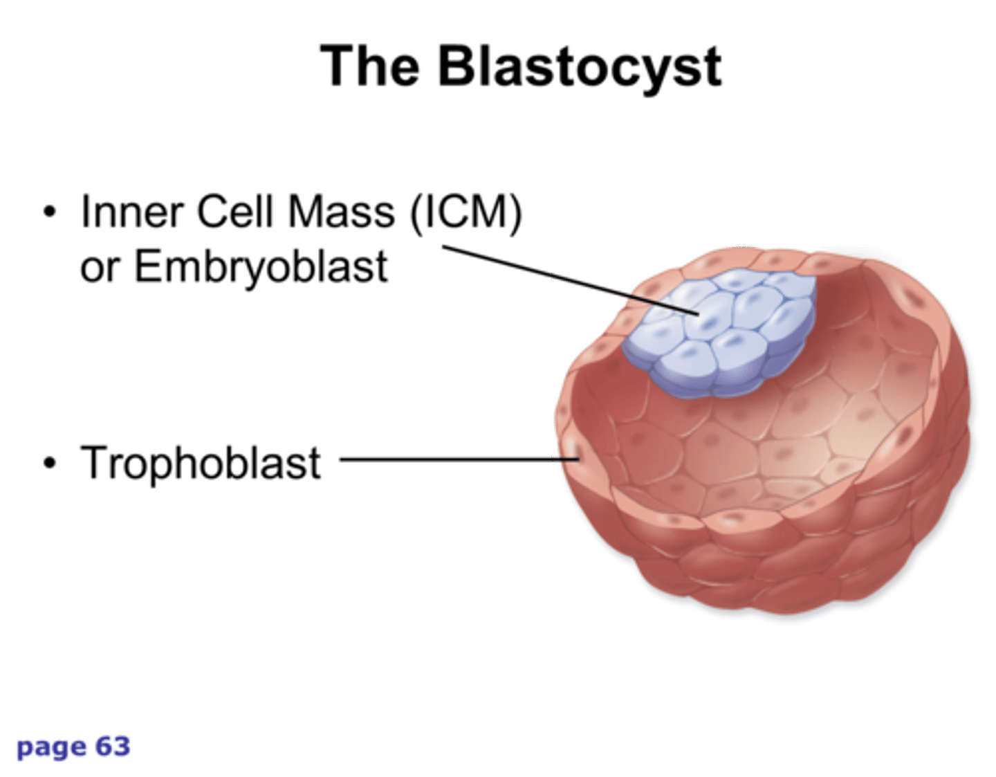
key features of a blastocyst and the significance
-The blastocyst has an inner cell mass and an outer cell layer known as the trophoblast
-The inner cell mass forms into a foetus
- the trophoblas forms the placenta
-The blastocyst will undergo a process called gastrulation
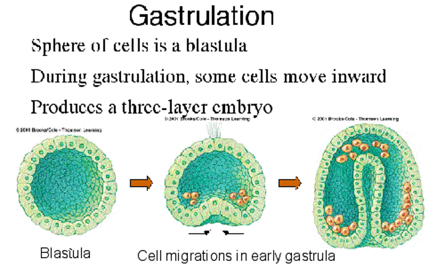
Gastrulation: germ layer development:
-Three separate germ layers are formed - endoderm, mesoderm and ectoderm, which are made of multipotent stem cells
-Each layer is responsible for developing specific components of the body
gastrulation
-A single-layered blastocyst develops into a three-layered gastrula
ectoderm (outer layer)
mesoderm (middle layer)
endoderm (inner layer).
-The amnion - gives rise to the amniotic sac that is filled with liquid that surrounds the baby
-The yolk sac - surrounds the yolk, filled with nutrients for the baby
endoderm
•Lungs
•Pancreas
•Liver
•Stomach
mesoderm
Heart muscle
•Blood cells
•Kidneys
•Bone cells
ectoderm
•Skin
•Neurons
•Brain
•Mouth
•Pigment cells
the role of stem cells in the formation of the three primary germ layers
stem cells are crucial for forming the three primary germ layers (ectoderm, mesoderm, and endoderm) during early embryonic development, ultimately giving rise to all tissues and organs.
stem cells
-They are undifferentiated cells with the capability of differentiating into specialised cells
-A stem cell's POTENCY is its capacity to differentiate into different cell types, or by the stage of development they exist in
-Their potency reduces
-Every cell in our body begins as a stem cell
-They go through differentiation to develop into specific cells with a particular function
-They are self-renewal, which means they can replicate themselves and give rise to more of the same type of cell
Critical periods of development:
1)Germinal stage: lasts until the blastocyst implants into the uterine wall, about 2 weeks later
2)Embryonic stage: Week 3-8, all major organs form and cell mass is considered embryo, the central nervous system is being developed
3)Foetal stage: Week 8-38, embryo turns into a foetus and develops functioning
-Exposure to chemicals, drugs, alcohol can result in birth abnormalities or death
embryonic stem cells
- They can differentiate into any cell type in the body (e.g., muscle, nerve, or blood cells).
- Self-renewal - They can divide indefinitely while maintaining their undifferentiated state.
- They are derived from the inner cell mass of a blastocyst (a very early-stage embryo, typically 3-5 days old).
-They rapidly multiply
-The use of ESCs is debated because extracting them typically involves destroying an embryo.
two properties of stem cells
self renewal: stem cells have the capacity to replicate without disrupting their ability to differentiate, by producing both a differentiated cell and a copy of themselves when they replicate
potency: stem cells are undifferentiated cells, which can give rise to differentiated cells with a specialised function
potency
-Potency: a measure of a stem cell's capacity to differentiate into different cell types
-Not all stem cells can differentiate into any cell
Some can only differentiate to a handful of cell types, depending on their capabilities, which means they have different types of potency
-The more cells it can differentiate into, the greater the potency
difference between unipotent, multipotent, pluripotent, totipotent
Totipotent: Can differentiate into ALL cell types. They are Cells that form the zygote. They are the first cell made after fertilisation of the egg and the sperm
Pluripotent: Can differentiate into almost any type of cell. Embryonic stem cells are in the early stages of the embryo and form the inner cell mass of the blastocyst.
Multipotent: Can differentiate into a variety of closely related types of cells. Stem cells found in bone marrow and cells of the differentiated germ layers
Unipotent: ONE type of cell produced. All somatic cells are of this type, such as skin, muscle, stem cells, etc., which help with repairing tissue
how are stem cells used in medicine
Cell-based therapies: Stem cells are introduced to people suffering from diseases caused by cell damage/death, so new/healthy cells can replace them. Drugs can be developed to stimulate adult stem cells already in a person's body to divide and replace dead cells
Drug discovery:Create cell lines of specific cell types in a lab and use them to test and refine their drug formulations as a prelude to animal testing
Enhancement of Biological knowledge: Scientists can study different diseases and determine how they function, leading to better treatments
adult stem cells
-Undifferentiated cells that are found in certain tissues
-Found in the brain, the bone marrow, the skin, the blood vessels, etc
-Adult stem cells repair and maintain damaged or old body tissue
-Give rise to a more limited variety of cells
Application of stem cell therapy
Tissue Regeneration - In burn victims, the skin has the ability to repair itself using epithelial stem cells. These cells can be grown in a lab and then grafted onto the damaged area to help restore lost skin.
Cell Deficiency Treatment - Scientists aim to develop stem cells into specific cell types that can be introduced into damaged organs to repair tissue and restore function.
Organ Transplants - In the future, stem cells could be used not only to grow tissue but also entire organs. Doctors may be able to use a patient's own stem cells to grow a new organ, reducing the risk of rejection.
ethics and stem cell therapy
Pluripotent Stem Cells and Ethics - Using pluripotent stem cells helps address ethical concerns since adult cells are reprogrammed to behave like embryonic stem cells, avoiding the need to use embryos.
Human-Animal Hybrid Concerns - Human cells are often inserted into animals, especially mice, raising ethical concerns about the possibility of creating organisms that are part human and part another species.
Ethical Use of Adult Stem Cells - Adult stem cells are only collected with full consent, eliminating ethical objections.
DNA structure
-DNA is a double helix with two antiparallel strands of nucleic acids that wrap around each other
-Made up of monomers called nucleotides
-Sides of the DNA ladder are formed by the alternating phosphate and deoxyribose sugar groups
-Hydrogen bonds between bases
nucleotides
-Phosphate group
-Five carbon deoxyribose sugar
-One of four nitrogenous bases - adenine, thymine, guanine and cytosine
-Can be either purines or pyrimidines
-A and G = purines
-T and C = pyrimidines
purpose of cell division
-Cell division in eukaryotic cells is called mitosis (used for growth and repair)
-Prokaryotic cells undergo binary fission. Their purpose is purely reproduction and they divide asexually. (makes more prokarytoes)
-Cutting the cell in half and then multiplying the cell
role of mitosis
GROWTH: Mitosis is responsible for division of cells that results in growth and the development of the 37 trillion cells into 210 specialised cells
REPAIR: cells have different life spans, so through mitosis, damaged or worn out cells are replaced
role of binary fission
-Prokaryotic cells like bacteria, reproduce rapidly via binary fission
-This is where 2 genetically identical copies of a cell are made (exactly the same)
-This is called ASEXUAL REPRODUCTION - a method of reproduction that produces genetically identical cells without the involvement of gametes
-There is no need to spend time looking for a suitable mate, so division can occur very quickly
-The offspring are genetically identical to each other and the parent, which is disadvantageous as it limits genetic variability, makes the population susceptible to changes in the environment and risks being wiped out (no mutations)
stages of binary fission
1. duplication of the chromosome and separation of the copies
2. continued elongation of the cell and movement of the copies
3. division into two daughter cells
3 stages of the cell cycle- mitosis
Interphase: cellular growth and duplication of chromosomes
Mitosis: separation of sister chromatids and the formation of 2 new nuclei
Cytokinesis: division of cytoplasm and formation of 2 daughter cells
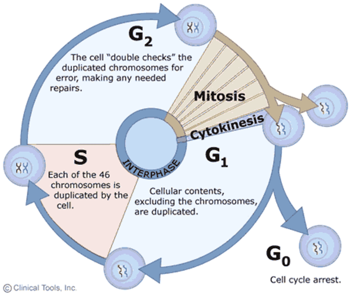
interphase - cell cycle
-First and longest stage, where the cell grows, replicates DNA, and has multiple organelles before moving onto mitosis
-Cell begins to synthesise the necessary DNA, protein and organelles
-Made of 3 substages:
G1,S, G2, G0 (phase is found in some cells)
G1 phase
In the G1 phase, the cell grows by:
-increasing the volume of its cytosol
-synthesising proteins for DNA replication
-replicating its organelles.
At the end of the G1 phase, cells either proceed to the S phase or exit the cell cycle and enter the G0 phase.
G0 phase is where cells are not required to replicate
G0 phase:
Not all cells progress through the whole cell cycle
Cells can exit in the G0 phase or resting state
Not a permanent state, can re-enter the cycle and divide normally
Synthesis (s) phase:
Cell replicates its DNA
One chromosome into two genetically identical chromatids
The centromere helps the sister chromatids, each chromatid is a single chromosome
the replication of DNA during S phase
During the S phase of the cell cycle, DNA replication occurs, ensuring each daughter cell receives a complete copy of the genetic material, doubling the DNA content from 2n to 4n
G2 phase
The G2 phase is the final stage of interphase where the cell continues to grow and prepare itself for mitosis.
The G2 phase is similar to the G1 phase in that it involves:
-increasing the volume of the cytosol
-synthesising proteins in preparation for mitosis
mitosis
Mitosis involves the separation of newly replicated chromosomes into two nuclei
Before division, the parent cell contains a DIPLOID number of chromosomes (2n). It has 2 sets of chromosomes, one from each parent.
This pair is called homologous chromosomes
The daughter cells will also have the same number of chromosomes
Mitosis produced daughter cells that are diploid and have a full amount of chromosomes
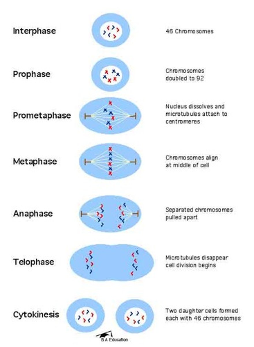
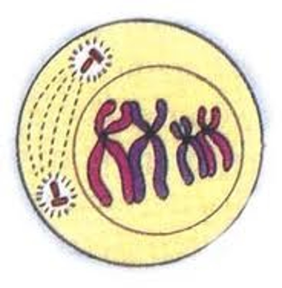
prophase
Condensation of chromatin into distinct chromosomes,
Centrioles migrate towards the opposite ends/poles
Spindle fibres form
Nuclear membrane breaks down, nucleolus disappears
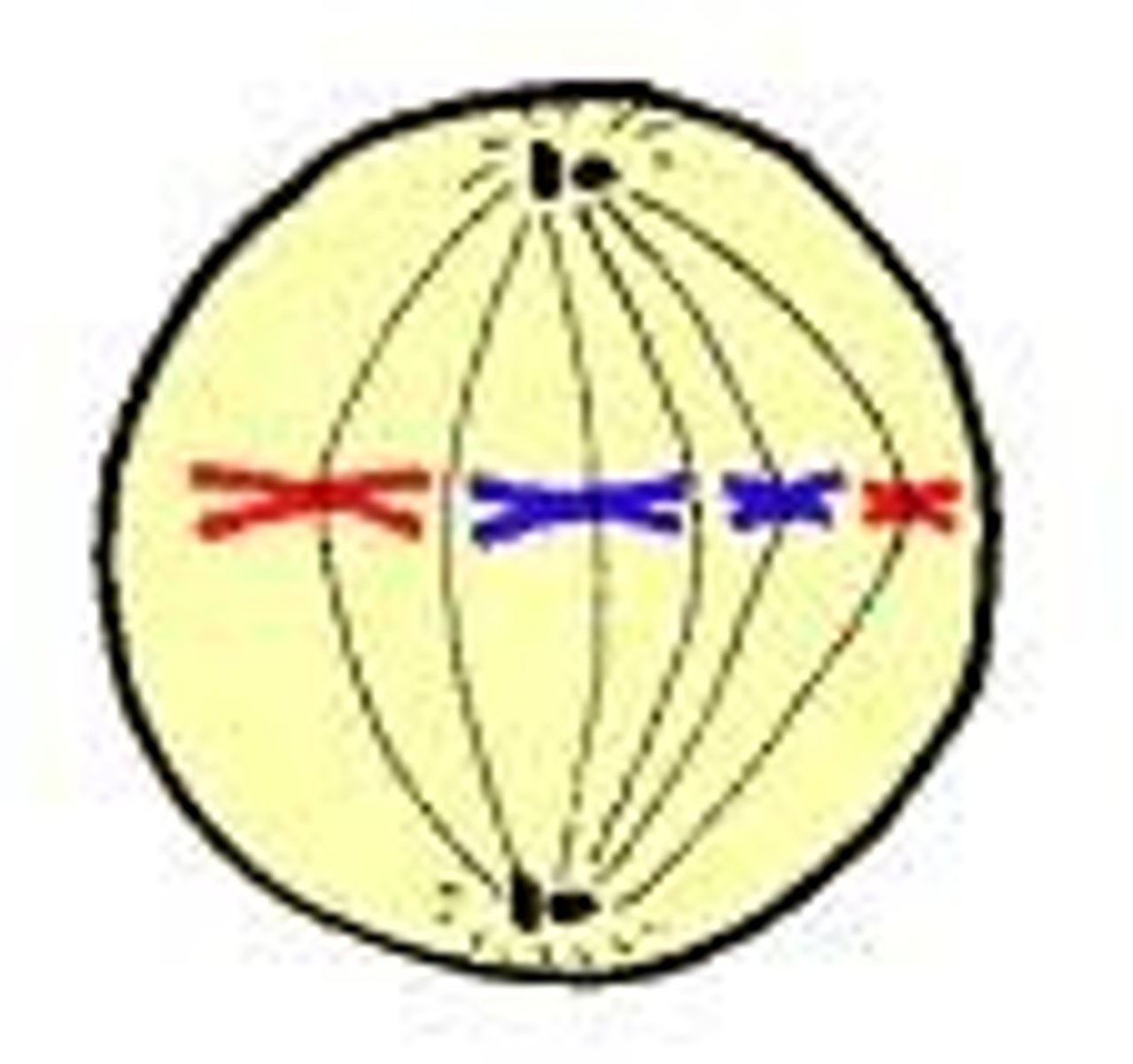
metaphase
Spindle fibres fully form and attach to the centromere of each chromosome
Chromosomes lined up at the equator of the cell
The nucleus is disassembled
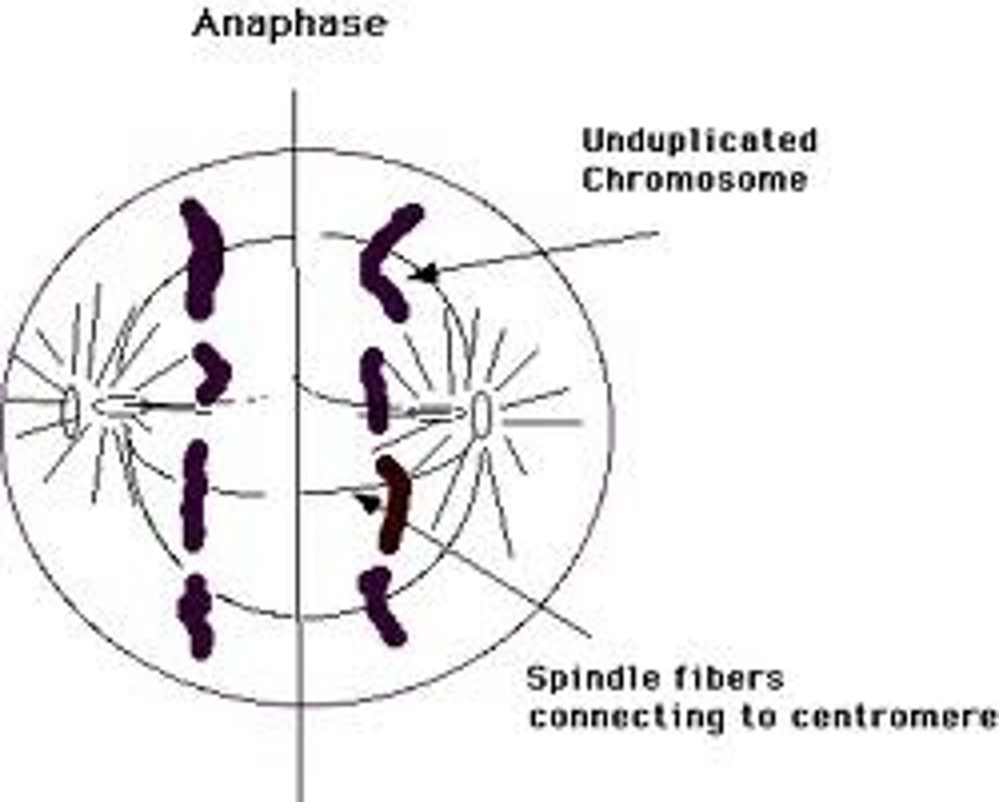
anaphase
Spindle fibres contract
Centromere is split
Sister chromatid is pulled to opposite ends
telophase
Chromosomes densely pack together
A new nuclear membrane forms, making 2 genetically identical nuclei
Spindle fibres disintegrate
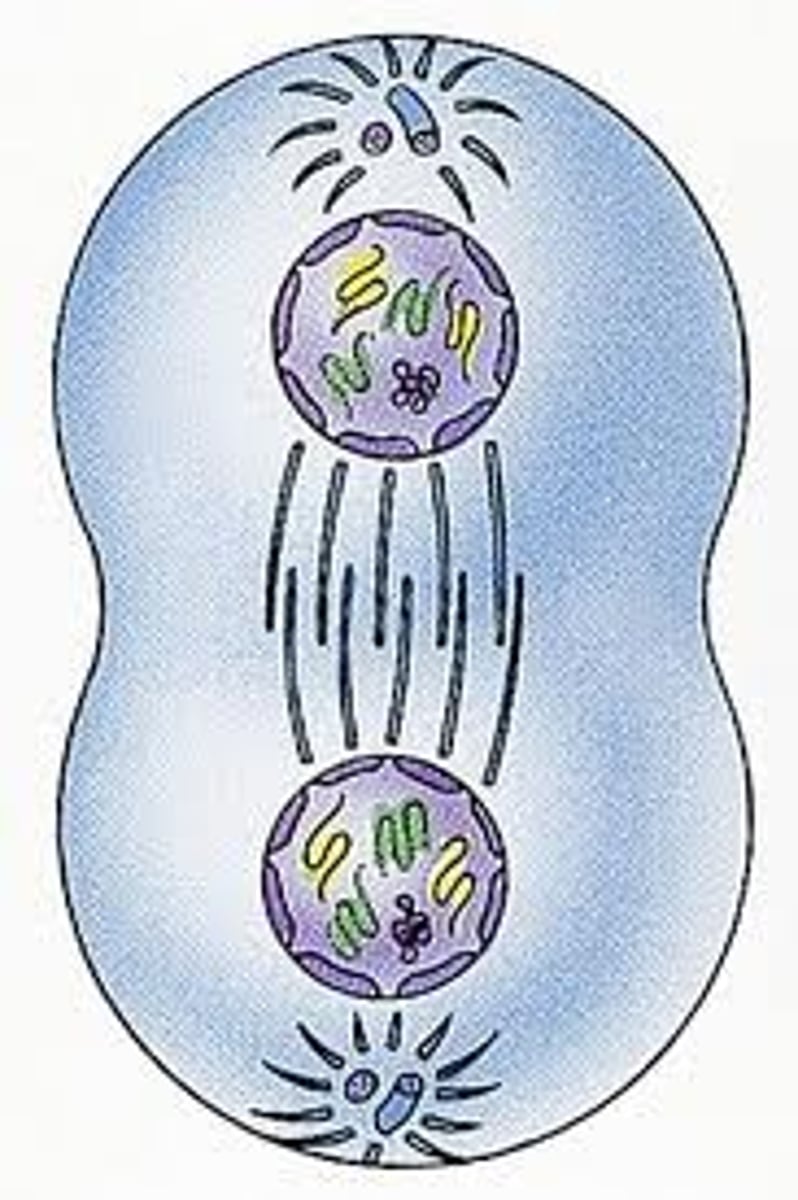
cytokinesis
The cytoplasm divides and the organelles evenly distribute themselves
Animal cells: a cleavage furrow develops and pinches the plasma membrane into two cells
Plant cells: a cell plate forms at the equator
Not part of mitosis, it is after mitosis
G1 checkpoint
Checks for - cell size, nutrient/energy stores, DNA damage and growth factors
Ensure all conditions are favourable for division
Monitors internal and external signals
For the cell to go to the next phase, it needs to be large enough to divide, have perfect DNA and enough nutrients and energy to sustain the cell
If the cell can't meet these requirements, it will attempt to rectify the error or pass into the G0 state of the cell cycle
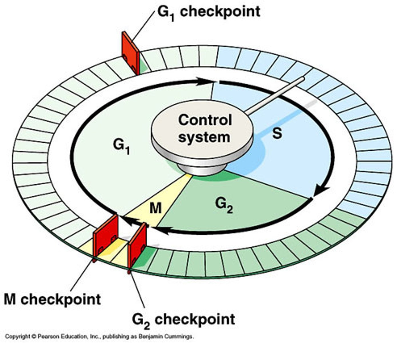
G2 checkpoint
Checks for DNA damage, DNA replication accuracy and cell size
Prior to this, the cell would have undergone the S phase of DNA replication
If damage is found, it will halt the cell's progression through the cycle until DNA replication is completed or damage is repaired
If it can't be fixed, the cell will be tagged and destroyed via apoptosis
M checkpoint
Checks spindle attachment
Occurs between metaphase and anaphase
Ensure that the double chromosomes are lined up along the metaphase plate and are attached to the spindle microtubules
The attachment of spindles to a chromosome's centromere gives rise to the kinetochore
M checkpoint scans the cell for loose chromosomes not in the correct position
If so, the cell will cease mitosis and correctly align the chromosomes. If not, apoptosis occurs
checkpoint proteins
In the cell, there are a set of regulatory proteins
The proteins keep the cell cycle in order by responding to internal and external signals
If a problem occurs, regulatory proteins become inactive and block the progression of the cell to the next stage of the cycle
apoptosis
apoptosis: Programmed cell death that is highly controlled
Results in cell committing 'cellular suicide'
The death of cells is important to maintain the overall health and balance of the organism
Apoptosis performs three key functions
functions of apoptosis: protection
The cell cycle has checkpoints that can halt the progression of a cell through the cycle and detect when the cell is faulty
If the cell can't fix the issue, it will be tagged for destruction and apoptosis will occur
Unhealthy or damaged cells do not divide and pass on their defect to the daughter cells
Helps in the removal of cells that could be cancerous
When cells are infected with a virus, they will also undergo apoptosis, which means the viral infection can be contained and the threat is eliminated
functions of apoptosis: development
During embryonic development, hands and feet start like blocks of tissues
Apoptosis occurs to sculpt them by removing the webbing between the digits
If it fails to occur, the fingers can be fused
functions of apoptosis: balence
Old cells need to make way for new cells
Organs and tissues retain their desired size and that
unnecessary cells don't create 'traffic' and slow down important reactions
pathway of apoptosis
1) the cell responds to signals that trigger apoptosis and it begins to shrink
2) chromatin inside the cell's nucleus is irreversibly condensed, DNA fragments
3) a class of enzymes known as CASPASES are activated. A specific type of caspase breaks down the mitochondria, which releases a protein complex known as Cytochrome c.
4) the release of cytochrome c activates other caspase enzymes which commence the breakdown of the nucleus and other organelles. this trigger the release of stress signal's that attract phagocytic cells
5) bubble like structures, known as BLEBS, form on the membrane of the cell
6) the cell cytoskeleton breaks up, resulting in the blebs forming membrane-bound vesicles known as apoptotic bodies. these bodies contain the cell's cytoplasm and tightly packed organelles
7) phagocytic immune cells are attracted to the area and at like a vacuum cleaner by 'engulfing' the apoptotic bodies (this step is not classified as part of the pathway, but instead the end result)
functions of apoptosis: pathways
For cell to undergo apoptosis, it needs to receive correct combination of signals from its internal and external environment
Positive signals - necessary for the continued survival of a cell
Negative signals - indicate the need to active the apoptosis pathway
Apoptosis can happen with the withdrawal of the positive signals it would usually receive
With the correct signals, the cells will enter apoptosis
There are specific steps that cells need to follow
intrinsic pathways
Activated when intracellular stress signals received by the mitochondria
Stress signals can cause radiation or toxic chemicals damaging the cell's DNA or proteins
In healthy cell, the mitochondria has a regulatory protein that inhibits apoptosis
When it gets the stress signal, another protein moves into the mitochondrial membrane and punctures holes
The release of cytochrome c leaks into the cell and leads to formation of apoptotic bodies
Extrinsic pathway
External signals bind to the death receptors on the plasma membrane of target cell that is about to be destroyed
This signal can be the result of extreme heat exposure or an immune response when the cell is diseased
Creates a signal in the cytoplasm
The caspase enzyme detects it, cytochrome c is released and apoptosis occurs
Necrosis
Premature/unprogrammed cell death
Result of trauma or injury
Not controlled and results of swelling of the cell before it bursts
The spillage of the cells inside causes inflammation which can also be detrimental to neighbouring tissue
P53 protein: master regulatory protein
A regulatory protein that operates at the G1 and G2 cell cycle checkpoints
It detects damaged DNA before mitosis and dividing, initiates enzyme repair of DNA and activation of apoptosis
If p53 is damaged, the above processes wont occur
Cells with damaged DNA will divide, leading to increasing number of mutated cells leading to cancer
Skin cancers due to UV radiation affects the expression of P53
Also can be caused by smoking and exposure to pollution
examples of factors that can contribute to the development of abnormal regulatory proteins
Abnormal regulatory protein development can be caused by various factors, including genetic mutations, environmental exposures, and epigenetic modifications, all of which can disrupt normal protein function and lead to cellular dysfunction
cancer development
It is a disease that occurs due to UNCONTROLLED cell division
For cancer to develop, regulatory proteins must be faulty or defective cells must acquire mechanisms that allow them to bypass the protective measures of regulatory proteins
Cancer development takes a series of steps
cancer
Can develop by a series of mutations that occur in DNA
Faulty cells pass through checkpoints undetected
Increase division of faulty cells can lead to a tumour
Tumour can be benign (not cancerous or invading neighbouring tissue) or malignant (cancerous; can spread to surrounding tissue or elsewhere in the body)
As tumour progresses, the accumulation of DNA mutations become greater and quickly
People in later stages of cancer will have significant changes in their genomes and even complete loss of chromosome
key differences between cancer cells and normal cells
- variation in shape and size of cells
- darker and larger than normal nucleus
-abnormal number of diagnosed chromosomes
- cells clustered without regular shapes or membrane
- cells continue to grow and divide in a disorganised fashion