Unit 5A.1: General Overview & SKIN Assessment | Skin, Hair, Nails, Head, Ears
1/159
There's no tags or description
Looks like no tags are added yet.
Name | Mastery | Learn | Test | Matching | Spaced | Call with Kai |
|---|
No analytics yet
Send a link to your students to track their progress
160 Terms
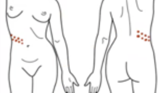
Cephalocaudal
Another name for the Head-to-Toe Assessment
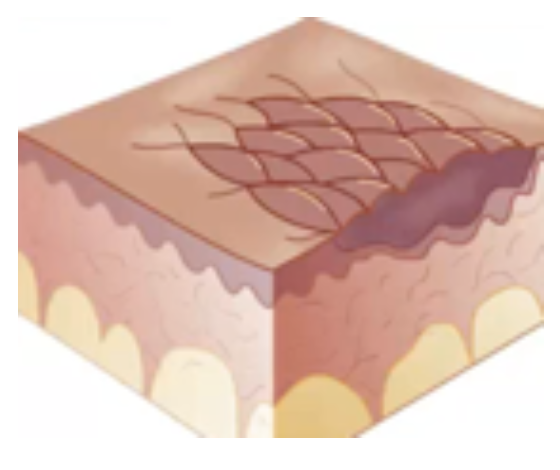
Dermis
Epidermis
Stratum Corneum
Stratum Lucidum
Stratum Granulosum
Stratum Germinativum
2 Layers of Skin (and Sublayers if applicable)
Sebum
Oily substance that lubricates hair and skin and reduces water loss through skin which also has some fungicidal and bactericidal effects
Ask client to remove all clothing and jewelry
Have client sit comfortably
Ensure privacy
Maintain comfortable room temperature
How to Prepare the Client Prior to Collecting Objective Data
INSPECTION:
General skin colorations
Odors emanating from the skin
Color Variations
PALPATION:
Moisture
Temperature
Texture
Thickness
Mobility & Turgor
Edema
INSPECTION:
Lesions
What is the procedure for skin assessment?
Examination Light, Penlight
Mirror for Client’s Self-Examination of Skin
Magnifying Glass, Centimeter Ruler
Gloves, Wood’s Light
Examination Gown or Drape
Equipment Needed During Skin, Hair, Nails, Head, Ears Assessment
Evenly colored skin tone s̅ unusual or prominent discoloration.
Normal Finding for General Skin Coloration
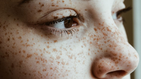
Freckles
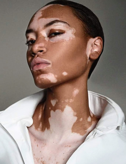
Vitiligo
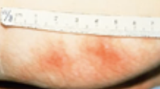
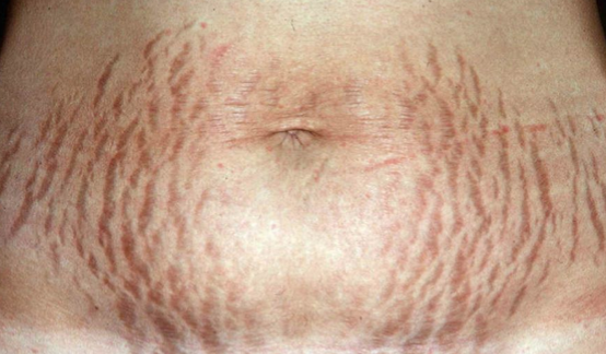
Striae
Seborrheic Keratosis
Scar
Mole (Nevus)
Cutaneous Tags
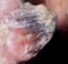
Cutaneous Horns
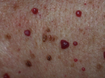
Cherry Angiomas
Common Skin Variations
Freckles
Common Skin Variation
Sun exposed skin
Common in Caucasians
Vitiligo
Common Skin Variation
Uneven patches on the skin
Common in African Americans
Striae
Common Skin Variation
“Stretch marks”
Usually permanent unless given professional treatment
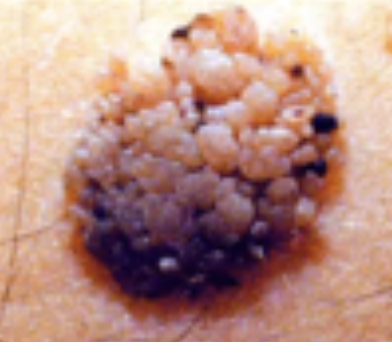
Seborrheic Keratosis
Common Skin Variation
Common in elderly patient
Warty & crusty pigmented skin/lesion
Can be removed by minor surgery
Scar
Common Skin Variation
Remnant of the wound
Common for patient who undergoes surgery
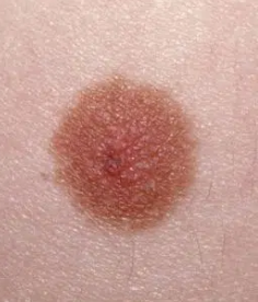
Mole (Nevus)
Common Skin Variation
Pigments on the skin
Usually flat or raised
Cutaneous Tags
Common Skin Variation
Raised papule but with depressed center
Common with elderly clients; normal
Cutaneous Horns
Common Skin Variation
Big portion of dead skin
Common in area with thin skin (ex: ear)
Normal
Cherry Angiomas
Common Skin Variation
Round macule of the skin, usually flat
Usually seen in elderly
Albinism
Erythema
Pallor
Cyanosis (Central & Peripheral)
Jaundice
Acanthosis nigricans
Abnormal Skin Pigmentation Findings
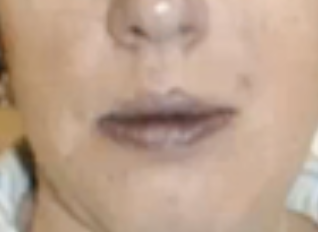
Malar Rash
Abnormal Skin Pigmentation Finding
Butterfly across the bridge of the nose and cheeks in patients with systemic lupus erythematosus
Systemic Lupus Erythematosus
Apatient is seen with a rash across the bridge of their nose and cheeks? What condition does this patient most likely have?
Females
African People
Hispanic People
In what groups is Systemic Lupus Erythematosus most commonly seen in?
Albinism
Abnormal Skin Pigmentation Finding
Total absence pigment on the skin
No melanocytes (which are responsible for skin color - melanin)
Congenital (patient is born to have the condition) but not hereditary

Erythema
Abnormal Skin Pigmentation Finding
In dark-skinned client may be difficult to see. However, the affected skin feels swollen and warmer than the surrounding skin.
Reddish discoloration of skin usually caused by inflammation or trauma
Heated skin temperature
Pallor
Abnormal Skin Pigmentation Finding
Seen in arterial insufficiency, decreased blood supply, and anemia
Vary from pale to ashen without underlying pink
Results from poor circulation
Commonly seen on anemic patients dur to decreased hemoglobin and red blood cells

Arterial Insufficiency
Decreased blood supply
Anemia
What conditions is pallor most likely seen in?
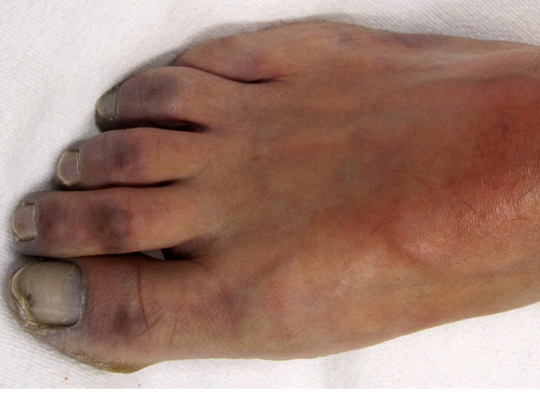
Cyanosis
Abnormal Skin Pigmentation Finding
May cause white skin to appear blue-tinged, especially in the perioral, nail bed, and conjunctival areas
Dark skin may appear blue, dull, and lifeless in the same areas
Either central or peripheral
Due to poor oxygenation
Central Cyanosis
Results from a cardiopulmonary problem; found in the labia of the patient
Cardiopulmonary Problem
A patient exhibits a blue-tinged labia. What condition dos the patient most likely have?
Peripheral Cyanosis
May be a local problem resulting from vasoconstriction
Blueish discoloration of skin or extremities due to vasoconstriction (poor blood flow/oxygenation)
Vasoconstriction
A patient exhibits blueish discoloration of the skin or extremities. What condition does the patient most likely have?
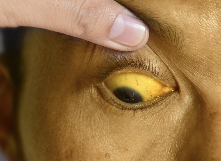
Jaundice
Abnormal Skin Pigmentation Finding
In light- and dark-skinned people is characterized by yellow skin tones, from pale to pumpkin, particularly in the sclera, oral mucosa, palms, and soles
Associated with hepatic dysfunction
Usually found in skin and/or sclera (for African-American patients)
Found in mucous membranes
Some with jaundice may have Hepatitis A, C which can infect other people through blood, sexual contact, and food
Sclera
Oral Mucosa
Palms
Soles
What parts of the body can jaundice be seen?
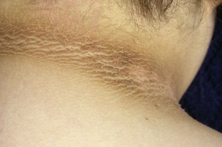
Acanthosis Nigricans
Abnormal Skin Pigmentation Finding
Roughening and darkening of skin in localized areas, especially the posterior neck
Suggests diabetes mellitus or PCOS
Diabetes Mellitus
PCOS
What conditions does Acanthosis Nigricans suggest?
“no lesions noted”
Normal Finding for Skin Lesions
Macule
Patch
Papule
Plaque
Nodule
Tumor
Vesicle
Bulla
Wheal
Pustule
Cyst
Primary Skin Lesions
Macule
Primary Skin Lesion
Any flat discolored lesion of less than 1 cm with circumscribed border
E.g., cherry angioma

Cherry Angioma
Example of a macule
Patch
Primary Skin Lesion
Flat discolored lesion of more than 1 cm with irregular border
E.g., vitiligo
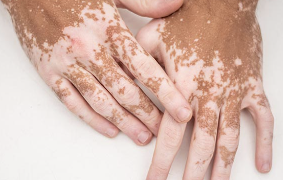
Vitiligo
Example of a patch
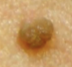
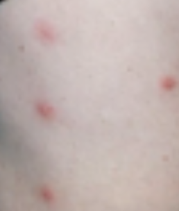
Papule
Primary Skin Lesion
Raised lesion of less than 0.5 cm with circumscribed border
E.g. wart
Wart
Example of a papule
Plaque
Primary Skin Lesion
Raised lesion of more than 0.5 cm
May coalesce or merge
E.g., seborrheic keratosis
Seborrheic Keratosis
Example of plaque
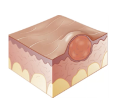
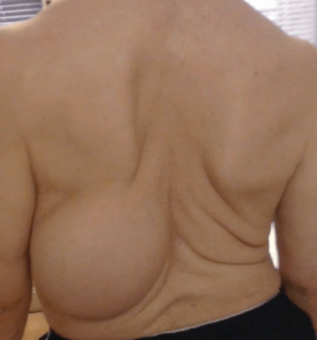
Nodule
Primary Skin Lesion
Solid mass of 0.5-2 cm with circumscribed border under dermis
E.g. lipoma
Lipoma
Example of nodule
Tumor
Primary Skin Lesion
Solid mass of tissue under dermis of more than 1-2cm but does not always have sharp borders
E.g., large lipoma
Large Lipoma
Example of a tumor
Vesicle
Primary Skin Lesion
Fluid-filled lesion of less than 0.5 cm
E.g. herpes simplex
Herpes Simplex
Example of a Vesicle
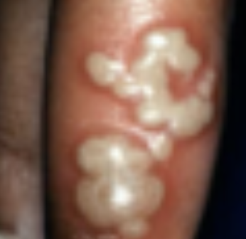
Bulla
Primary Skin Lesion
Fluid-filled lesion of more than 0.5 cm
E.g., large burn blister

Large Burn Blister
Example of a Bulla
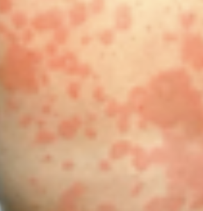
Wheal
Primary Skin Lesion
Rises from allergic reactions
E.g., urticaria/Hives
No fluid and not a raised lesion
Warm to touch
Urticaria/Hives
Example of a Wheal
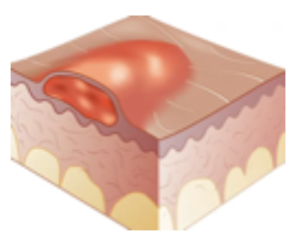
Pustule
Primary Skin Lesion
Pus filled lesion (yellow)
E.g., acne vulgaris
Presence means there is bacterial infection
Acne Vulgaris
Example of a Pustule
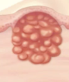
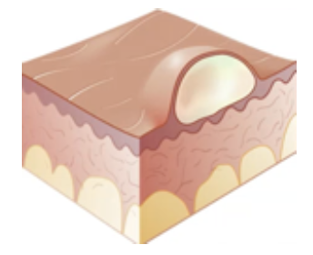
Cyst
Primary Skin Lesion
Semi-solid mass of skin and rubbery in texture
Mostly sacs containing fluids, air, etc
E.g., epidermoid cyst
More superficial and evident
Will increase in size
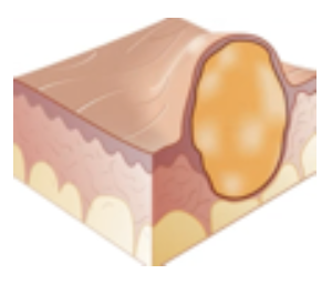
Epidermoid Cyst
Example of a Cyst
Erosion
Ulcer
Scar (Cicatrix)
Fissure
Scales
Crust
Keloid
Atrophy
Lichenification
Secondary Skin Lesions
Erosion
Secondary Skin Lesion
Eroded area of skin
Usually superficial
E.g., aphthous ulcer/cankersores, popper chicken pox
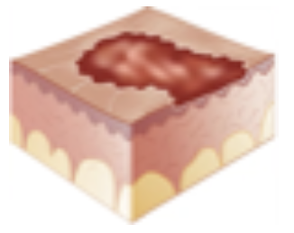
Aphthous Ulcer/Cankersores
Popper Chicken Pox
Examples of Erosion
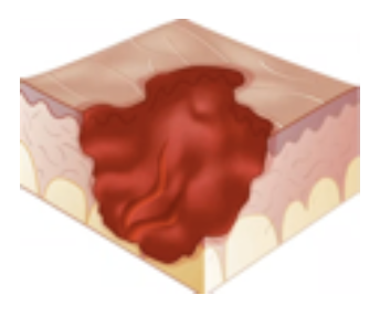
Ulcer
Secondary Skin Lesion
Deeper than erosion with many stages
Can extend to the bones
E.g., pressure __, advanced stage
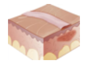
Scar (Cicatrix)
Secondary Skin Lesion
Remnant of wound, usually found after surgery
E.g., surgical site
Fissure
Secondary Skin Lesion
Linear crack on skin
E.g. tinea pedis/Athlete’s foot, Cheilosis
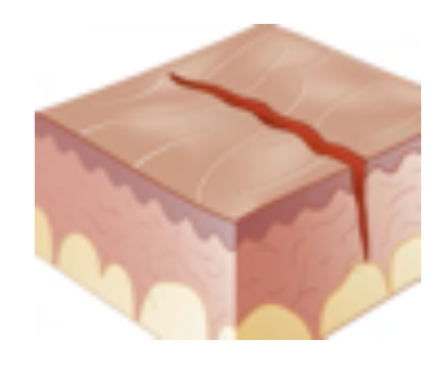
Tinea Pedis/Athlete’s Foot
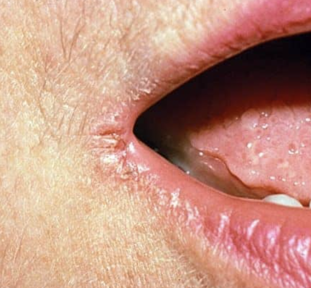
Cheilosis
Examples of a Fissure
Cheilosis (Fissure)
Secondary Skin Lesion
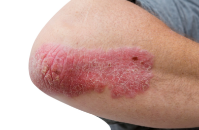
Scales
Secondary Skin Lesion
Portion of dead skin or desquamation of epidermis
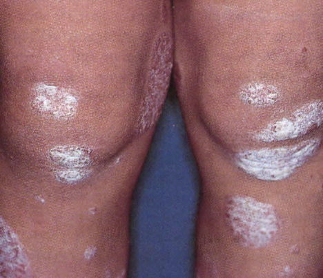
E.g., psoriasis
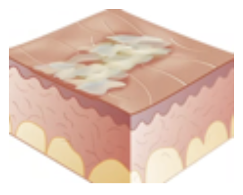
Psoriasis
Example of Scales
Scales
Secondary Skin Lesion
Crust
Secondary Skin Lesion
Dried serum/blood
Ruptured fluid filled lesion
E.g., ruptured vesicles of herpes simplex
Ruptured Vesicles of Herpes Simplex
Ruptured & Dried Chicken Pox Vesicles
Example of Crust
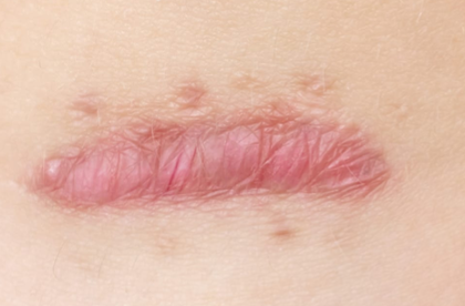
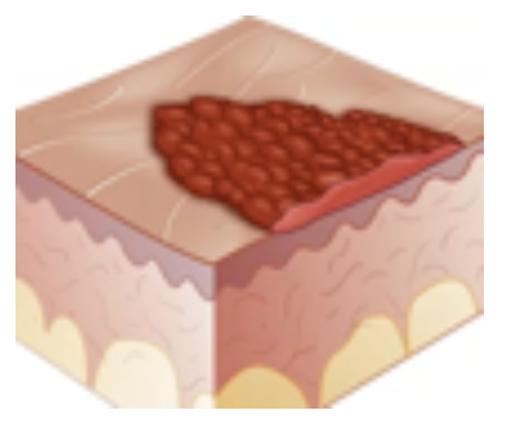
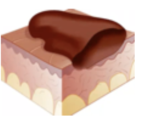
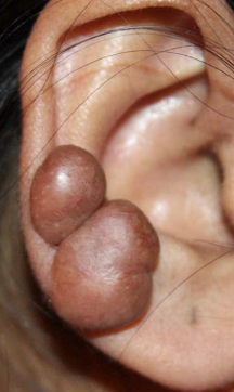
Keloid
Secondary Skin Lesion
Hypertrophic enlarged scar
Increase growth of muscle skin
E.g. ___ of ear piercing, injection, hereditary
Sca'r’s size won’t change, but ___ will hypertrophy
Keloid
Secondary Skin Lesion
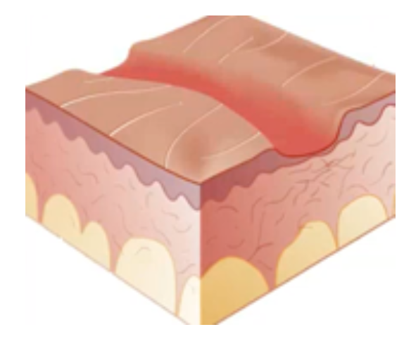
Atrophy
Secondary Skin Lesion
Degeneration of skin
Loss of elasticity and texture of skin normally
Common to elderly only (e.g., aged skin)
Presence on young px indicates dehydration
Aged Skin
Example of Atrophy
Atrophy

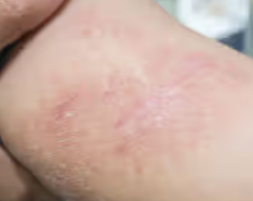
Lichenification
Secondary Skin Lesion
Degeneration of skin
Loss of elasticity and texture brought about by friction (e.g., contact dermatitis)
Contact Dermatitis (Lichenification)
Secondary Skin Lesion
Petechiae
Ecchymosis
Hematoma
Cherry Angioma
Spide Angioma
Telangiectasis (Venous Star)
Vascular Skin Lesions
Petechiae
Vascular Skin Lesion
FIne pinpoint lesions
Caused by rupture of capillaries
Common in dengue patients
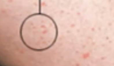
Ruptured Capillaries
What is petechiae consisted of?
Ecchymosis
Vascular Skin Lesion
Rupture of fine blood vessels from trauma or clotting problem
Reddish to purplish color (in comparison with hematoma)
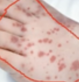
Ruptured Fine Blood Vessels
What is ecchymosis consisted of?
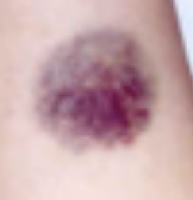
Hematoma
Vascular Skin Lesion
Due to trauma
Raised ecchymosis
Aka Bun-og; blue, purple, or black in color
Cherry Angioma
Vascular Skin Lesion
Common in elderly clients
angio means engorged arteriole
a macule
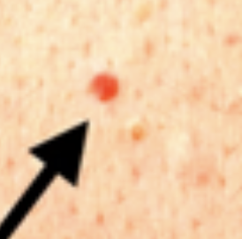
Engorged Arteriole
What is Cherry Angioma consisted of?
Spider Angioma
Vascular Skin Lesion
Dilated arterioles
Common to patients with liver problems, Vitamin B deficiency, and pregnant clients
Usually in abdomen or cheeks
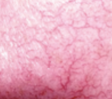
Dilated Arterioles
What is Spider Angioma consisted of?
Liver Problems
Vitamin B Deficiency
Pregnant Clients
What conditions have Spider Angioma?
Telangiectasis (Venous Star)
Vascular Skin Lesion
Dilated veins and venules commonly found in the popliteal area
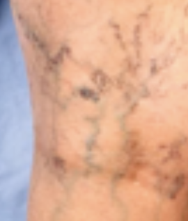
Dilated veins and venules
What does telangiectasis consist of?
Linear
Annular
Zosteriform
Discrete
Polycyclic
Confluent
Configurations of Skin Lesion
Linear
Configuration of Skin Lesion
Follows a straight line pattern

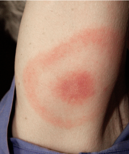
Annular
Configuration of Skin Lesion
Circular pattern
Usually common to ring worm patients or those with allergies

Ring worm patients
Allergies
What conditions usually have Annular Skin Lesions?
Zosteriform
Configuration of Skin Lesion
Spinal nerve root pattern
Horizontal or tranverse
Discrete
Configuration of Skin Lesion
Skin lesions are isolated with no pattern
Polycyclic
Configuration of Skin Lesion
Itchiness
Tends to merge together
E.g., pruritic ___ macular lesion
Pruritus itchiness
Confluent
Configuration of Skin Lesion
Localized in one small body area, merge
Stage 1 Unblanched Erythema
Pressure Ulcer Stage
Intact skin with non-blanchable redness of a localized area usually over a bony prominence
Darkly pigmented skin may not have visible blanching; its color may differ from the surrounding area