Looks like no one added any tags here yet for you.
bones are ________________.
multifunctional
bone functions
support/protect softer tissues, points of attachment for muscles, blood production, store inorganic salts
bones are classified by __________.
shape
what are the classifications of bones?
long, short, flat, irregular
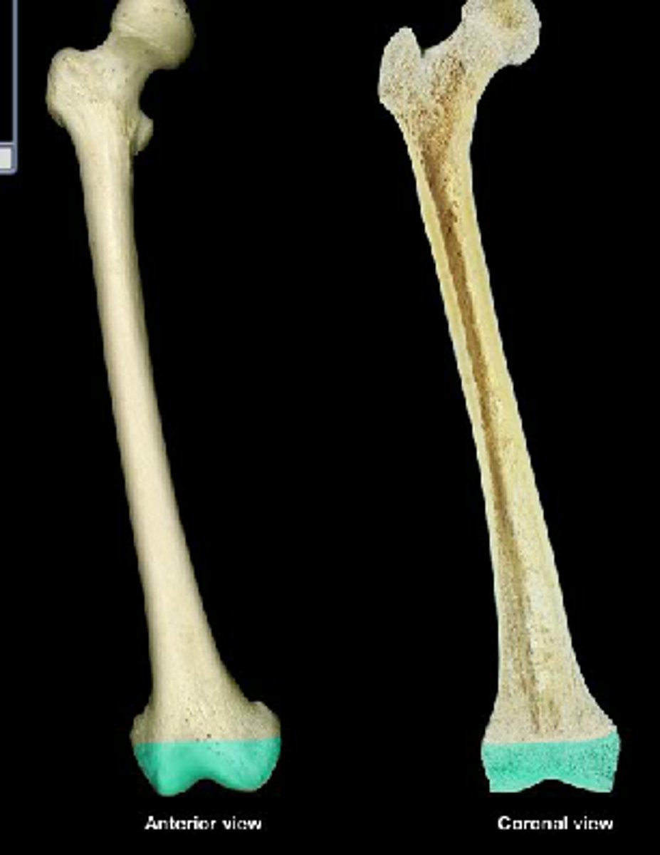
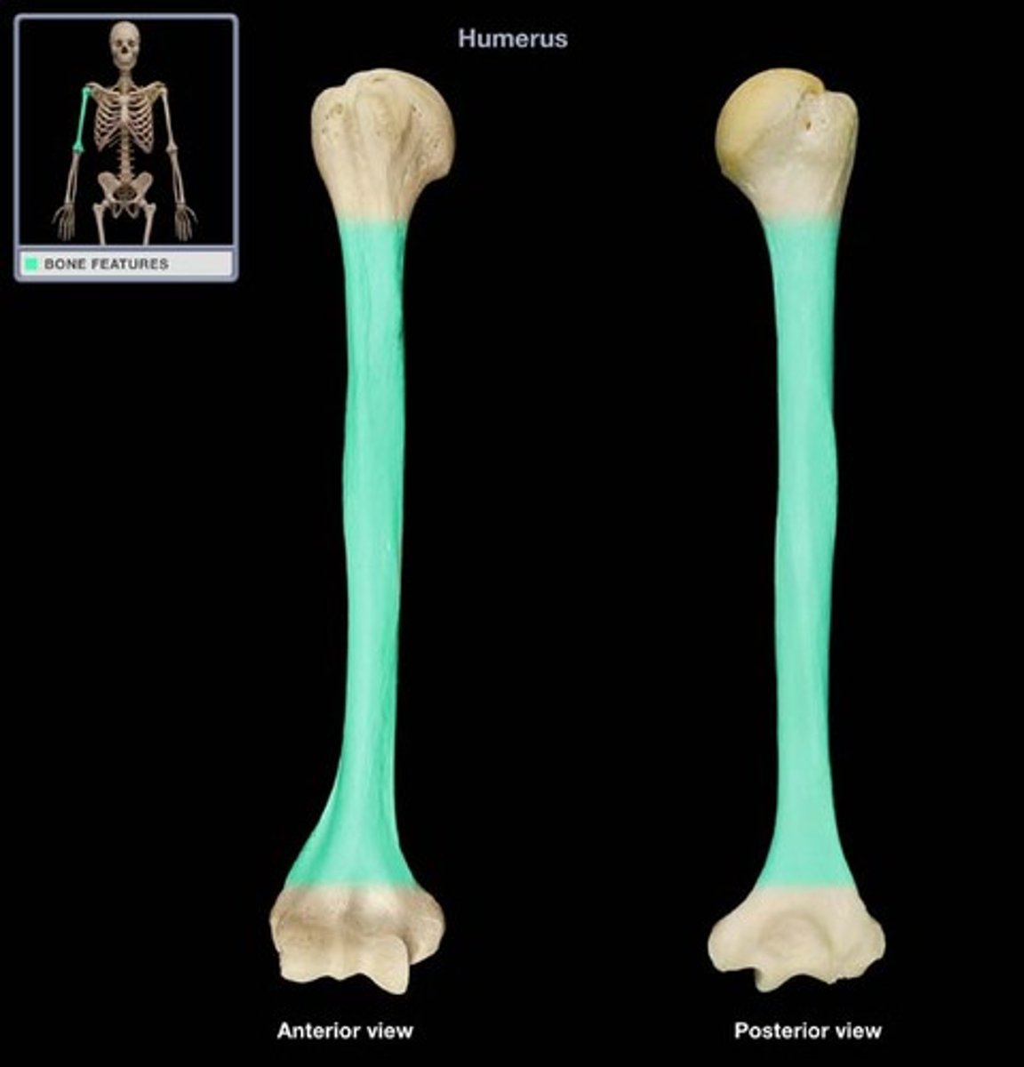
long bones
long longitudinal axes and expanded ends
what is an example of a long bone?
femur, forearm
short bones
equal in length and width
what are examples of short bones?
wrist and ankle bones
sesamoid bone
short bones that form within tendons ex: patella
flat bones
platelike structures with broad surfaces
what are examples of flat bones?
ribs, scapulae, sternum, parts of skull
irregular bones
variety of shapes, most are connected to other bones
what are examples of irregular bones?
vertebrae and facial bones
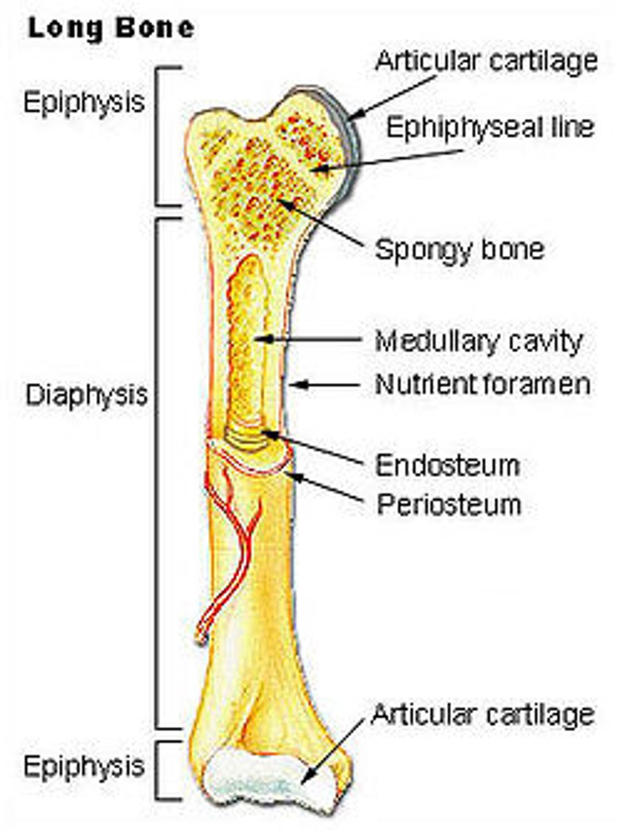
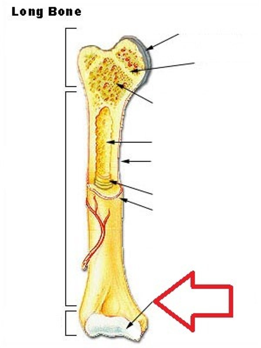
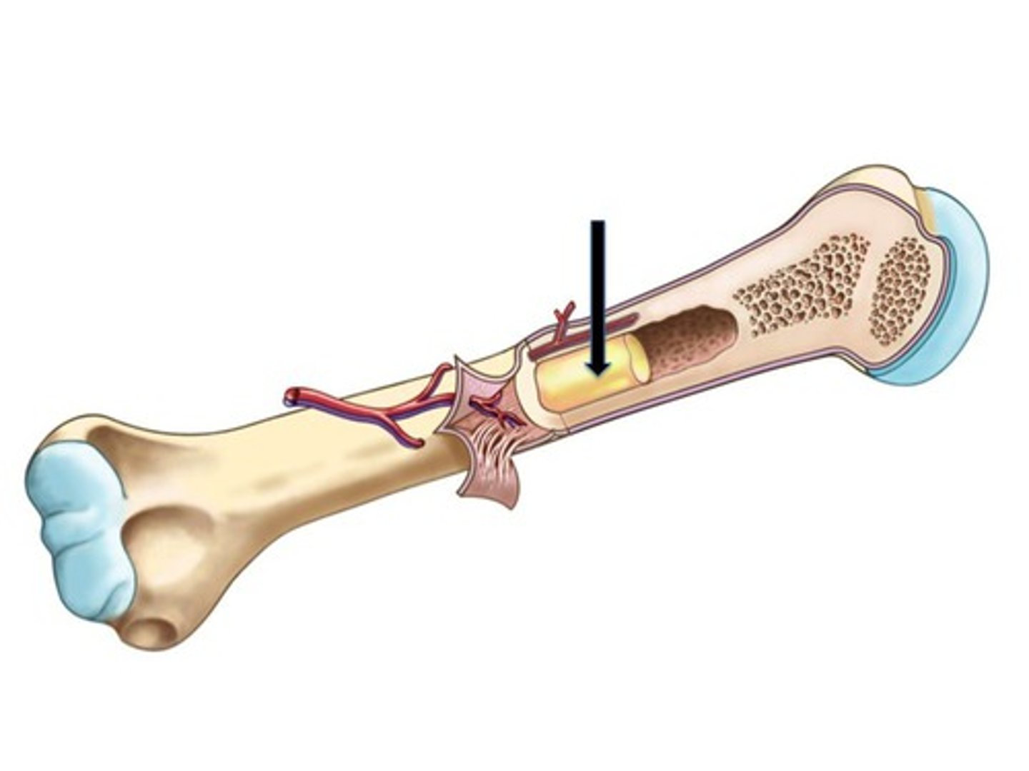
parts of long bone
epiphysis, diaphysis, compact bone, spongy bone, articular cartilage, periosteum, endosteum, medullary cavity, trabeculae, marrow
epiphysis
expanded end of long bone; articulates another bone, proximal and distal ends
articular cartilage
hyaline cartilage that covers end of epiphysis
diaphysis
bone shaft
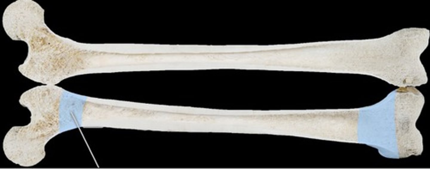
metaphysis
widened part of bone between diaphysis and epiphysis
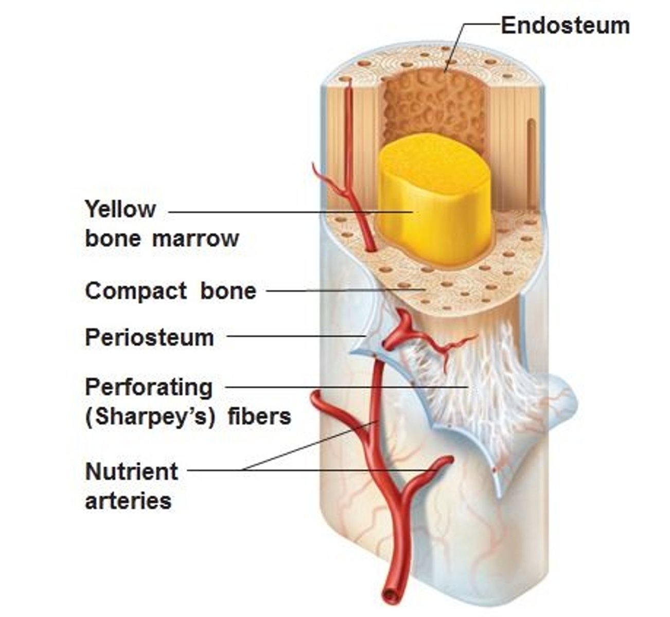
periosteum
vascular covering of dense connective tissue that encloses entire bone; helps form and repair bone tissue
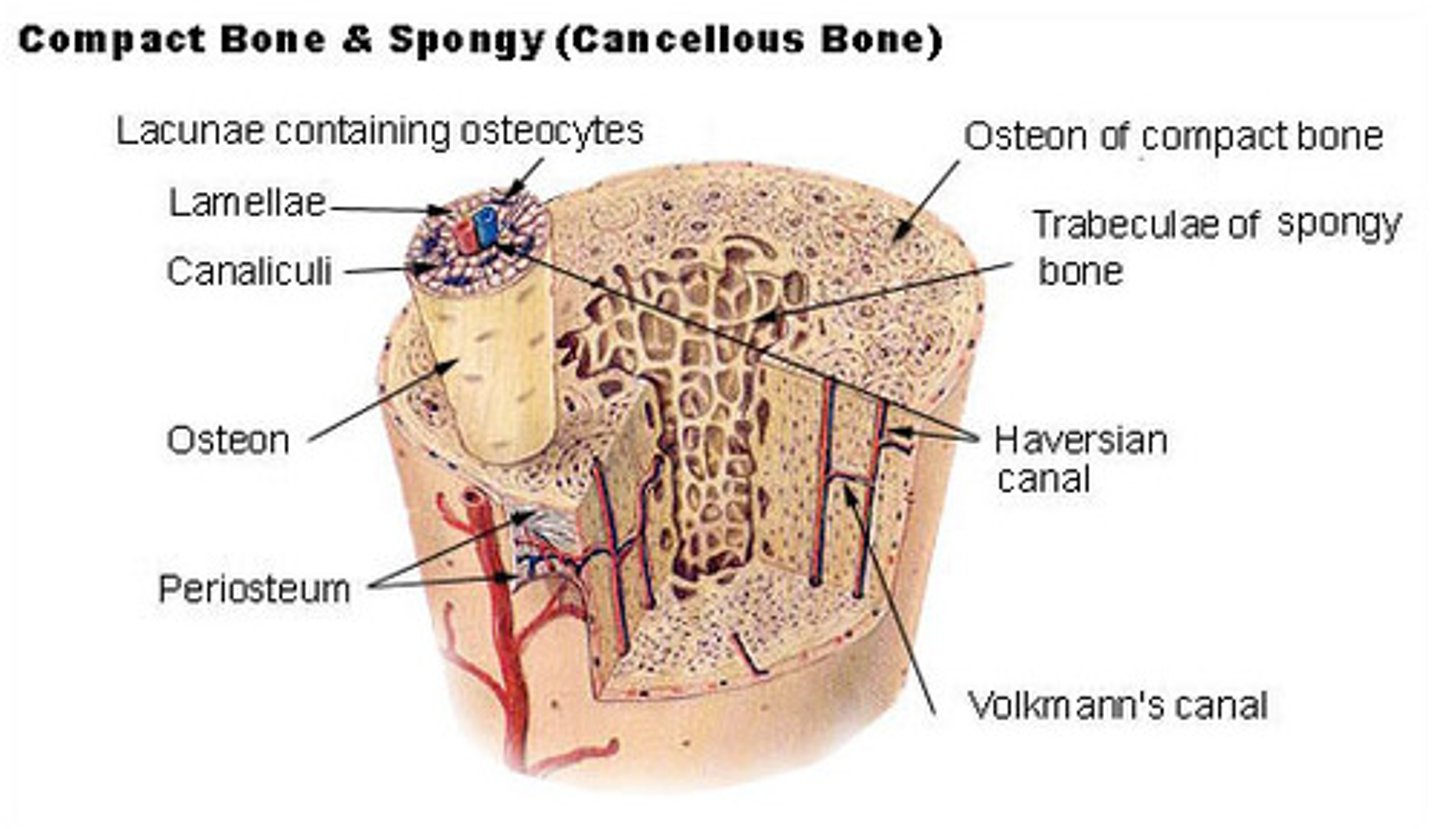
compact bone
tightly packed tissue, continuous ECM with no gaps, makes up wall of diaphysis
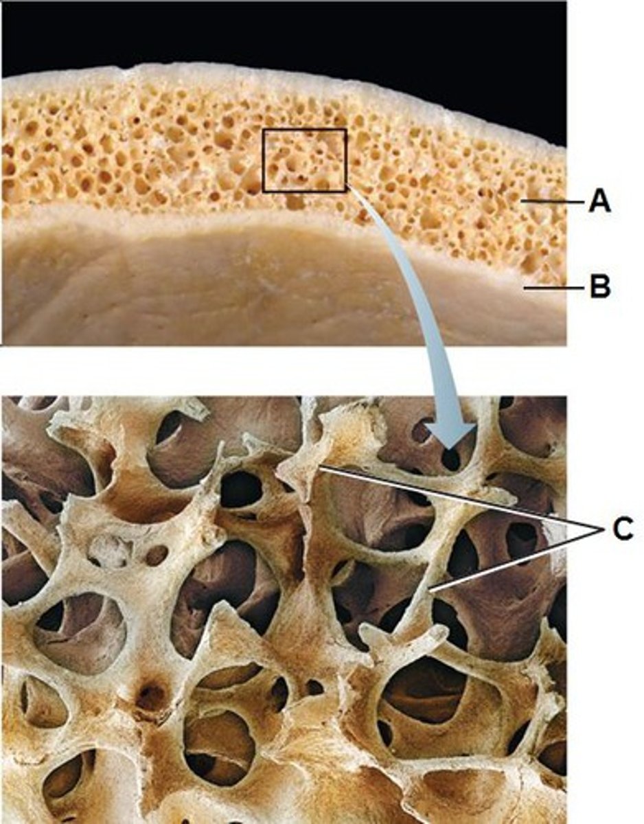
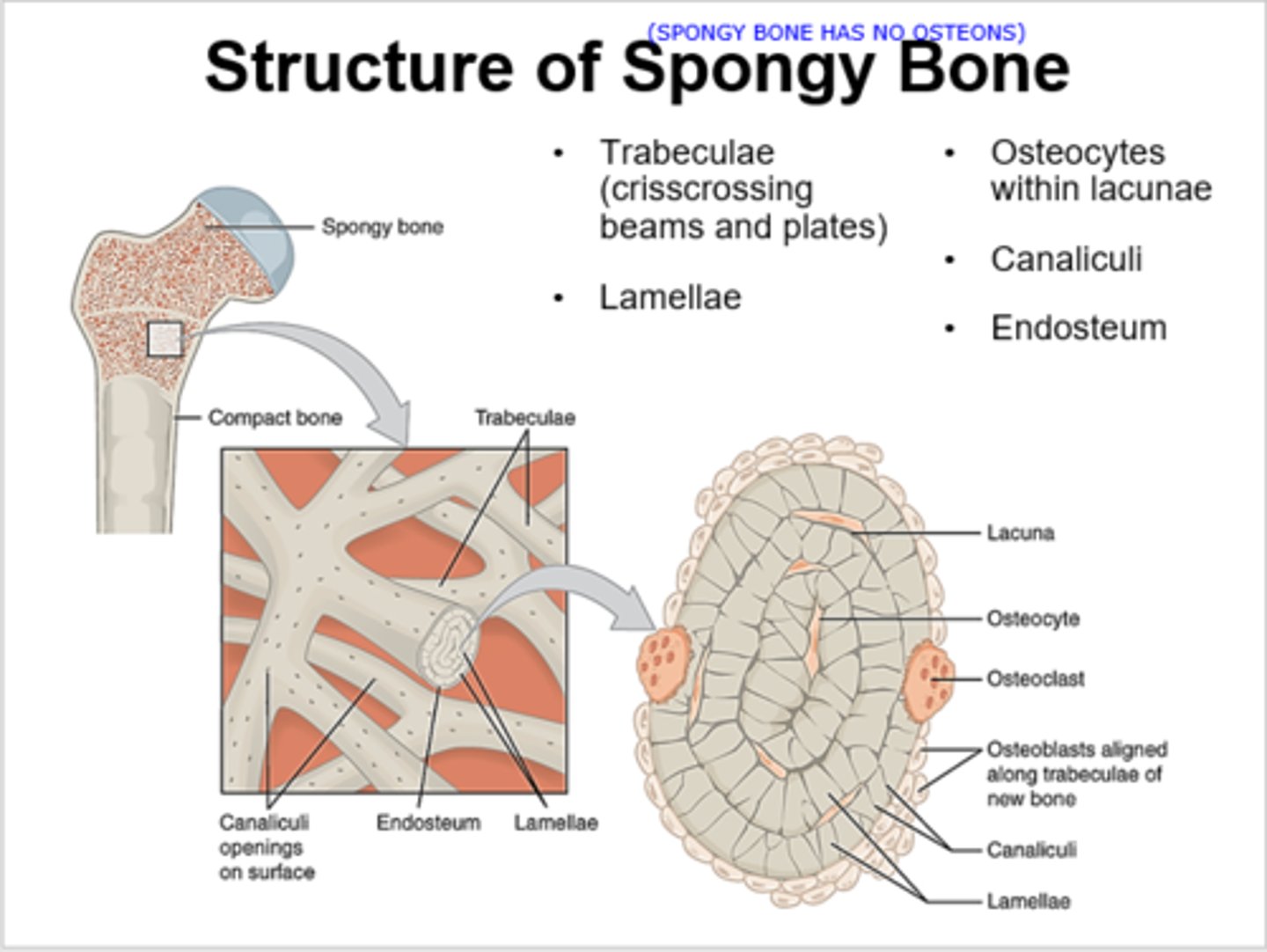
spongy bone
branching bony plates called trabeculae, spaces help reduce weight of bone, makes up most epiphyses
trabeculae
branching bony plates that make up spongy bone
spongy bone is found in regions that are prone to _______________.
compression
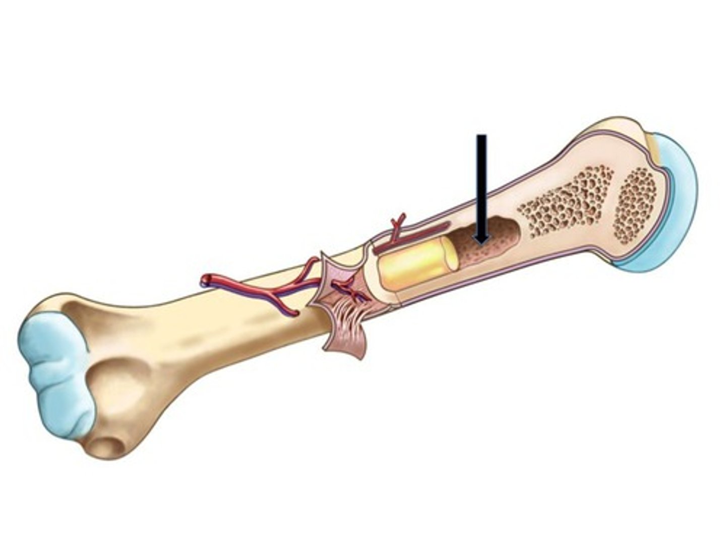
medullary cavity
tube with hollow chamber of compact bone in diaphysis of long bone; continuous with some spaces of spongy bone
endosteum
thin membrane of bone forming cells that line medullary cavity and spaces within spongy bone
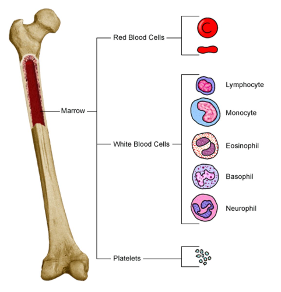
marrow
specialized soft connective tissue that fills spongy bone spaces and medullary cavity
yellow marrow
soft, fatty material found in the medullary cavity of long bones; stores fat
red marrow
location of blood cell formation; usually found near epiphysis in long bones
bone cells are called ___________.
osteocytes
lacunae
small cavities in bone that contain osteocytes
canaliculi
channels that connect lacunae; allows neighboring osteocytes to exchange substances with each other
ECM of bone tissues is composed of __________ and ___________.
collagen and inorganic salts
haversian canal
central canal in compact bone containing blood vessels and nerves; forms shape of osteon
volkmans canal
horizontal canals between osteons; contain a nerve and a blood vessel
collagen gives bones _________ and resilience.
strength
inorganic salts make bones resistant to ___________.
crushing
why do osteons run longitudinally with the axis of a bone?
it allows the bone to bear weight and resist compression
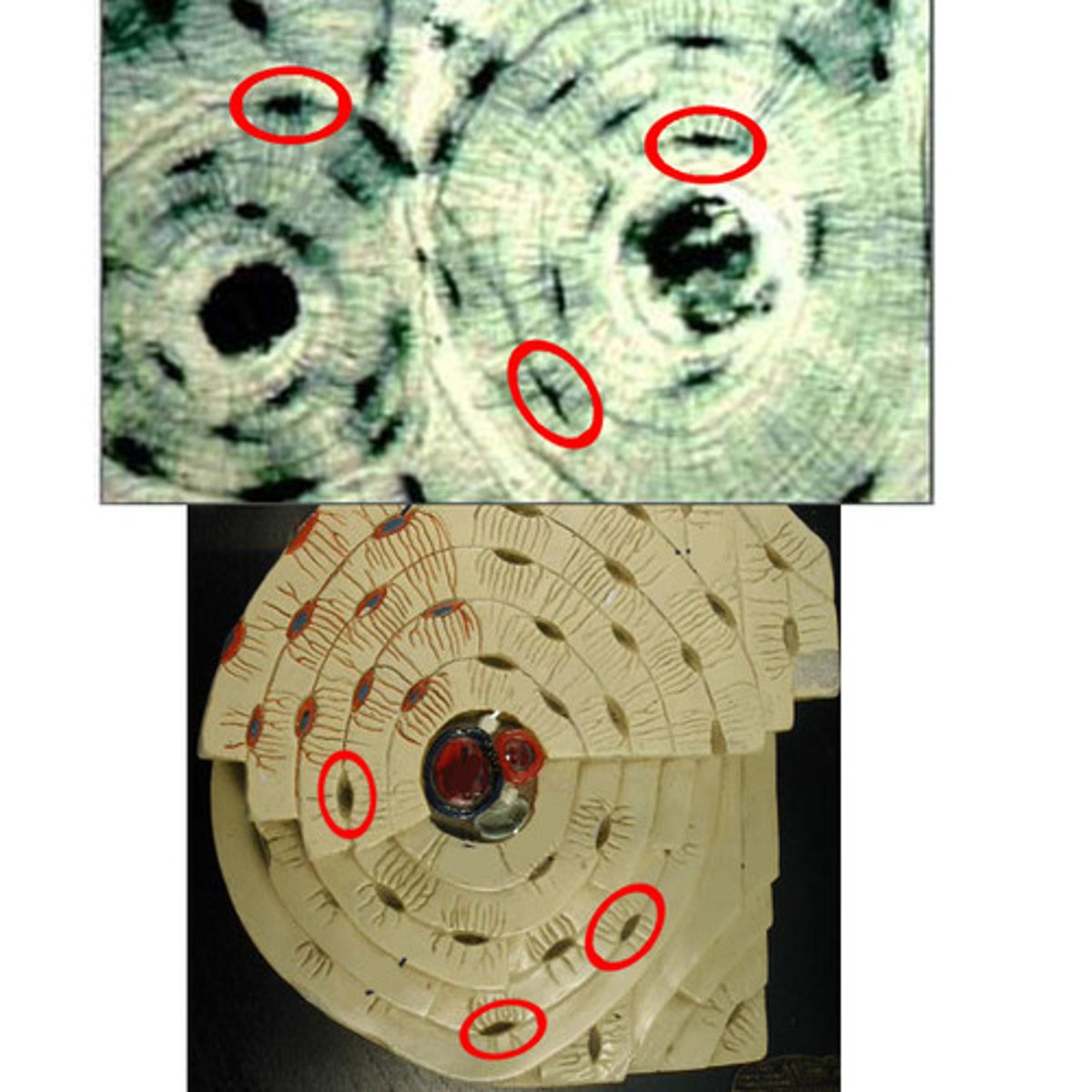
compact bone is made of many _________ together.
osteons
compact bone diagram
do spongy bone cells surround the haversian canal?
no
spongy bone cells are in the ________________.
trabeculae
how do spongy bone cells receive nutrients?
from the diffusion of nutrients through the canaliculi
spongy bone diagram
ossification
formation of bone
intramembranous bones
sheet like layers of connective tissue
when do parts of the skeletal system begin to develop in fetuses?
during the first few weeks of pregnancy
what are the 2 types of bone formation?
intramembranous and endochondral
intramembranous ossification
- bones develop as sheet like layers of connective tissue
- produces broad/flat bones
what are examples of bones that go through intramembranous ossification?
skull bones (not mandible, clavicles, sternum, facial bones
endochondral ossification
- bones develop from hyaline cartilage
- occurs in most bones of the skeleton
intramembranous ossification replaces _____________________________.
embryonic connective tissue
process of intramembranous ossification
- mesenchymal cells in primitive tissue differentiate into osteoblasts
- once osteoblasts are completely surrounded by matrix, they are called osteocytes in lacunae
- mesenchyme on outside forms periosteum
mesencyme
embryonic connective tissue
process of endochondral ossification
- begins as hyaline cartilage
- chondrocytes in large, lacunae grows
- matrix breaks down, chondrocytes die
- osteoblasts invade and deposit bone matrix
- osteoblasts from spongy then compact bone
- osteoblasts are called osteocytes once in matrix
primary ossification center
center of diaphysis where bone tissue first starts to replace cartilage
examples of endochondral bones
Femur, humerus, radius, tibia, phalanges, vertebrae
secondary ossification center
area of epiphyses; spongy bone forms later in development
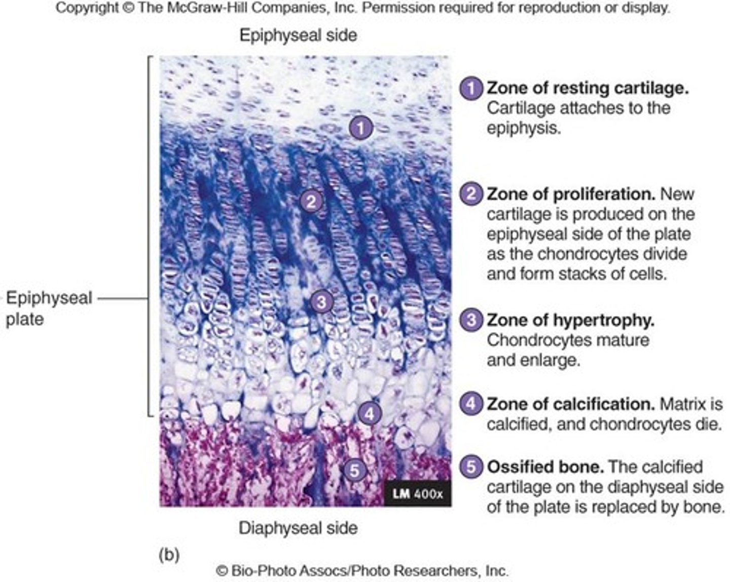
epiphyseal plate
a band of cartilage that remains between the two ossification centers; remains as spongy bone is deposited into the diaphysis and epiphysis
when does a fetus begin to show development of intramembranous and endochondral bones?
at 14 weeks
osteogenesis imperfecta
hereditary disease that involves a collagen defect; bones are extremely brittle
the epiphyseal plate is also called the ______________.
growth plate
in long growing long bones, the growth plate separates the diaphysis from the ___________.
epiphysis
metaphysis
growth zone between the epiphysis and the diaphysis during development of a long bone
there are ___ layers of the epiphyseal plate.
4
what are the 4 layers of the epiphyseal plate?
resting zone, proliferating zone, hypertrophic zone, calcified zone
zone of resting cartilage
- nearest the epiphysis and contains randomly arranged chondrocytes that do not divide rapidly
- anchors epiphyseal plate to tissue of epiphysis
zone of proliferating cartilage
- rows of young cells undergoing mitosis; rapid cell division
- cartilaginous plate thickens as new cells grow and ECM forms
zone of hypertrophic cartilage
- consists of large, maturing chondrocytes arranged in columns
- thickens epiphyseal plate and lengthens bone
- osteoblasts secrete calcium salts to calcify the matrix
zone of calcified cartilage
thin layer of dead cartilage cells and calcified matrix
at the growth plate, osteoclasts break down ________________.
calcified matrix
osteoblasts invade the epiphyseal plate and deposit _________ tissue in place of calcified cartilage.
bone
how does bone thicken?
deposition of compact bone on outside
when does bone remodeling occur?
throughout life
bone resorption
removal of bone by osteoclasts
bone deposition
formation of bone by osteoblasts
where does bone resorption and bone deposition occur?
surfaces of endosteum and periosteum
bone remodeling is not ____________ and depends on bone type.
uniform
bone remodeling is tightly ___________.
regulated
what percent of bone tissue is replaced per year?
10-20%
what factors effect bone development, repair, and growth?
nutrition, sunlight exposure, hormonal secretions, physical exercise
vitamin D role in bones
calcium absorption; deficiency causes rickets (children) and osteomalacia (adults)
vitamin A role in bones
osteoblast and osteoclast activity; deficiency hinders bone development
vitamin C role in bones
collagen synthesis; deficiency causes slender and fragile bones
proteins role in bones
make up large percentage of bones
collagen makes up ______ percent of bones.
90
growth hormone role in bones
stimulates cartilage cell division in epiphyseal plates
growth hormone is secreted by the __________ gland.
pituitary
pituitary dwarfism
insufficient production of growth hormone in children
pituitary giantism
hypersecretion of growth hormone in childhood
acromegaly
enlargement of the extremities in adults
thyroid gland role in bone
- secretes thyroxine (T4) hormone
- stimulates osteoblasts
parathyroid gland role in bone
- secretes parathyroid hormone
- stimulates osteoclasts
sex hormones role in bones
- promote bone formation at puberty
- stimulate ossification of epiphyseal plates to stop bone lengthening
fractures are classified by
cause and nature of break
fracture classifications by cause
traumatic and spontaneous/pathologic
traumatic fractures
caused by injury
pathologic/spontaneous fractures
caused by disease
fracture classifications by break
simple and compound
simple fractures
fracture protected by uninjured skin
compound fractures
bone penetrates skin and is exposed through outside