Looks like no one added any tags here yet for you.
Hormones
Can you label, describe and explain what this diagram is/shows?
Endocrine system: overview
How is the endocrine system related to the nervous system?
How does the endocrine system maintain homeostasis?
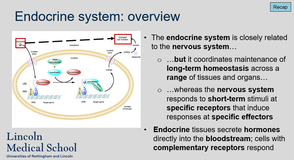
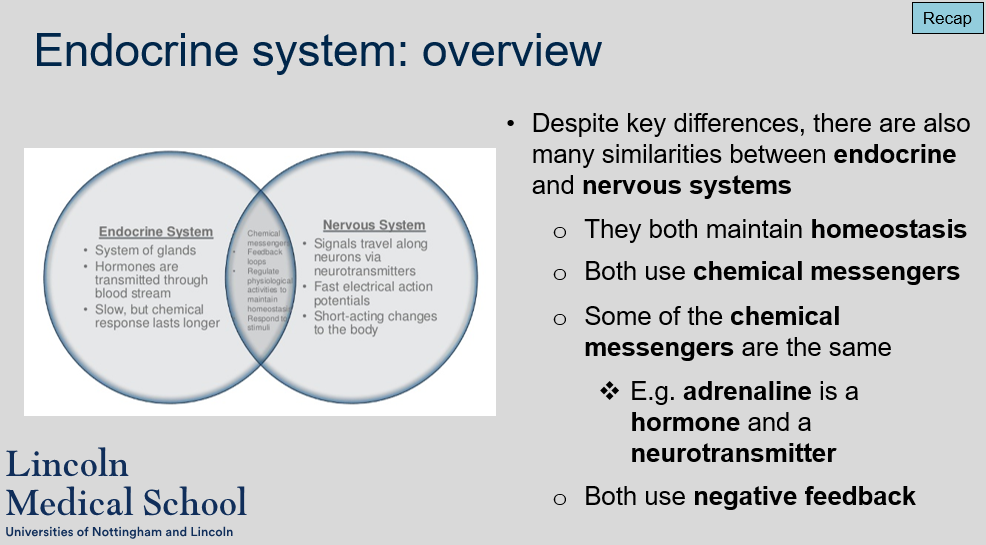
The endocrine system is closely related to the nervous system, as both systems are involved in coordinating the body's physiological responses. However, while the nervous system responds to short-term stimuli at specific receptors that induce responses at specific effectors, the endocrine system coordinates the maintenance of long-term homeostasis across a range of tissues and organs.
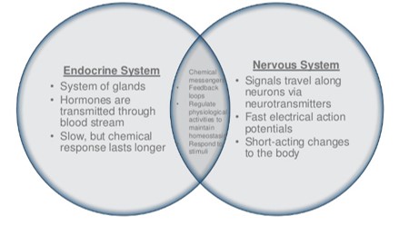
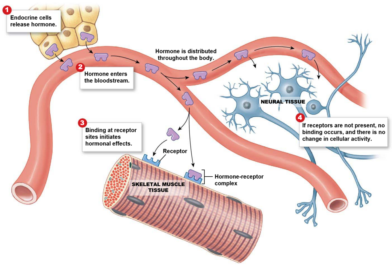
The endocrine system maintains homeostasis by secreting hormones directly into the bloodstream. Cells with complementary receptors respond to these hormones, allowing the endocrine system to coordinate physiological responses across multiple tissues and organs.
Endocrine system: overview
Can you label, describe and explain what this diagram is/shows?
Endocrine system: overview
What are some similarities between the endocrine and nervous systems?
Despite key differences, there are many similarities between the endocrine and nervous systems. Both systems play a critical role in maintaining homeostasis, use chemical messengers to transmit signals, and use negative feedback mechanisms to regulate physiological processes. Additionally, some of the chemical messengers used by the two systems are the same, such as adrenaline, which functions as both a hormone and a neurotransmitter.
Endocrine system: overview
Can you label, describe and explain what this diagram is/shows?
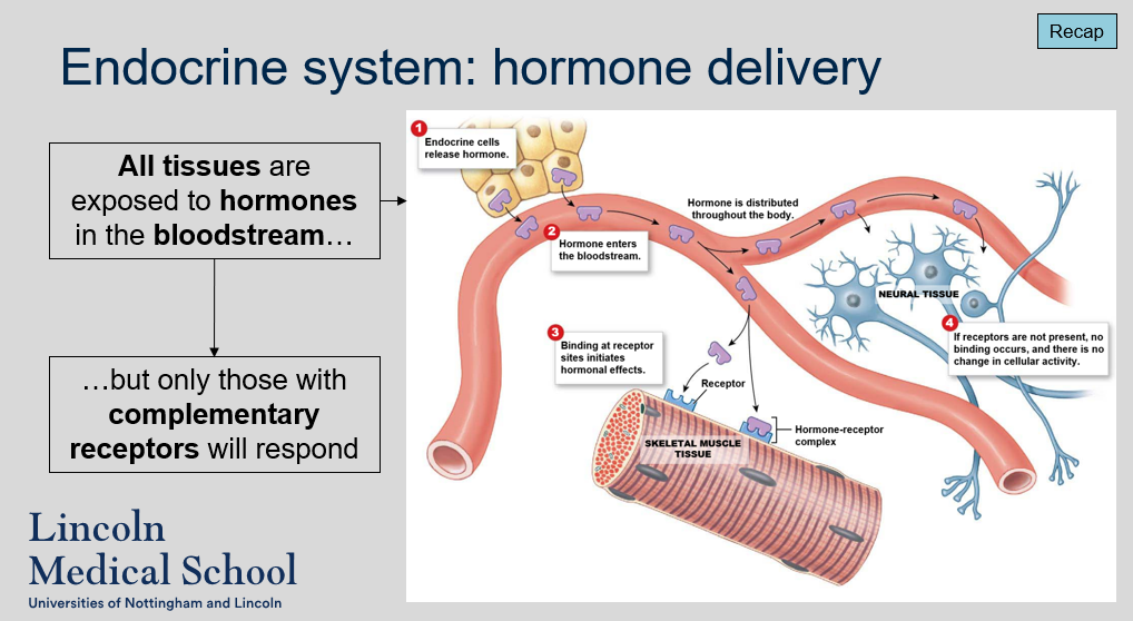
Endocrine system: hormone delivery
Do all tissues in the body respond to hormones in the bloodstream?
Yes, all tissues in the body are exposed to hormones in the bloodstream. However, only those tissues that have complementary receptors for a specific hormone will respond to that hormone.
Endocrine system: hormone delivery
Can you label, describe and explain what this diagram is/shows?
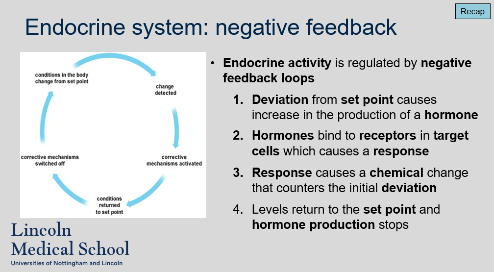
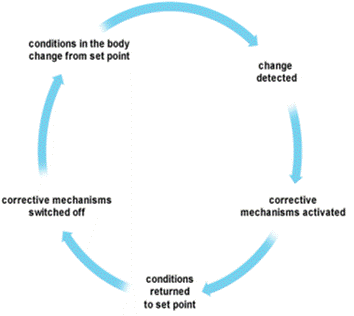
Endocrine system: negative feedback
How is endocrine activity regulated?
Endocrine activity is regulated by negative feedback loops. When there is a deviation from the set point of a physiological parameter, such as blood glucose levels, it causes an increase in the production of a hormone. The hormone then binds to receptors in target cells, triggering a response. This response causes a chemical change that counters the initial deviation, bringing the parameter back to the set point. Once the levels return to the set point, hormone production stops, completing the negative feedback loop.
Endocrine system: negative feedback
Can you label, describe and explain what this diagram is/shows?
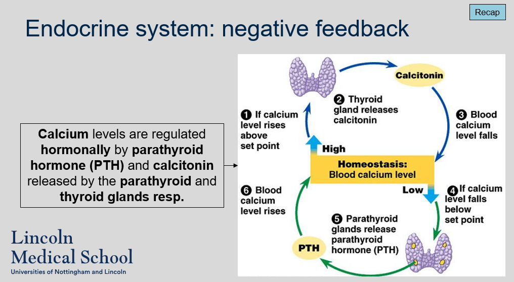
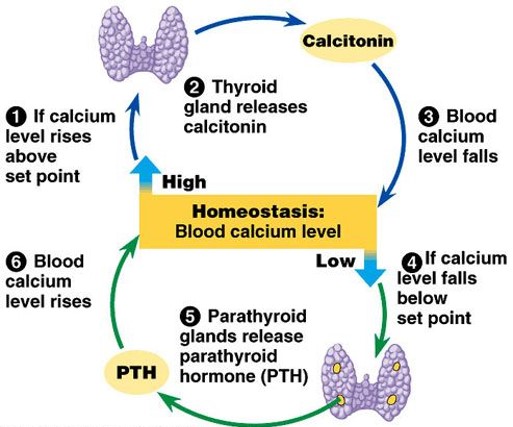
Endocrine system: negative feedback
How are calcium levels in the body regulated hormonally?
Calcium levels in the body are regulated hormonally by two hormones: parathyroid hormone (PTH) and calcitonin. PTH is released by the parathyroid glands, while calcitonin is released by the thyroid gland.
Endocrine system: negative feedback
Can you label, describe and explain what this diagram is/shows?
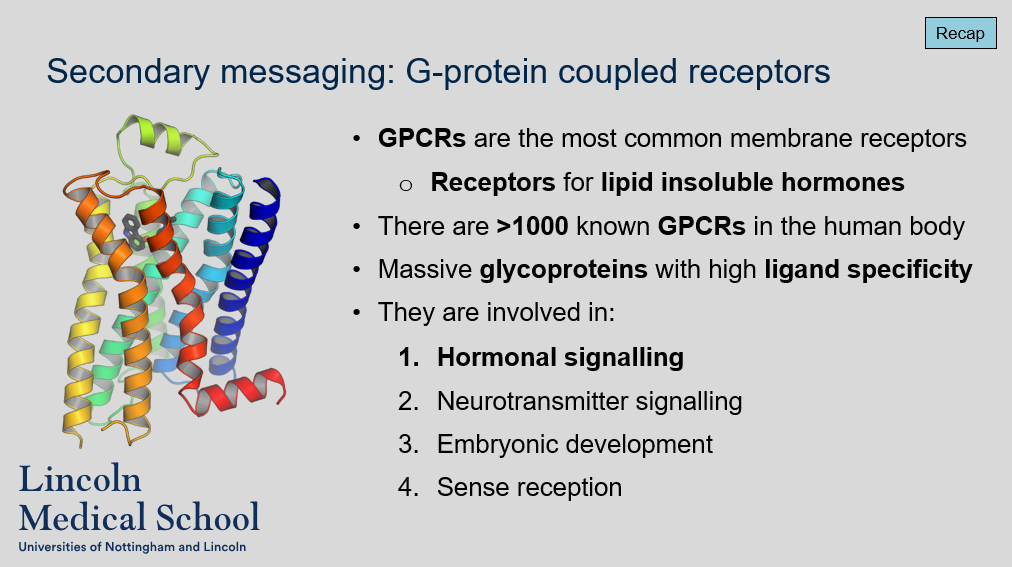
Secondary messaging: G-protein coupled receptors
What are GPCRs?
Are GPCRs the most common membrane receptors?
What type of hormones do GPCRs bind to?
How many GPCRs are there in the human body and what are their functions?
What is the structure of GPCRs?
GPCRs (G protein-coupled receptors) are a type of membrane receptor that are involved in transmitting signals from the external environment to the inside of the cell.
Yes, GPCRs are the most common type of membrane receptor in the human body.
GPCRs primarily bind to lipid insoluble hormones, such as adrenaline and noradrenaline.
There are over 1000 known GPCRs in the human body, which are involved in a wide range of physiological processes, including hormonal signaling, neurotransmitter signaling, embryonic development, and sense reception.
GPCRs are massive glycoproteins with high ligand specificity.
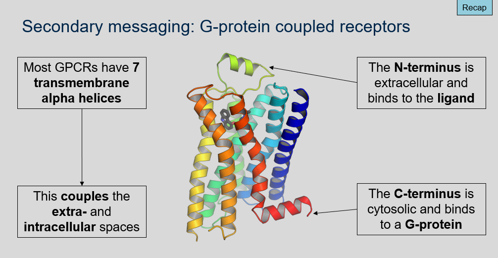
Secondary messaging: G-protein coupled receptors
What is the typical structure of a G protein-coupled receptor (GPCR)?
Where is the N-terminus located in relation to the cell membrane in a GPCR, and what is its function?
Where is the C-terminus located in relation to the cell membrane in a GPCR, and what is its function?
Most GPCRs have seven transmembrane alpha helices, which span the cell membrane and couple the extracellular and intracellular spaces.
The N-terminus of a GPCR is located on the extracellular side of the cell membrane. It is responsible for binding to the ligand (such as a hormone or neurotransmitter) that activates the receptor.
The C-terminus of a GPCR is located on the cytosolic side of the cell membrane. It is responsible for binding to a G protein, which activates intracellular signaling pathways in response to ligand binding at the receptor.
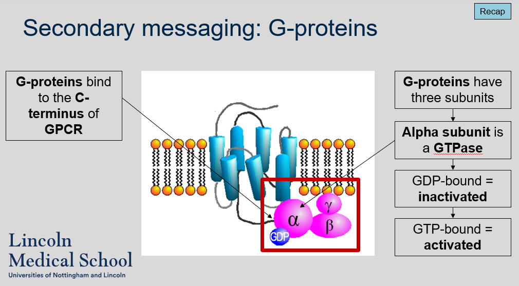
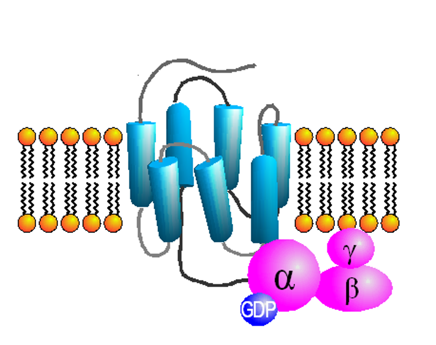
Secondary messaging: G-proteins
What is the role of G-proteins in G protein-coupled receptor (GPCR) signaling?
How many subunits do G-proteins have, and what are their names?
What is the function of the alpha subunit of a G-protein, and what happens to it when it is bound to GTP or GDP?
G-proteins are intracellular signaling molecules that bind to the C-terminus of activated GPCRs and transmit signals to downstream effector molecules.
G-proteins have three subunits, which are called alpha, beta, and gamma.
The alpha subunit of a G-protein is a GTPase, meaning it can hydrolyze GTP (guanosine triphosphate) to GDP (guanosine diphosphate) and release energy. When the alpha subunit is bound to GDP, it is inactivated and unable to transmit signals downstream. However, when it is bound to GTP, it is activated and can dissociate from the beta and gamma subunits to interact with downstream effector molecules.
Secondary messaging: G-proteins
Can you label, describe and explain what this diagram is/shows?
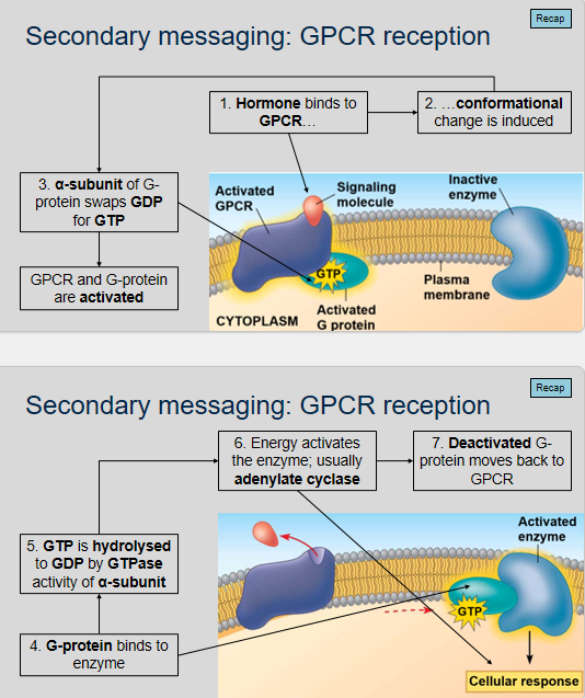
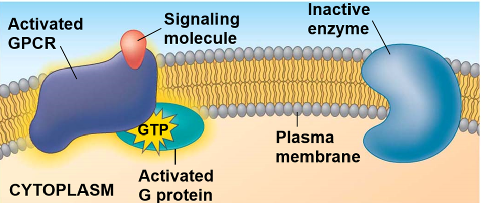
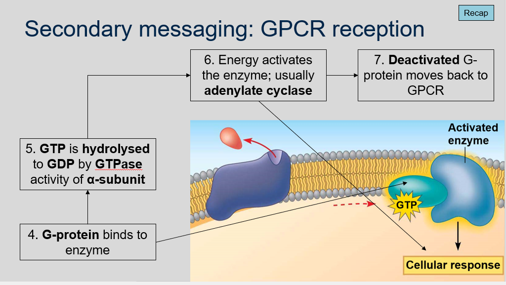
Secondary messaging: GPCR reception
What is GPCR reception?
What happens when the α-subunit of the G-protein swaps GDP for GTP?
What is the role of the G-protein in GPCR signaling?
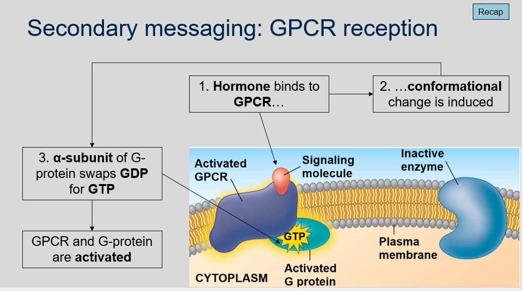
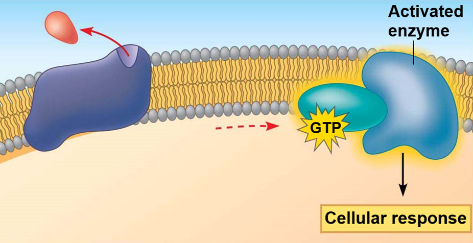
What happens after the G-protein binds to the enzyme?
What happens when the G-protein is deactivated?
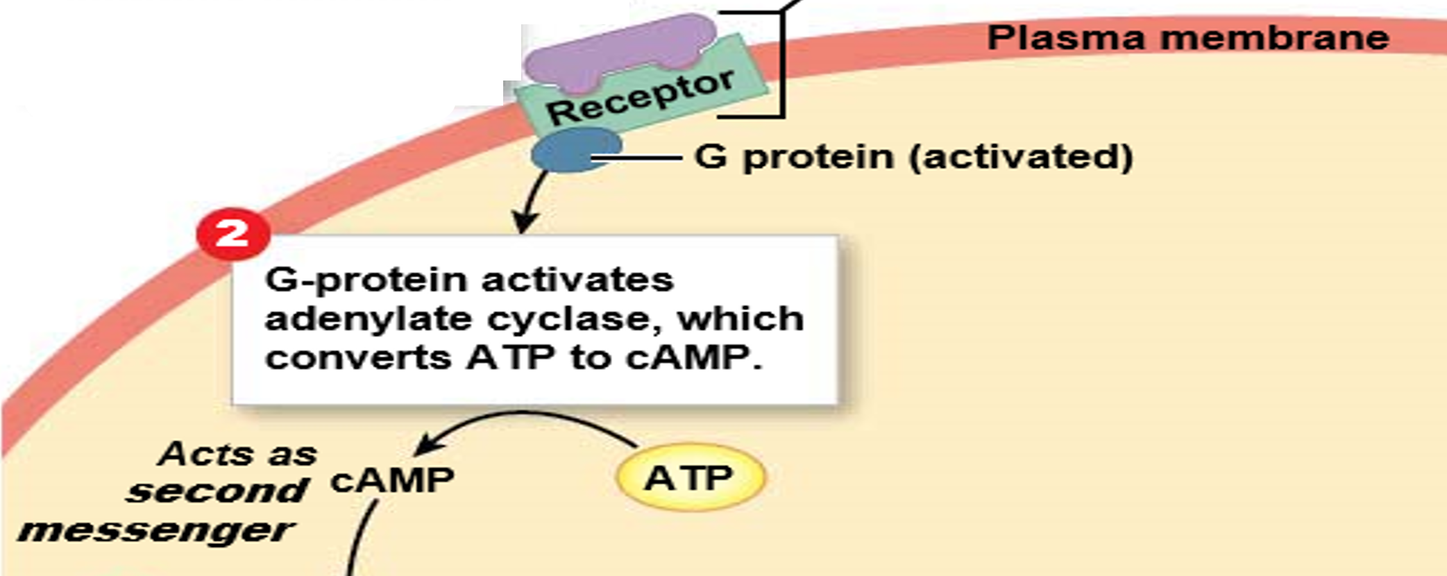
GPCR reception is the process by which a hormone or ligand binds to a G protein-coupled receptor (GPCR), inducing a conformational change in the receptor.
When the α-subunit of the G-protein swaps GDP for GTP, the GPCR and G-protein become activated, initiating downstream signaling pathways.
The G-protein binds to an enzyme.
After the G-protein binds to the enzyme, GTP is hydrolyzed to GDP by the GTPase activity of the α-subunit. This releases energy that activates the enzyme, typically adenylate cyclase, which catalyzes the conversion of ATP to cyclic AMP (cAMP).
When the G-protein is deactivated, it dissociates from the enzyme and moves back to the GPCR, where it re-associates with the receptor in its inactive state, ready for another round of signaling.
Secondary messaging: GPCR reception
Can you label, describe and explain what this diagram is/shows?
Secondary messaging: GPCR reception
Can you label, describe and explain what this diagram is/shows?
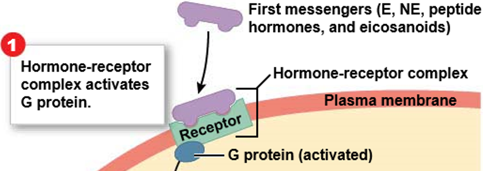
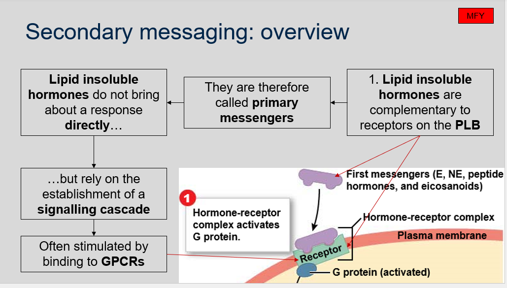
Secondary messaging: overview
What are lipid insoluble hormones, and how do they initiate cellular signaling pathways?
Lipid insoluble hormones are complementary to receptors on the PLB, therefore are called primary messengers. Lipid insoluble hormones do not bring about a response directly but rely on the establishment of a signaling cascade, often stimulated by binding to GPCRs.
Secondary messaging: overview
Can you label, describe and explain what this diagram is/shows?
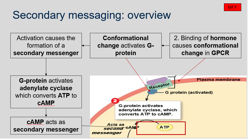
Secondary messaging: overview
How does binding of a hormone to a G protein-coupled receptor (GPCR) result in the activation of a secondary messenger and ultimately lead to cellular responses?
The binding of a hormone to a GPCR causes a conformational change in the GPCR, which in turn activates a G protein. The activation of the G protein leads to the formation of a secondary messenger, such as cyclic AMP (cAMP). The G protein then activates an enzyme called adenylate cyclase, which converts ATP to cAMP. The cAMP acts as a secondary messenger to activate downstream effector molecules and ultimately lead to cellular responses.
Secondary messaging: overview
Can you label, describe and explain what this diagram is/shows?
Secondary messaging: signal amplification
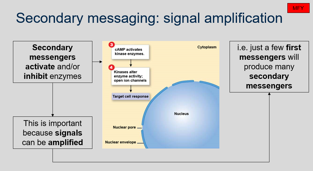
Why is the ability of secondary messengers to activate and/or inhibit enzymes important in cellular signaling pathways?
Secondary messengers are important in cellular signaling pathways because they can activate or inhibit enzymes, allowing for the amplification of signals. Even just a few primary messengers can trigger the production of multiple secondary messengers, which can then activate downstream enzymes and result in significant cellular responses. This amplification of signals is important for efficient and effective cellular signaling.
Secondary messaging: cAMP
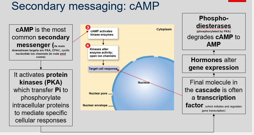
What is the role of cAMP in cellular signaling pathways and how is it regulated?
cAMP is a common secondary messenger that can activate protein kinases, such as PKA, to phosphorylate intracellular proteins and mediate specific cellular responses. Its downstream targets also include cyclic nucleotide ion channels in rods and cones of the eye. The final molecule in the signaling cascade is often a transcription factor, which initiates and regulates gene transcription and leads to changes in cellular behavior. Hormones can alter gene expression by activating cAMP-mediated signaling pathways. To regulate the level of cAMP, phosphodiesterases, which are phosphorylated by PKA, degrade cAMP to AMP.
Secondary messaging: cAMP
Can you label, describe and explain what this diagram is/shows?
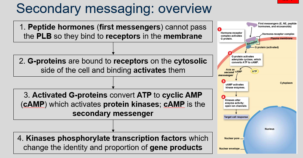
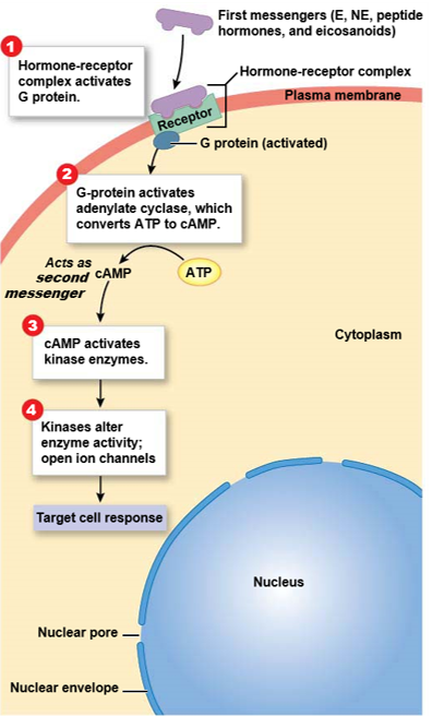
Secondary messaging: overview
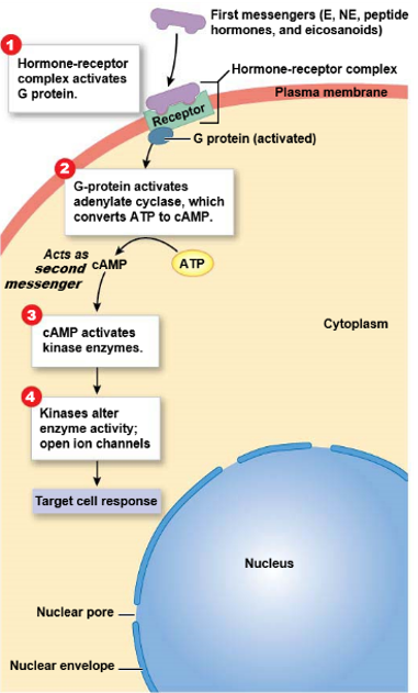
What are the steps involved in peptide hormone signaling?
The steps involved in peptide hormone signaling are:
Peptide hormones (first messengers) cannot pass the PLB so they bind to receptors in the membrane.
G-proteins are bound to receptors on the cytosolic side of the cell and binding activates them.
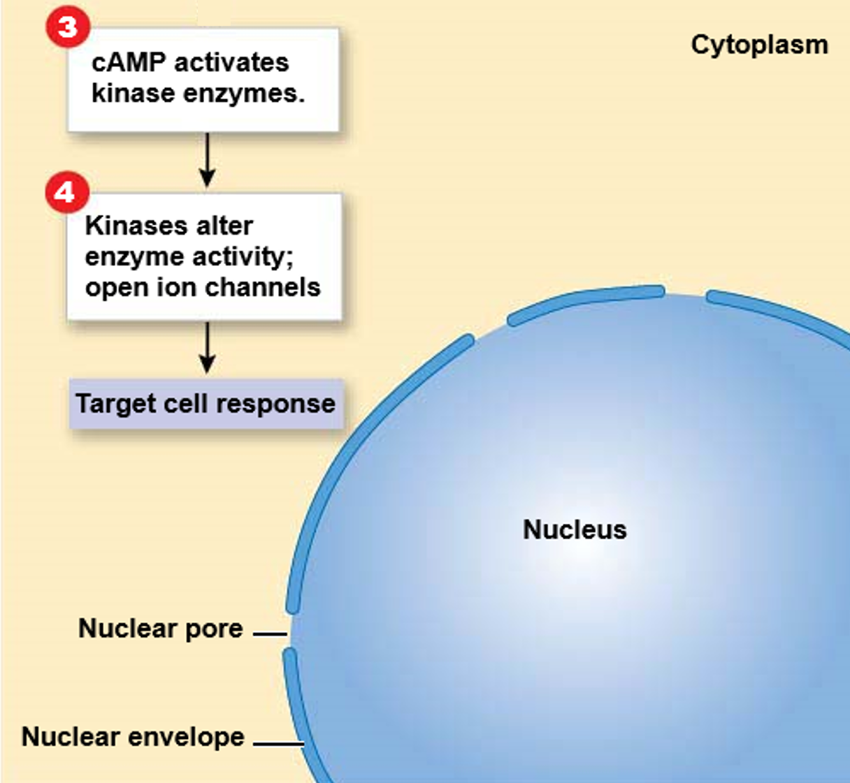
Activated G-proteins convert ATP to cyclic AMP (cAMP) which activates protein kinases; cAMP is the secondary messenger.
Kinases phosphorylate transcription factors which change the identity and proportion of gene products.
Secondary messaging: overview
Can you label, describe and explain what this diagram is/shows?
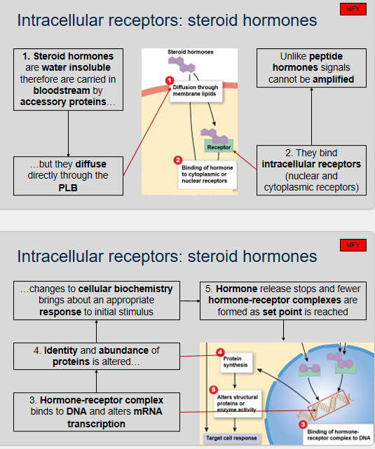
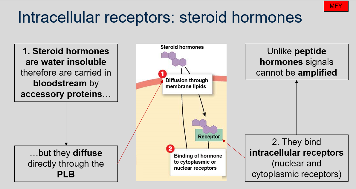
Intracellular receptors: steroid hormones
Why are steroid hormones carried in the bloodstream by accessory proteins?
How do steroid hormones enter cells?
What types of receptors do steroid hormones bind to?
Can signals from steroid hormones be amplified?
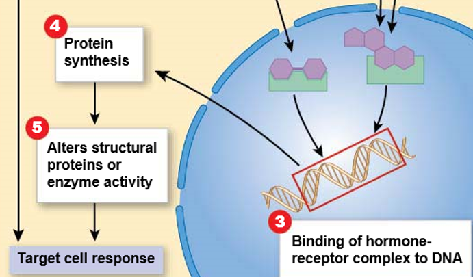
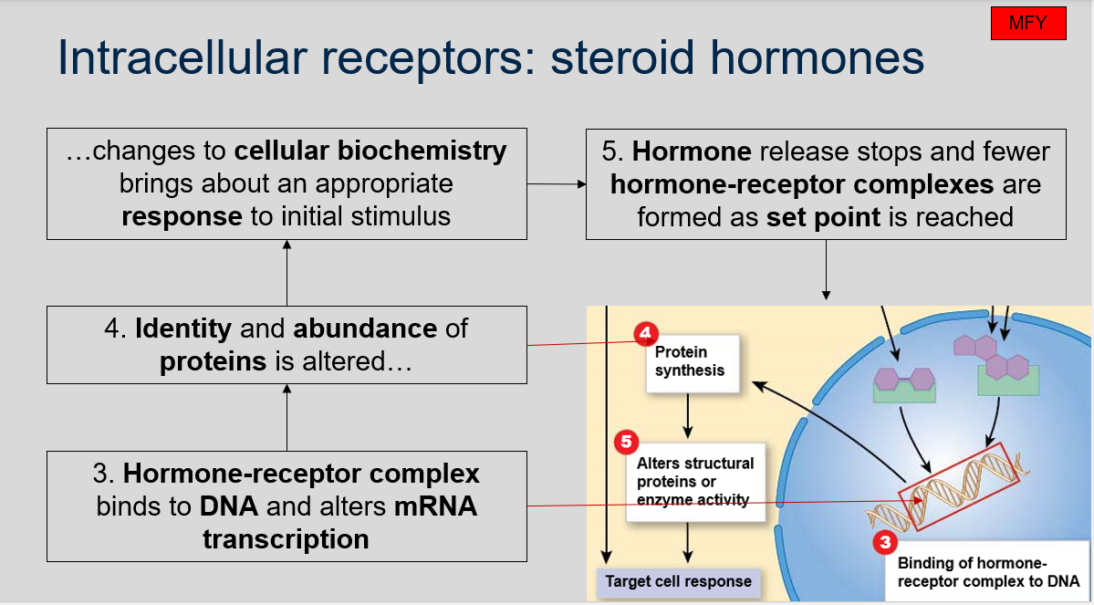
What happens when the hormone-receptor complex binds to DNA?
How does the identity and abundance of proteins change in response to steroid hormone signals?
What happens when the set point is reached in response to steroid hormone signals?
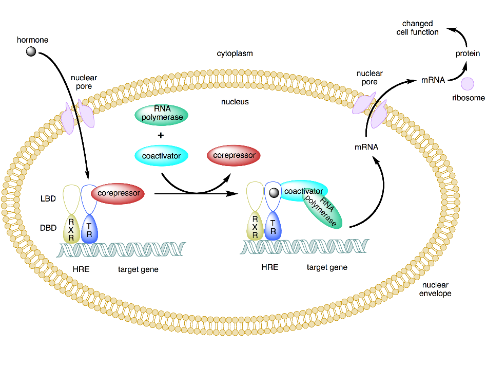
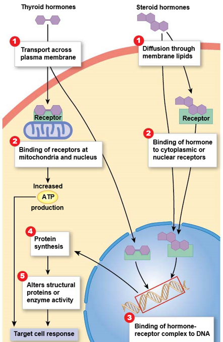
Steroid hormones are water insoluble, so they require accessory proteins to be carried in the bloodstream.
Steroid hormones diffuse directly through the plasma membrane (PLB) and enter cells.
Steroid hormones bind to intracellular receptors, including nuclear and cytoplasmic receptors.
No, signals from steroid hormones cannot be amplified, unlike peptide hormones.
When the hormone-receptor complex binds to DNA, it alters mRNA transcription.
The identity and abundance of proteins are altered in response to steroid hormone signals, leading to changes in cellular biochemistry and an appropriate response to the initial stimulus.
When the set point is reached, hormone release stops and fewer hormone-receptor complexes are formed.
Intracellular receptors: steroid hormones
Can you label, describe and explain what this diagram is/shows?
Intracellular receptors: steroid hormones
Can you label, describe and explain what this diagram is/shows?
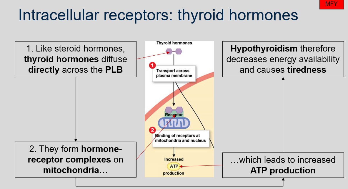
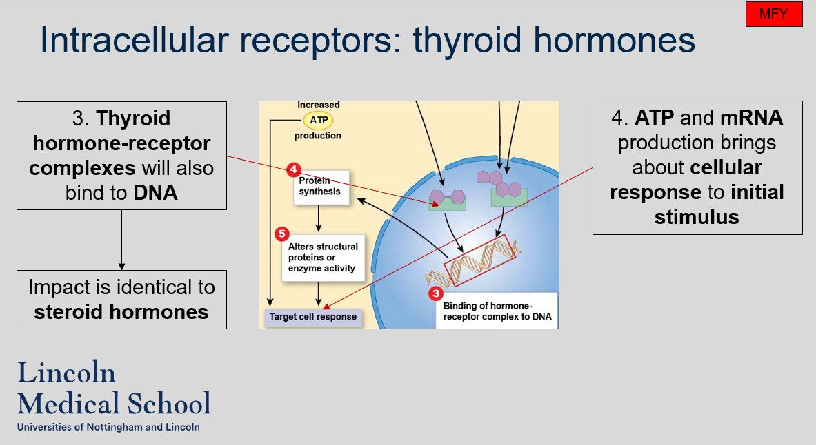
Intracellular receptors: thyroid hormones
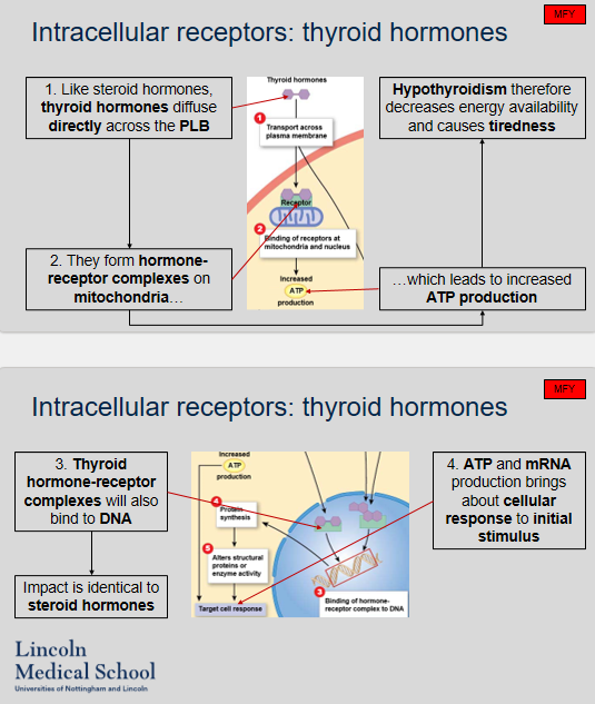
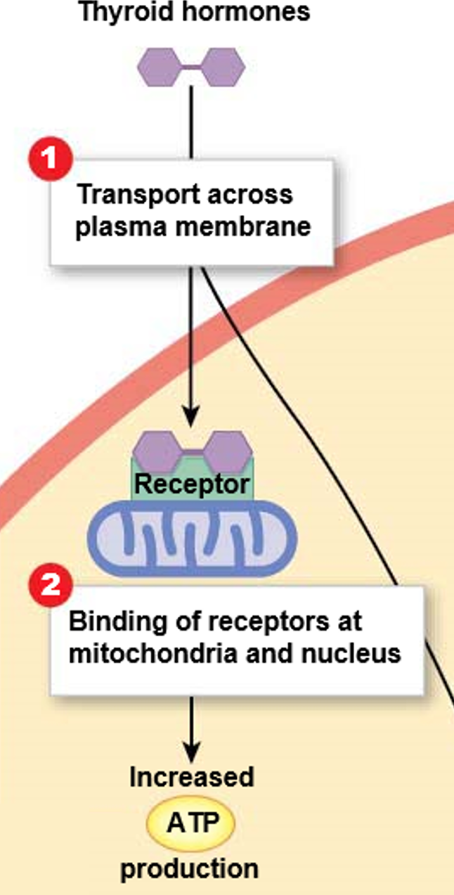
How do thyroid hormones enter cells?
What happens when thyroid hormones form hormone-receptor complexes on mitochondria?
How does hypothyroidism affect energy availability and cause tiredness?
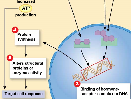
What happens when thyroid hormone-receptor complexes bind to DNA?
How does the cellular response to initial stimulus occur in response to thyroid hormone signals?
Similar to steroid hormones, thyroid hormones diffuse directly across the plasma membrane (PLB).
When thyroid hormones form hormone-receptor complexes on mitochondria, it leads to increased ATP production.
Hypothyroidism decreases energy availability due to decreased production of ATP, leading to tiredness.
When thyroid hormone-receptor complexes bind to DNA, the impact is similar to steroid hormones, resulting in altered mRNA transcription.
ATP and mRNA production brings about cellular response to the initial stimulus.
Intracellular receptors: thyroid hormones
Can you label, describe and explain what this diagram is/shows?
Intracellular receptors: thyroid hormones
Can you label, describe and explain what this diagram is/shows?
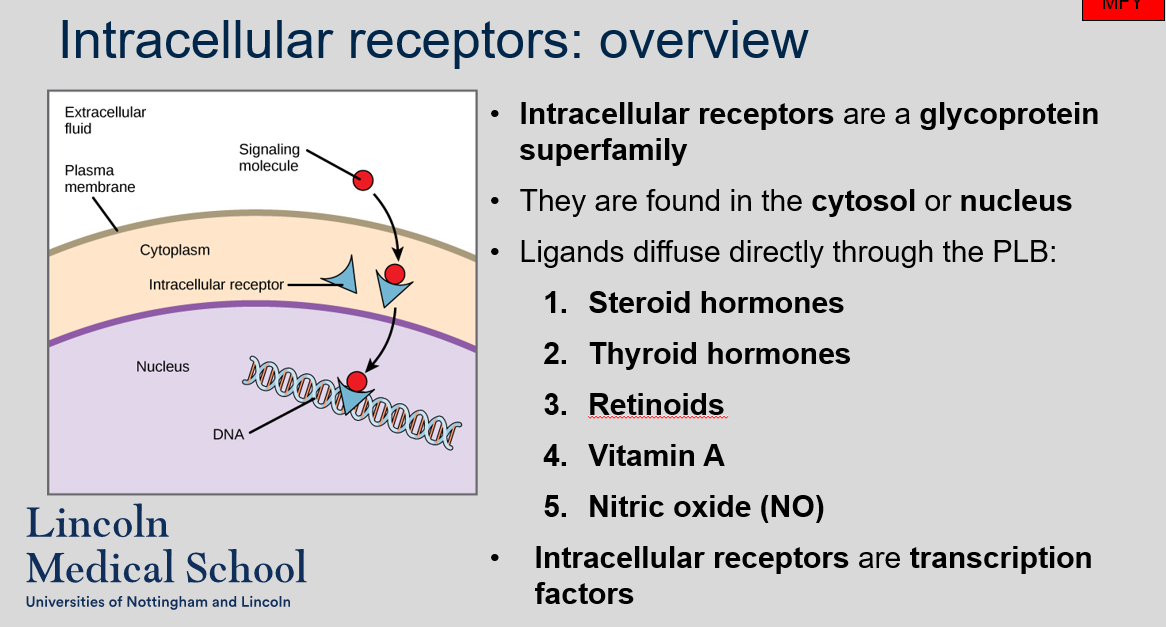
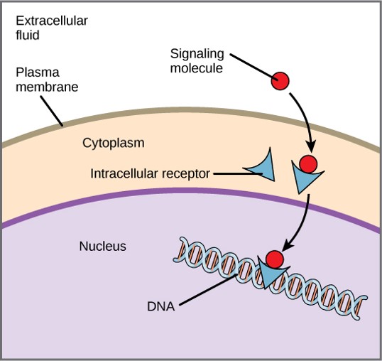
Intracellular receptors: overview
What is the composition of intracellular receptors?
Where are intracellular receptors typically found within the cell?
Which ligands can diffuse directly through the plasma membrane (PLB) to bind to intracellular receptors?
What is the function of intracellular receptors?
Intracellular receptors are a glycoprotein superfamily.
Intracellular receptors are found in the cytosol or nucleus.
Several ligands can diffuse directly through the PLB to bind to intracellular receptors, including steroid hormones, thyroid hormones, retinoids, vitamin A, and nitric oxide (NO).
Intracellular receptors are transcription factors that bind to specific DNA sequences and regulate gene expression in response to ligand binding.
Intracellular receptors: overview
Can you label, describe and explain what this diagram is/shows?
Intracellular receptors: overview
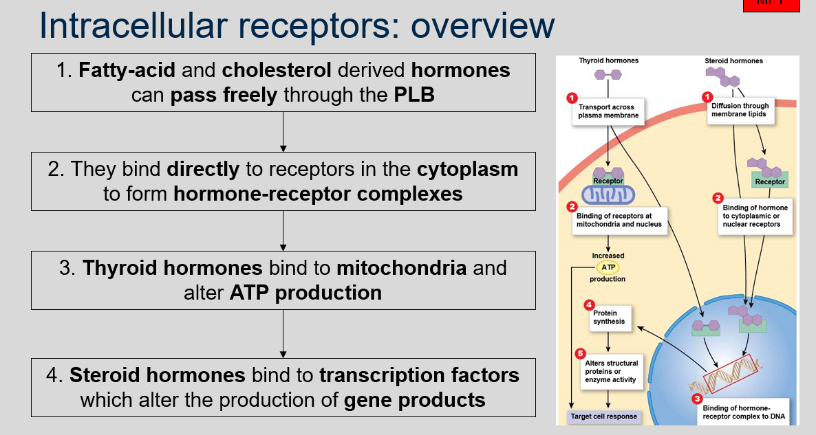
Which types of hormones can pass freely through the plasma membrane (PLB) to bind to intracellular receptors?
Where do fatty-acid and cholesterol derived hormones bind to receptors?
What is the impact of thyroid hormones binding to mitochondria?
What happens when steroid hormones bind to transcription factors?
Fatty-acid and cholesterol derived hormones can pass freely through the PLB to bind to intracellular receptors.
Fatty-acid and cholesterol derived hormones bind directly to receptors in the cytoplasm to form hormone-receptor complexes.
When thyroid hormones bind to mitochondria, they alter ATP production.
When steroid hormones bind to transcription factors, they alter the production of gene products.
Intracellular receptors: overview
Can you label, describe and explain what this diagram is/shows?
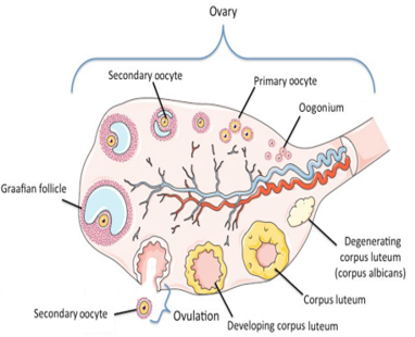
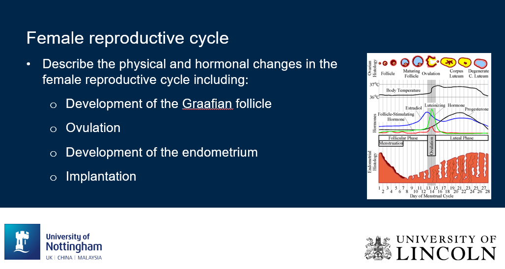
Female reproductive cycle
Can you label, describe and explain what this diagram is/shows?
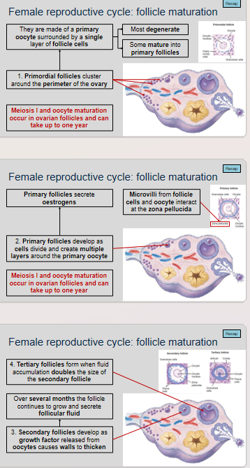
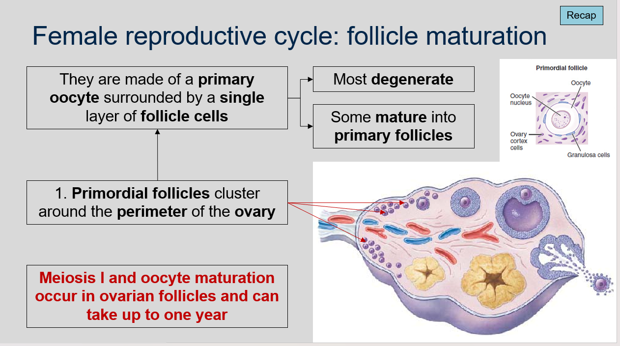
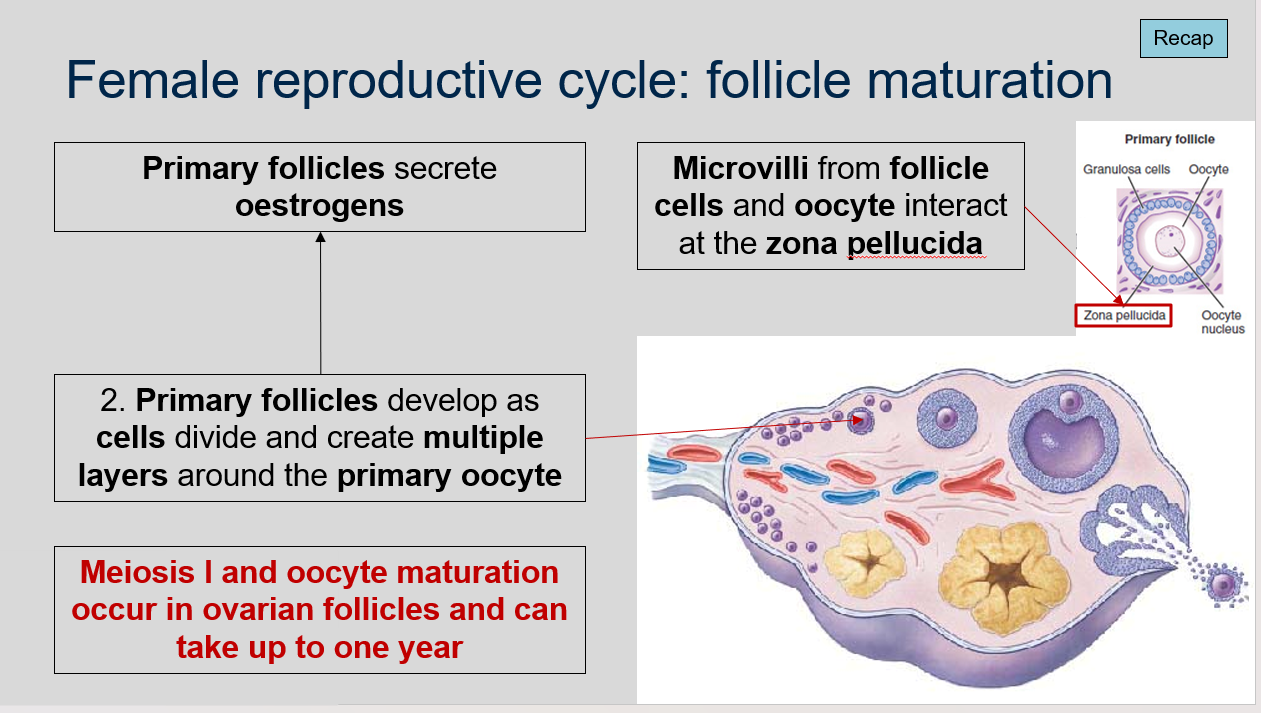
Female reproductive cycle: follicle maturation
Where do meiosis I and oocyte maturation occur?
How long can meiosis I and oocyte maturation take in ovarian follicles?
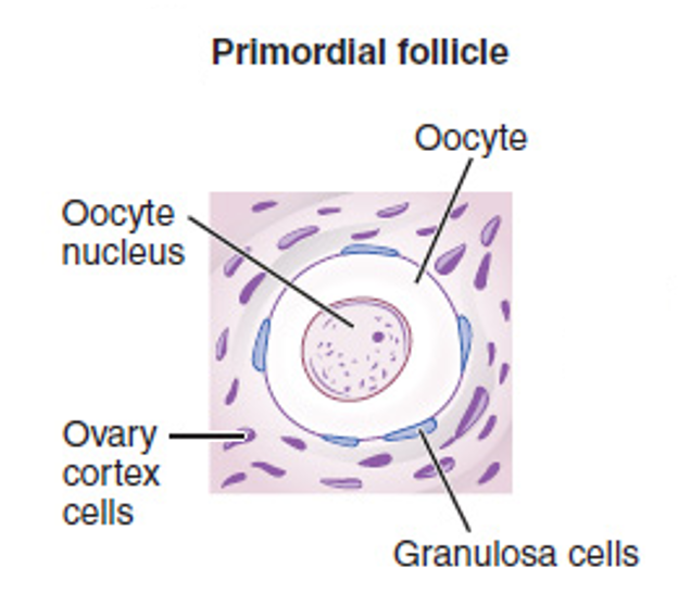
What is the structure of primordial follicles?
Where do primordial follicles cluster?
What happens to most of the primordial follicles?
What happens to the primordial follicles that do not degenerate?
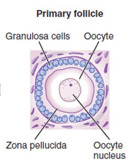
What is the structure of primary follicles?
What do primary follicles secrete?
What is the zona pellucida?
What happens when microvilli from follicle cells and oocyte interact at the zona pellucida?
What causes the walls of secondary follicles to thicken?
How long does it take for a secondary follicle to develop from a primary follicle?
What is the function of the follicular fluid?
What is the fourth stage of ovarian follicle development?
Meiosis I and oocyte maturation occur in ovarian follicles.
Meiosis I and oocyte maturation can take up to one year in ovarian follicles.
Primordial follicles are made of a primary oocyte surrounded by a single layer of follicle cells.
Primordial follicles cluster around the perimeter of the ovary.
Most of the primordial follicles degenerate.
The primordial follicles that do not degenerate some can mature into primary follicles.
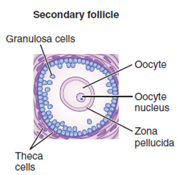
Primary follicles develop as cells divide and create multiple layers around the primary oocyte.
Primary follicles secrete estrogens.
The zona pellucida is a glycoprotein layer that surrounds the oocyte.
When microvilli from follicle cells and oocyte interact at the zona pellucida, they allow for communication and exchange of substances between the two cells.
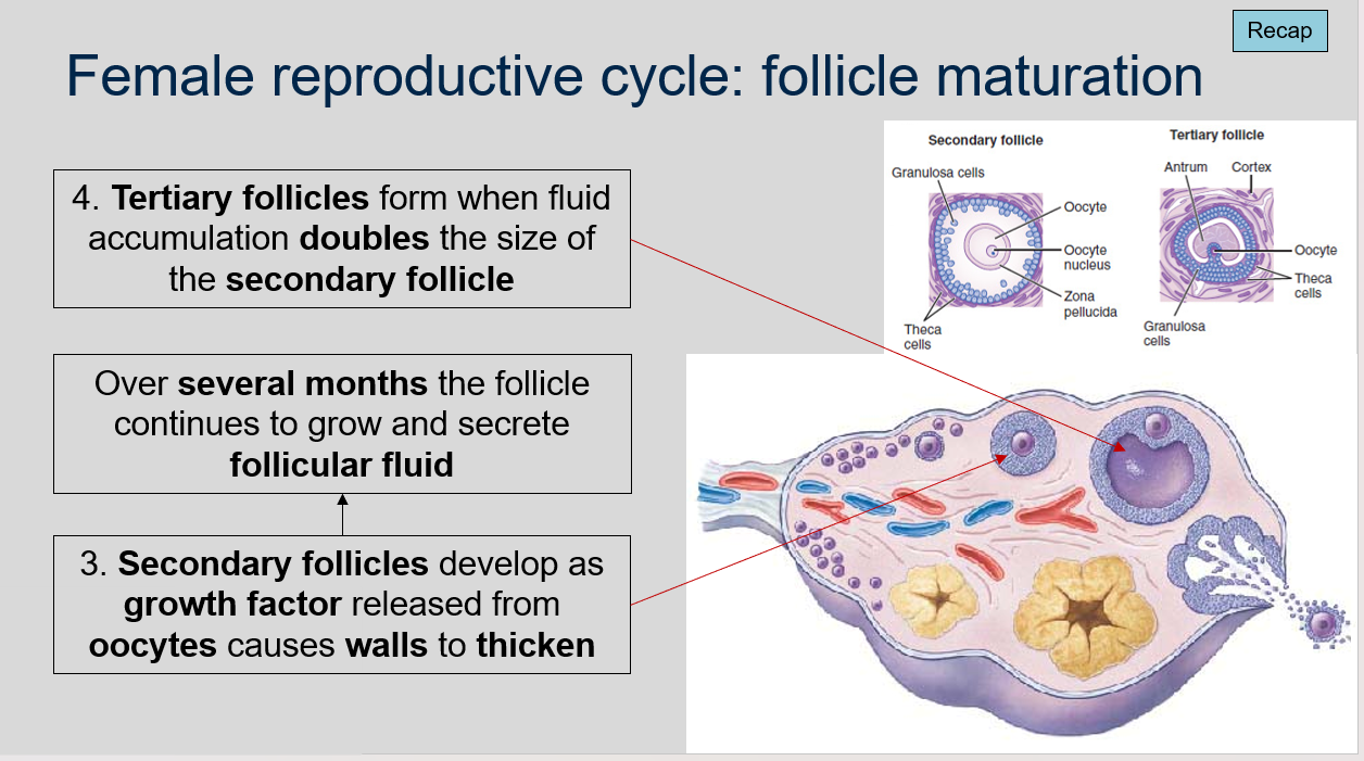
Growth factors released from the oocyte cause the walls of secondary follicles to thicken.
It takes several months for a secondary follicle to develop from a primary follicle.
The follicular fluid secreted by the follicle provides nutrients and support to the developing oocyte.
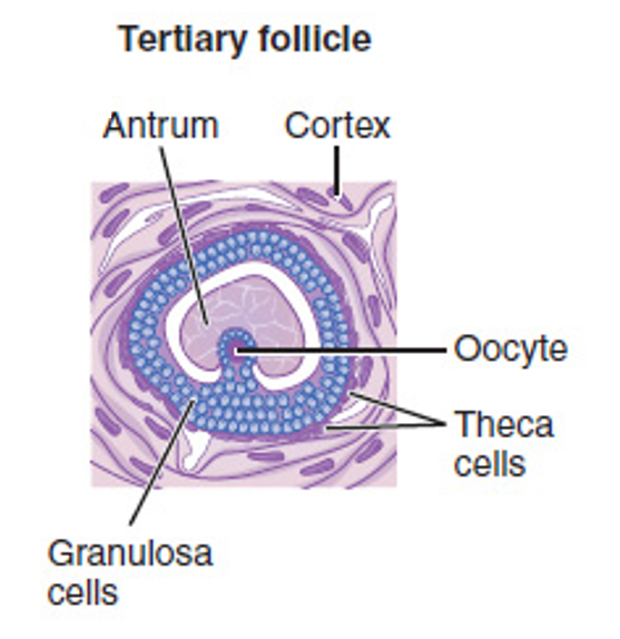
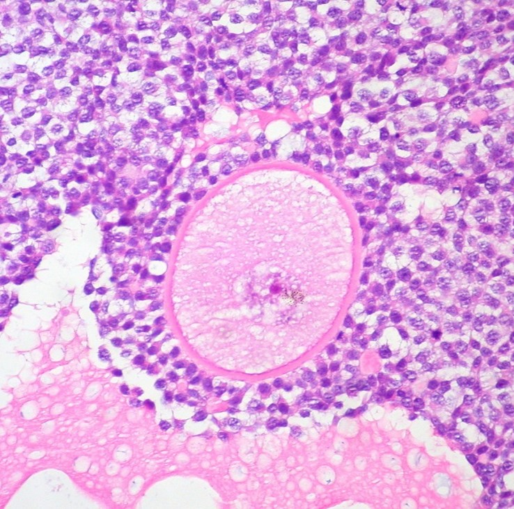
The fourth stage of ovarian follicle development is the formation of tertiary follicles. This occurs when the accumulation of follicular fluid doubles the size of the secondary follicle. The tertiary follicle has a fluid-filled cavity called an antrum, and it is at this stage that the oocyte resumes meiosis I and oocyte maturation begins. The follicle will continue to grow and mature until it reaches ovulation or atresia.
Female reproductive cycle: follicle maturation
Can you label, describe and explain what this diagram is/shows?
Female reproductive cycle: follicle maturation
Can you label, describe and explain what this diagram is/shows?
Female reproductive cycle: follicle maturation
Can you label, describe and explain what this diagram is/shows?
Female reproductive cycle: follicle maturation
Can you label, describe and explain what this diagram is/shows?
Female reproductive cycle: follicle maturation
Can you label, describe and explain what this diagram is/shows?
Female reproductive cycle: follicle maturation
Can you label, describe and explain what this diagram is/shows?
Female reproductive cycle: follicle maturation
Can you label, describe and explain what this diagram is/shows?
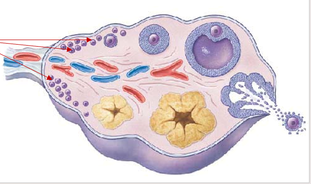
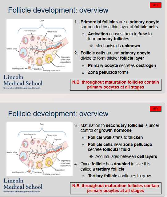
Follicle development: overview
What are the stages of follicle development, from primordial follicles to tertiary follicles?
Do follicles at all stages of maturation contain primary oocytes?
Stage 1: Primordial Follicles:
Consist of a primary oocyte surrounded by a thin layer of follicle cells
Activation causes them to fuse to form primary follicles
Mechanism is unknown
Stage 2: Primary Follicles:
Follicle cells around primary oocyte divide to form thicker follicle layer
Primary oocyte secretes estrogen
Zona pellucida forms
Stage 3: Secondary Follicles:
Maturation to secondary follicles is under the control of growth hormone
Follicle wall starts to thicken
Follicle cells near zona pellucida secrete follicular fluid
Accumulates between cell layers
Stage 4: Tertiary Follicles:
Once the follicle has doubled in size, it is called a tertiary follicle
Tertiary follicle continues to grow
Yes, throughout the process of follicle development and maturation, the follicles contain primary oocytes at all stages.
Follicle development: overview
Can you label, describe and explain what this diagram is/shows?
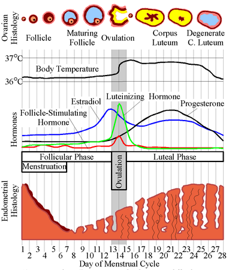
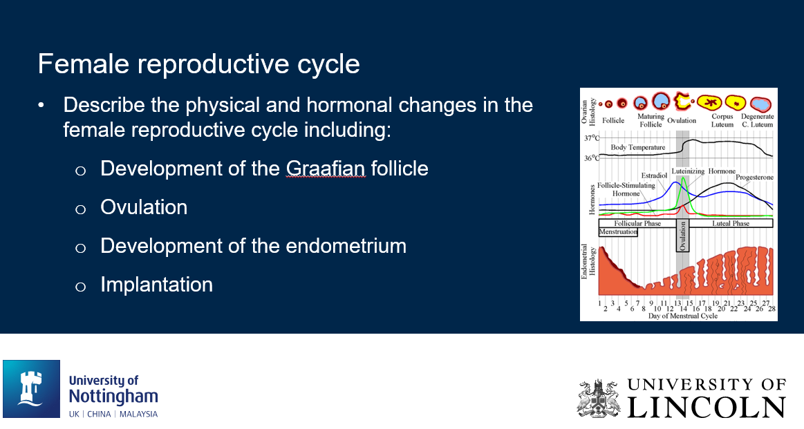
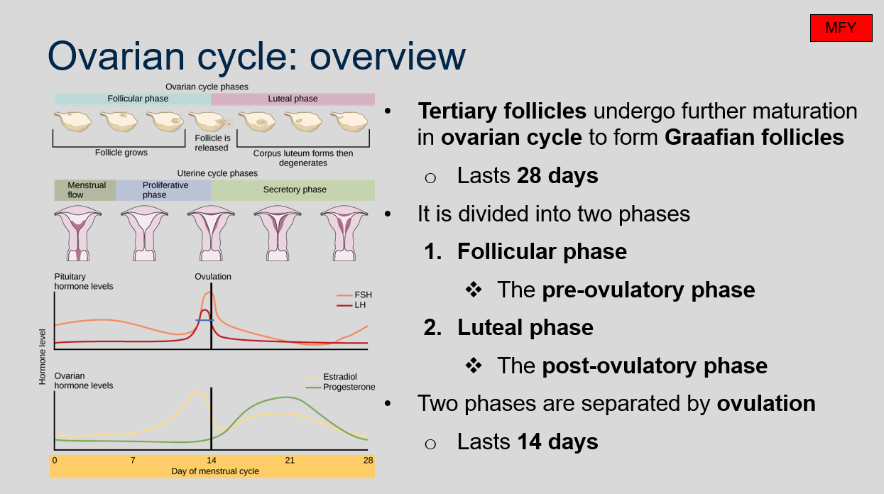
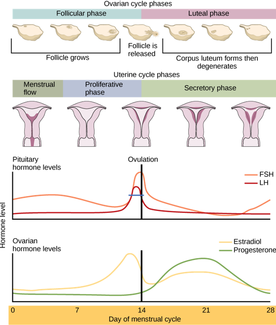
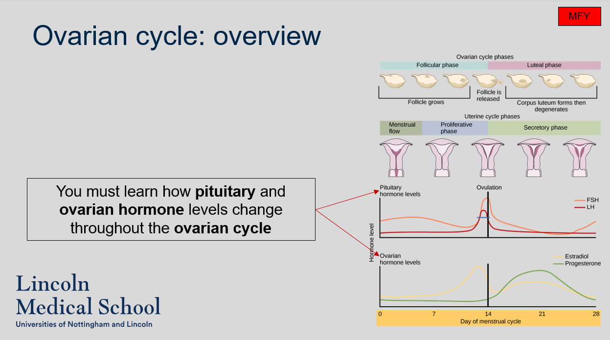
Ovarian cycle: overview
What is the overview of the ovarian cycle?
The ovarian cycle is a 28-day cycle that is divided into two phases: the follicular phase and the luteal phase. Tertiary follicles undergo further maturation in the ovarian cycle to form Graafian follicles. The two phases are separated by ovulation, which lasts for 14 days. The follicular phase is the pre-ovulatory phase, while the luteal phase is the post-ovulatory phase.
Ovarian cycle: overview
Can you label, describe and explain what this diagram is/shows?
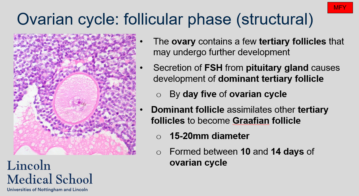
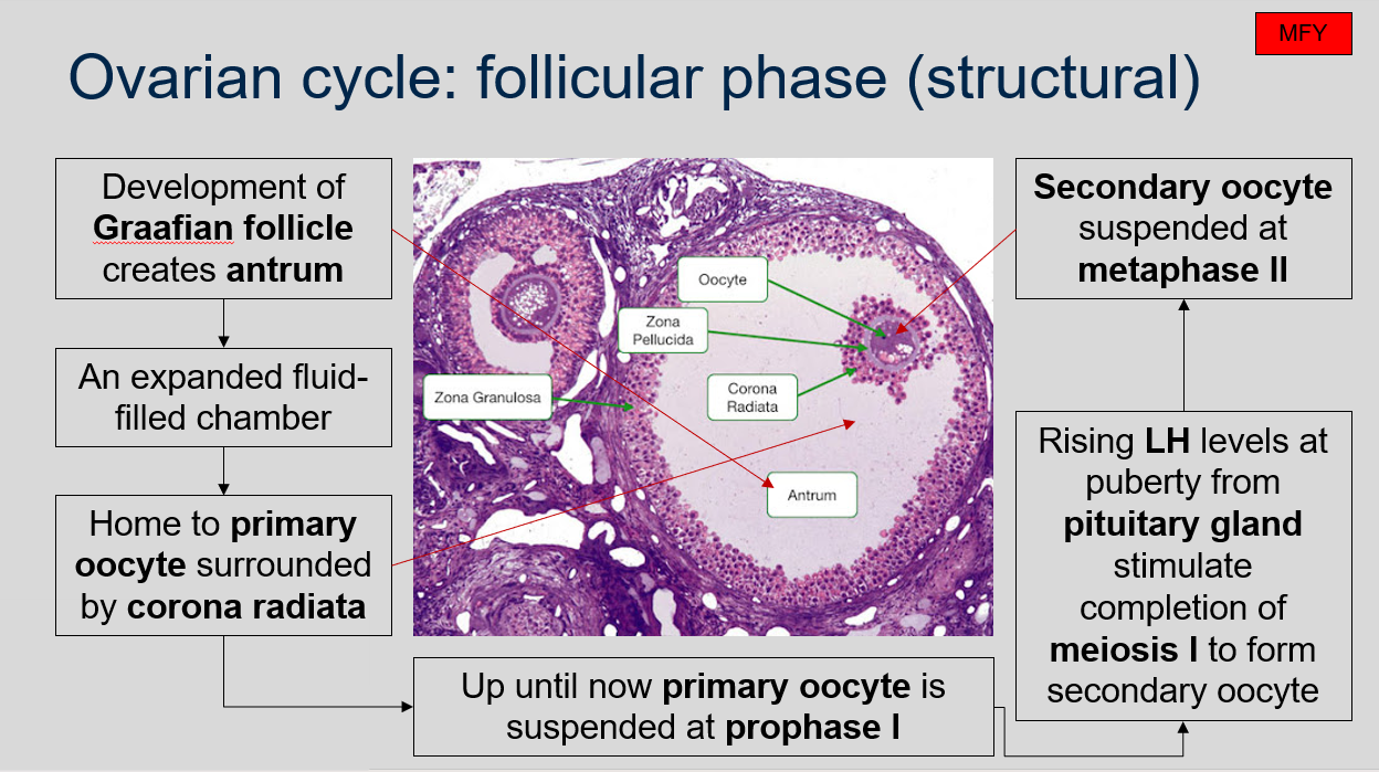
Ovarian cycle: follicular phase (structural)
What is the follicular phase of the ovarian cycle?
The follicular phase is the structural phase of the ovarian cycle, during which a few tertiary follicles in the ovary may undergo further development. The secretion of follicle-stimulating hormone (FSH) from the pituitary gland causes the development of a dominant tertiary follicle. By day five of the ovarian cycle, the dominant follicle assimilates other tertiary follicles to become a Graafian follicle with a diameter of 15-20mm, which is formed between days 10 and 14 of the ovarian cycle.
Ovarian cycle: follicular phase (structural)
Can you label, describe and explain what this diagram is/shows?
Ovarian cycle: follicular phase (structural)
What happens during the follicular phase of the ovarian cycle?
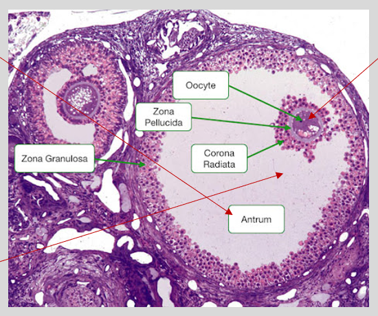
During the follicular phase of the ovarian cycle, a dominant tertiary follicle is developed in the ovary due to the secretion of follicle-stimulating hormone (FSH) from the pituitary gland. This follicle assimilates other tertiary follicles and becomes a Graafian follicle with a diameter of 15-20mm between 10 and 14 days of the ovarian cycle. The Graafian follicle creates antrum, which is an expanded fluid-filled chamber that is home to the primary oocyte surrounded by the corona radiata. The primary oocyte remains suspended at prophase I until the rising levels of luteinizing hormone (LH) from the pituitary gland at puberty stimulate the completion of meiosis I, resulting in the formation of a secondary oocyte. The secondary oocyte is then suspended at metaphase II.
Ovarian cycle: follicular phase (structural)
Can you label, describe and explain what this diagram is/shows?
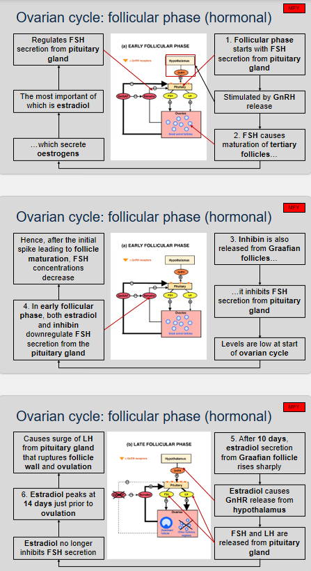
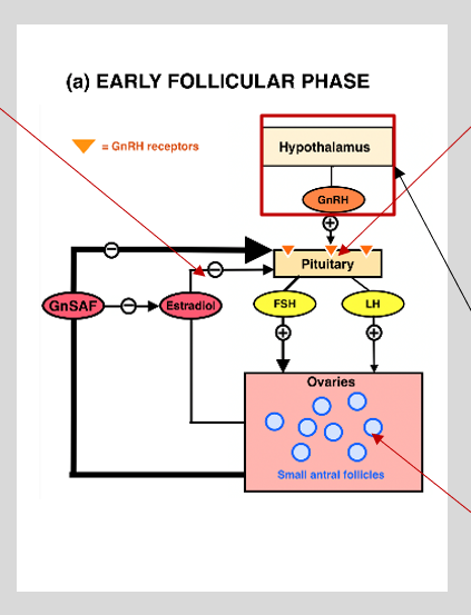
Ovarian cycle: follicular phase (hormonal)
What are the stages of the ovarian cycle during the follicular phase, and how do hormones such as FSH, estradiol, and inhibin regulate these stages?
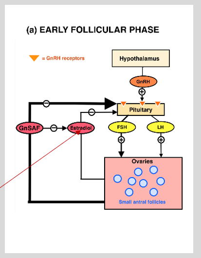
The ovarian cycle consists of several stages that are essential for the process of ovulation. The follicular phase is the first stage of the ovarian cycle, and it is characterized by the following six stages.
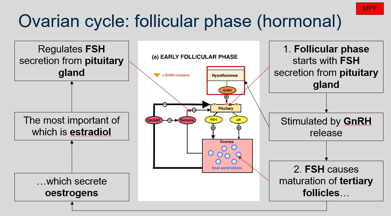
Stage 1: The follicular phase begins with the secretion of follicle-stimulating hormone (FSH) from the pituitary gland, which is stimulated by gonadotropin-releasing hormone (GnRH) release.
Stage 2: FSH causes the maturation of tertiary follicles, which then secrete oestrogens, the most important of which is estradiol. Estradiol regulates FSH secretion from the pituitary gland.
Stage 3: Inhibin is also released from Graafian follicles, and it inhibits FSH secretion from the pituitary gland. The levels of inhibin are low at the start of the ovarian cycle.
Stage 4: In the early follicular phase, both estradiol and inhibin downregulate FSH secretion from the pituitary gland, causing FSH concentrations to decrease after the initial spike that leads to follicle maturation.
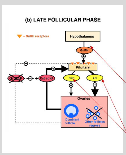
Stage 5: After 10 days, estradiol secretion from the Graafian follicle rises sharply, causing GnRH release from the hypothalamus. FSH and luteinizing hormone (LH) are then released from the pituitary gland, and estradiol no longer inhibits FSH secretion.
Stage 6: Estradiol peaks at 14 days just prior to ovulation, causing a surge of LH from the pituitary gland that ruptures the follicle wall and ovulation occurs.
Ovarian cycle: follicular phase (hormonal)
Can you label, describe and explain what this diagram is/shows?
Ovarian cycle: follicular phase (hormonal)
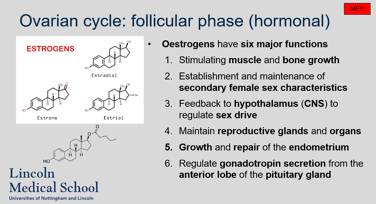
What are the six major functions of oestrogens?
The six major functions of oestrogens are:
Stimulating muscle and bone growth
Establishing and maintaining secondary female sex characteristics
Providing feedback to the hypothalamus (CNS) to regulate sex drive
Maintaining reproductive glands and organs
Promoting growth and repair of the endometrium
Regulating gonadotropin secretion from the anterior lobe of the pituitary gland.
Ovarian cycle: follicular phase (hormonal)
Can you label, describe and explain what this diagram is/shows?
Ovarian cycle: follicular phase (hormonal)
Can you label, describe and explain what this diagram is/shows?
Ovarian cycle: follicular phase (hormonal)
Can you label, describe and explain what this diagram is/shows?
Ovarian cycle: follicular phase (hormonal)
Can you label, describe and explain what this diagram is/shows?
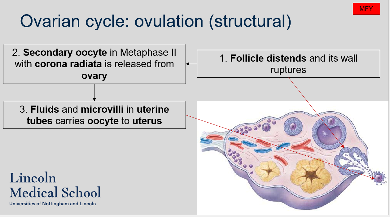
Ovarian cycle: ovulation (structural)
What are the stages of the ovarian cycle during ovulation (structural)?
The follicle distends and its wall ruptures.
The secondary oocyte in Metaphase II with corona radiata is released from the ovary.
Fluids and microvilli in the uterine tubes carry the oocyte to the uterus.
Ovarian cycle: ovulation (structural)
Can you label, describe and explain what this diagram is/shows?
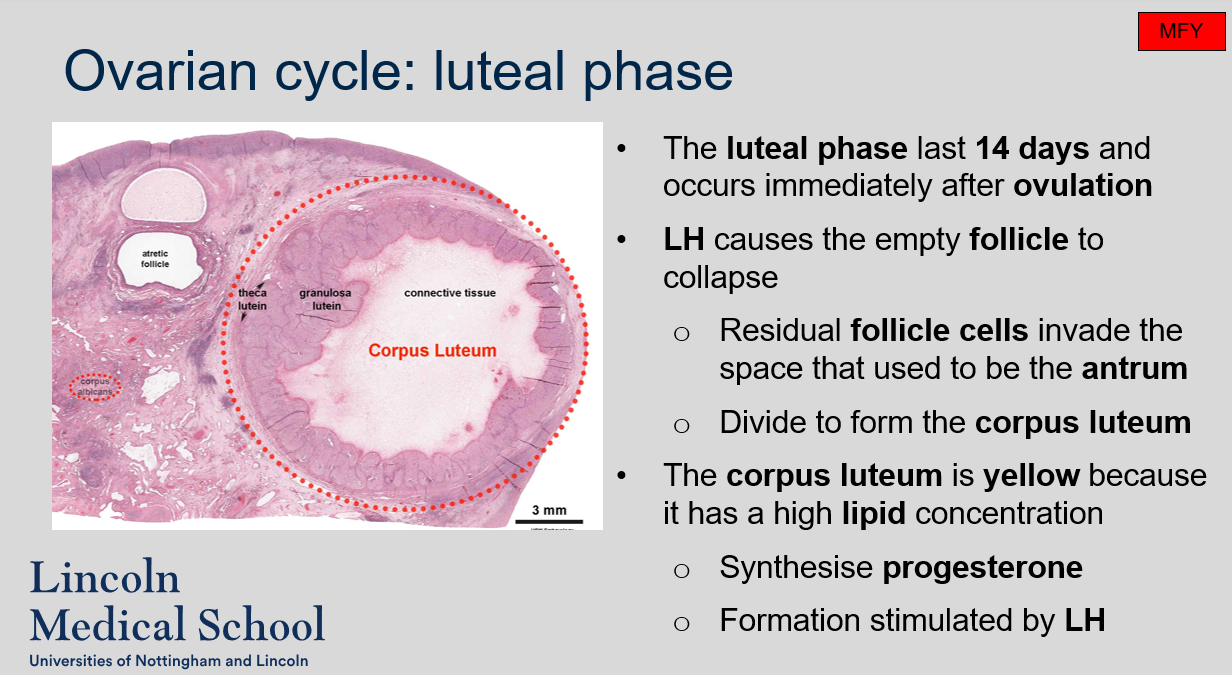
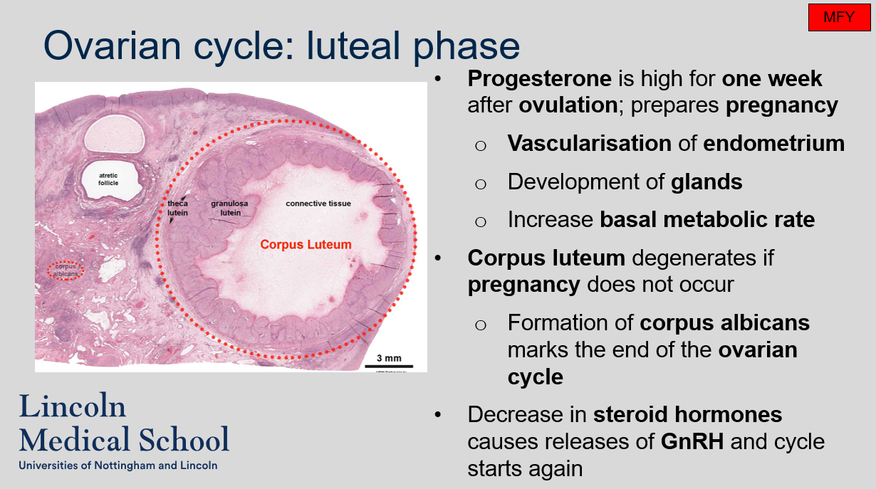
Ovarian cycle: luteal phase
How long does the luteal phase of the ovarian cycle last, and when does it occur?
What happens to the empty follicle after ovulation?
Why is the corpus luteum yellow in color?
What hormone is synthesized by the corpus luteum?
The luteal phase of the ovarian cycle lasts 14 days and occurs immediately after ovulation.
LH causes the empty follicle to collapse, and residual follicle cells invade the space that used to be the antrum and divide to form the corpus luteum.
The corpus luteum is yellow because it has a high lipid concentration.
The corpus luteum synthesizes progesterone, which is important for preparing the uterus for pregnancy. The formation of the corpus luteum is stimulated by LH.
Ovarian cycle: luteal phase
What is the role of progesterone during the luteal phase of the ovarian cycle?
What happens to the corpus luteum if pregnancy does not occur during the luteal phase of the ovarian cycle?
Progesterone is synthesized by the corpus luteum during the luteal phase of the ovarian cycle. It prepares the endometrium for pregnancy by causing vascularization of the endometrium, development of glands, and increase in basal metabolic rate.
If pregnancy does not occur during the luteal phase of the ovarian cycle, the corpus luteum will degenerate. The degenerated corpus luteum and the formation of corpus albicans marks the end of the ovarian cycle. The decrease in steroid hormones after corpus luteum degeneration causes the release of GnRH and the start of a new ovarian cycle.
Ovarian cycle: luteal phase
Can you label, describe and explain what this diagram is/shows?
Ovarian cycle: overview
Can you label, describe and explain what this diagram is/shows?
Female reproductive cycle
Can you label, describe and explain what this diagram is/shows?
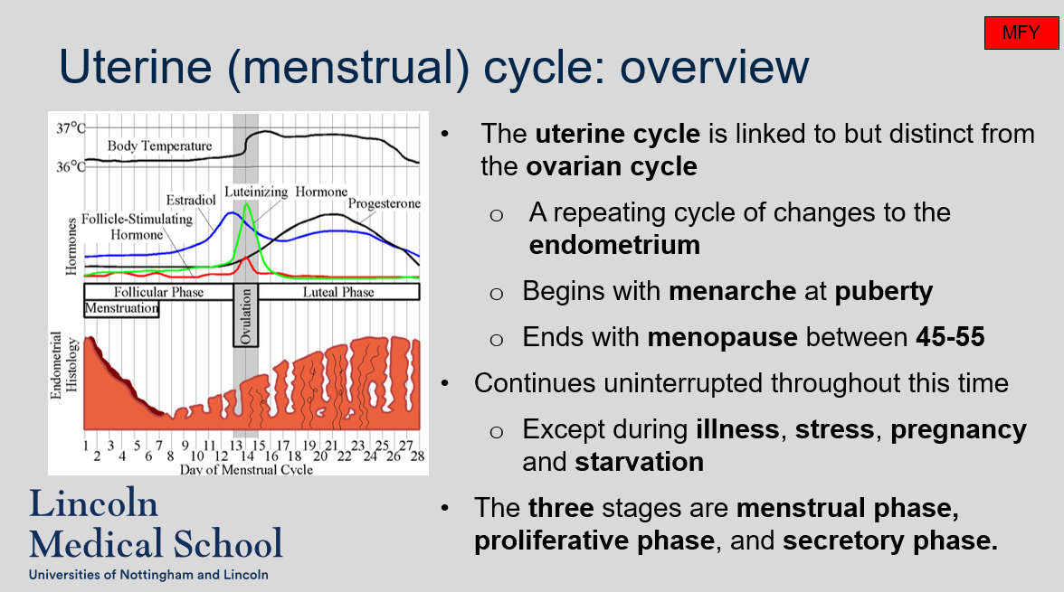
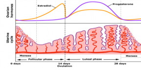
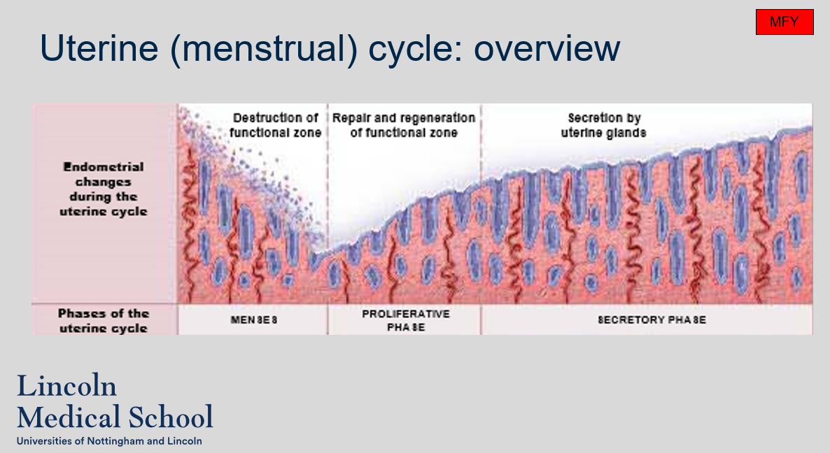
Uterine (menstrual) cycle: overview
What is the uterine cycle?
When does the uterine cycle begin and end?
Does the uterine cycle continue uninterrupted throughout this time?
What are the three stages of the uterine cycle?
The uterine cycle is a repeating cycle of changes to the endometrium that is linked to but distinct from the ovarian cycle.
The uterine cycle begins with menarche at puberty and ends with menopause between 45-55.
The uterine cycle continues uninterrupted throughout this time, except during illness, stress, pregnancy, and starvation.
The three stages of the uterine cycle are the menstrual phase, proliferative phase, and secretory phase.
Uterine (menstrual) cycle: overview
Can you label, describe and explain what this diagram is/shows?
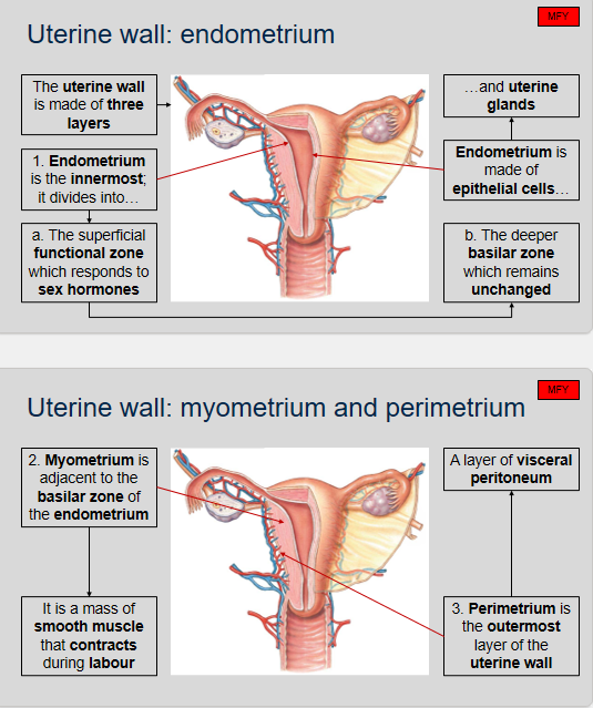
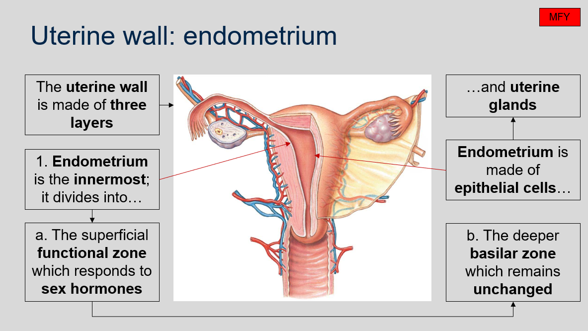
Uterine wall: endometrium
What are the three layers that make up the uterine wall?
What is the endometrium?
How is the endometrium divided?
What is the function of the superficial functional zone?
What is the function of the deeper basilar zone?
What is the composition of the endometrium?
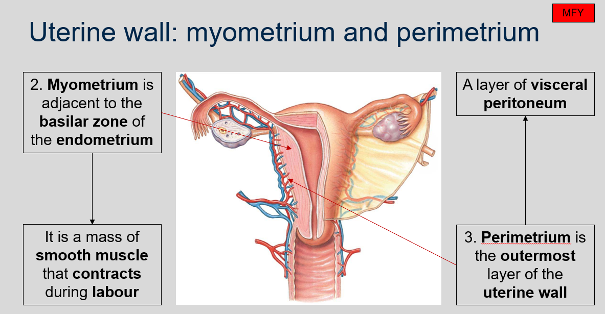
What is the myometrium in the uterine wall?
What is the perimetrium?
The uterine wall is made up of three layers: the endometrium, the myometrium, and the perimetrium.
The endometrium is the innermost layer of the uterine wall.
The endometrium is divided into two zones: the superficial functional zone and the deeper basilar zone.
The superficial functional zone responds to sex hormones.
The deeper basilar zone remains unchanged.
The endometrium is made of epithelial cells and uterine glands.
The myometrium is a mass of smooth muscle in the uterine wall that is adjacent to the basilar zone of the endometrium. It is responsible for contracting during labor to help expel the fetus and placenta from the uterus.
The perimetrium is the outermost layer of the uterine wall. It is a layer of visceral peritoneum.
Uterine wall: endometrium
Can you label, describe and explain what this diagram is/shows?
Uterine wall: myometrium and perimetrium
Can you label, describe and explain what this diagram is/shows?
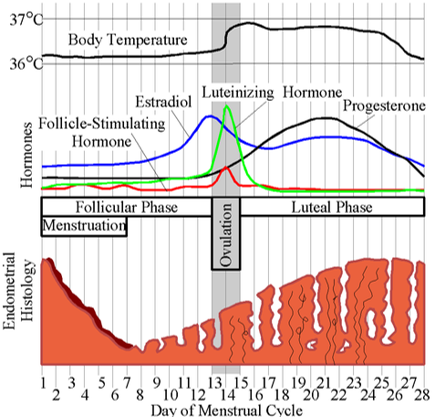
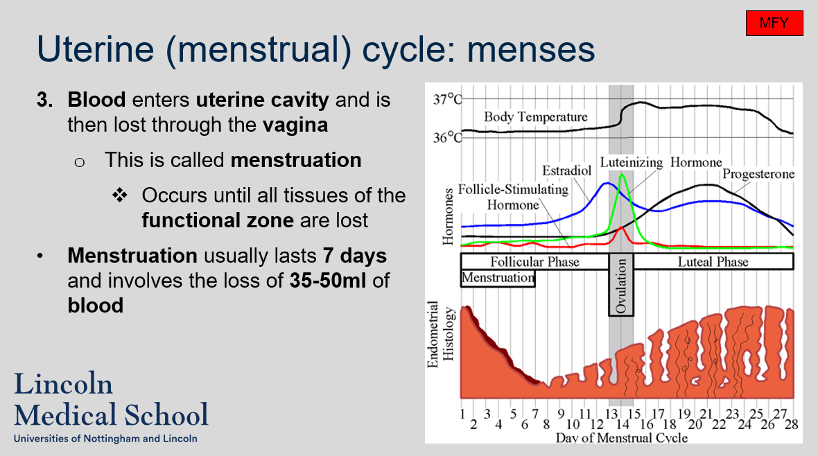
Uterine (menstrual) cycle: menses
What marks the start of the uterine cycle?
What happens during menses?
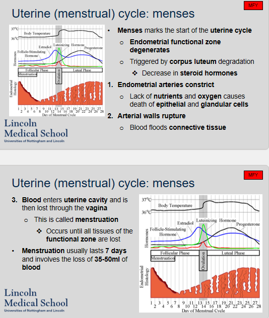
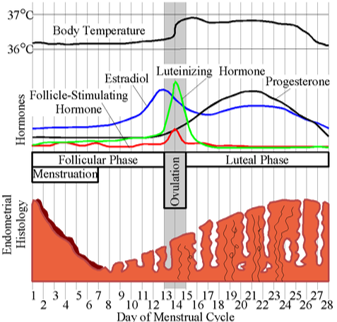
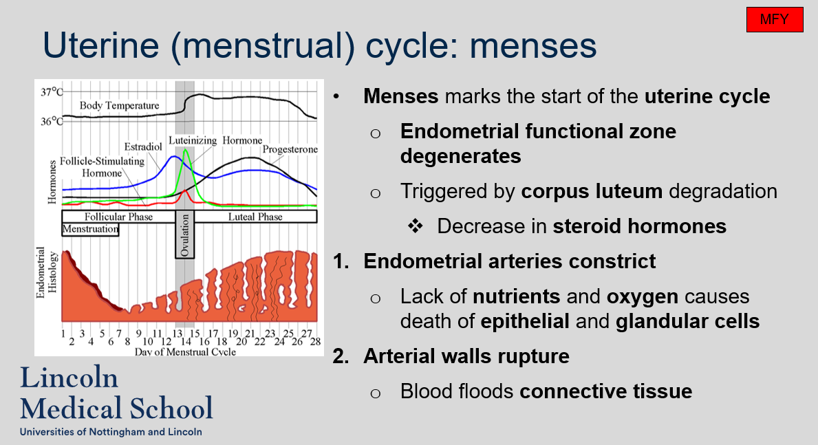
Menses marks the start of the uterine cycle.
During menses, the endometrial functional zone degenerates triggered by corpus luteum degradation and a decrease in steroid hormones. The endometrial arteries constrict, which leads to the lack of nutrients and oxygen causing the death of epithelial and glandular cells. The arterial walls rupture, and blood floods the connective tissue. Blood enters the uterine cavity and is then lost through the vagina. This is called menstruation, which occurs until all tissues of the functional zone are lost. Menstruation usually lasts 7 days and involves the loss of 35-50ml of blood.
Uterine (menstrual) cycle: menses
Can you label, describe and explain what this diagram is/shows?
Uterine (menstrual) cycle: menses
Can you label, describe and explain what this diagram is/shows?
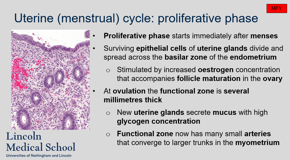
Uterine (menstrual) cycle: proliferative phase
When does the proliferative phase of the uterine cycle occur?
What happens during the proliferative phase?
How thick is the functional zone of the endometrium at ovulation during the proliferative phase?
What do new uterine glands secrete during the proliferative phase?
What is the vascularization like in the functional zone during the proliferative phase?
The proliferative phase starts immediately after menses.
Surviving epithelial cells of uterine glands divide and spread across the basilar zone of the endometrium. This process is stimulated by increased estrogen concentration that accompanies follicle maturation in the ovary.
At ovulation, the functional zone is several millimeters thick.
New uterine glands secrete mucus with a high glycogen concentration.
The functional zone now has many small arteries that converge to larger trunks in the myometrium.
Uterine (menstrual) cycle: proliferative phase
Can you label, describe and explain what this diagram is/shows?
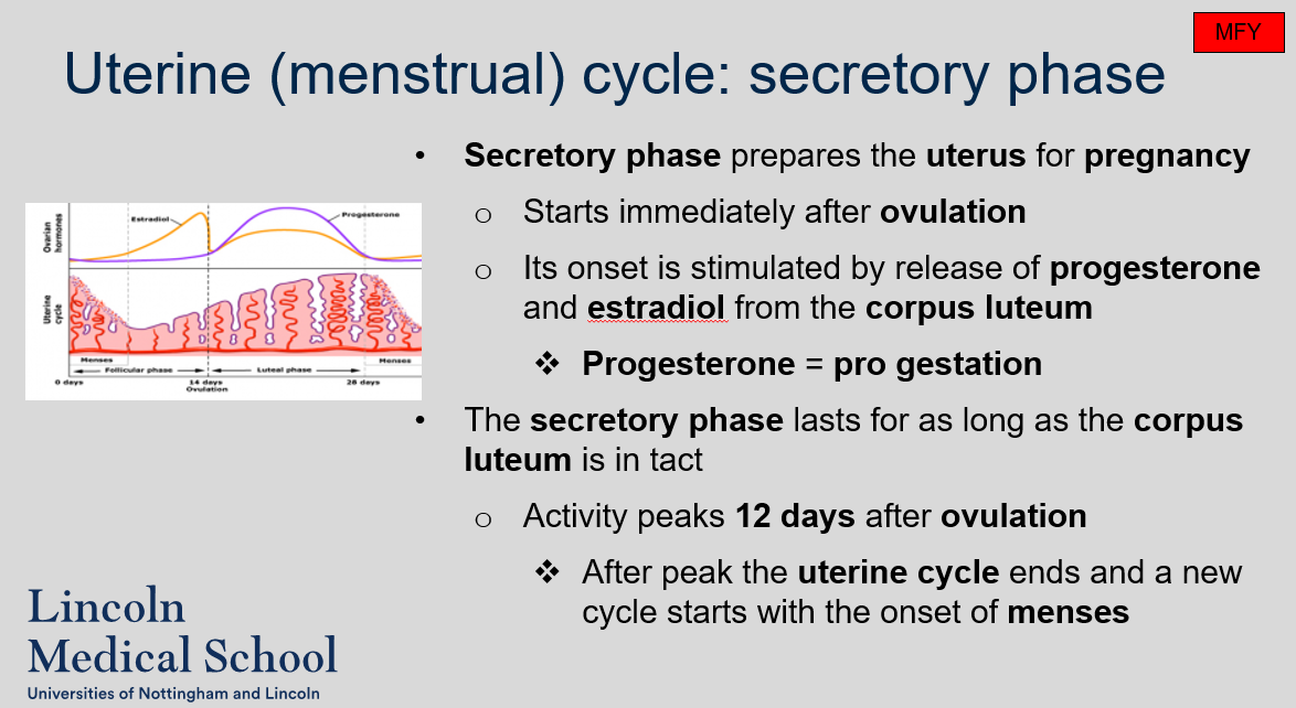
Uterine (menstrual) cycle: secretory phase
What is the purpose of the secretory phase of the uterine cycle?
When does the secretory phase start?
What stimulates the onset of the secretory phase?
What is the role of progesterone in the secretory phase?
How long does the secretory phase last?
When does the activity of the secretory phase peak?
What happens after the peak of the secretory phase?
The purpose of the secretory phase is to prepare the uterus for pregnancy.
The secretory phase starts immediately after ovulation.
The onset of the secretory phase is stimulated by the release of progesterone and estradiol from the corpus luteum.
Progesterone is known as "pro-gestation" and plays a key role in preparing the uterus for pregnancy during the secretory phase.
The secretory phase lasts for as long as the corpus luteum is intact.
The activity of the secretory phase peaks 12 days after ovulation.
After the peak of the secretory phase, the uterine cycle ends and a new cycle starts with the onset of menses.
Uterine (menstrual) cycle: secretory phase
Can you label, describe and explain what this diagram is/shows?
Uterine (menstrual) cycle: overview
Can you label, describe and explain what this diagram is/shows?
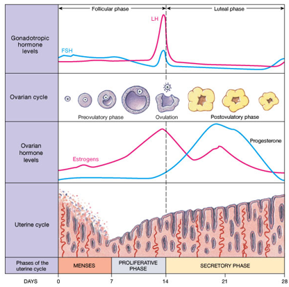
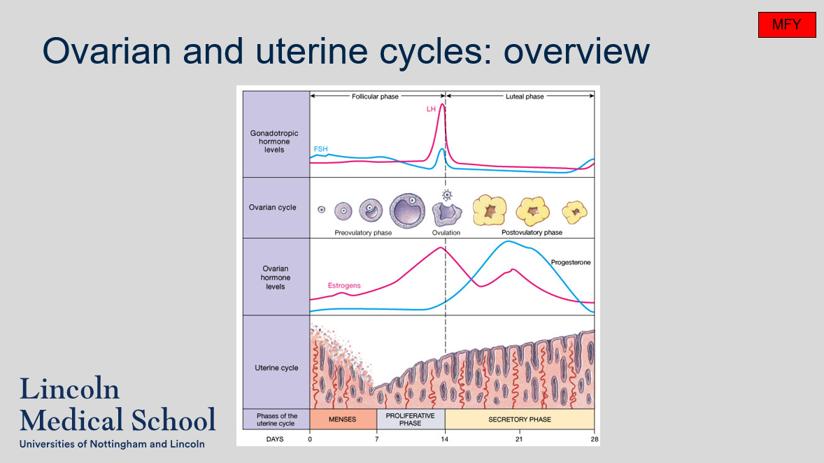
Ovarian and uterine cycles: overview
Can you label, describe and explain what this diagram is/shows?
Female reproductive cycle
Can you label, describe and explain what this diagram is/shows?
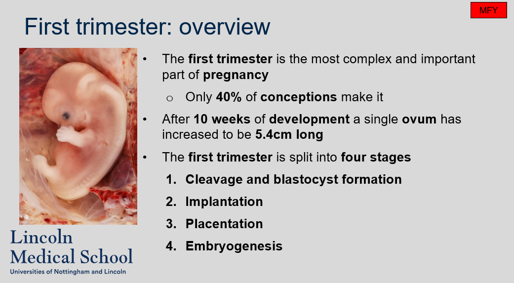
First trimester: overview
What is the first trimester?
Why is the first trimester important?
What is the success rate of conceptions during the first trimester?
How much does a single ovum grow during the first trimester?
What are the four stages of the first trimester?
The first trimester is the first three months of pregnancy.
The first trimester is the most complex and important part of pregnancy as it involves the development of major organs and structures of the fetus.
Only 40% of conceptions make it through the first trimester.
After 10 weeks of development, a single ovum has increased to be 5.4cm long.
The four stages of the first trimester are cleavage and blastocyst formation, implantation, placentation, and embryogenesis.
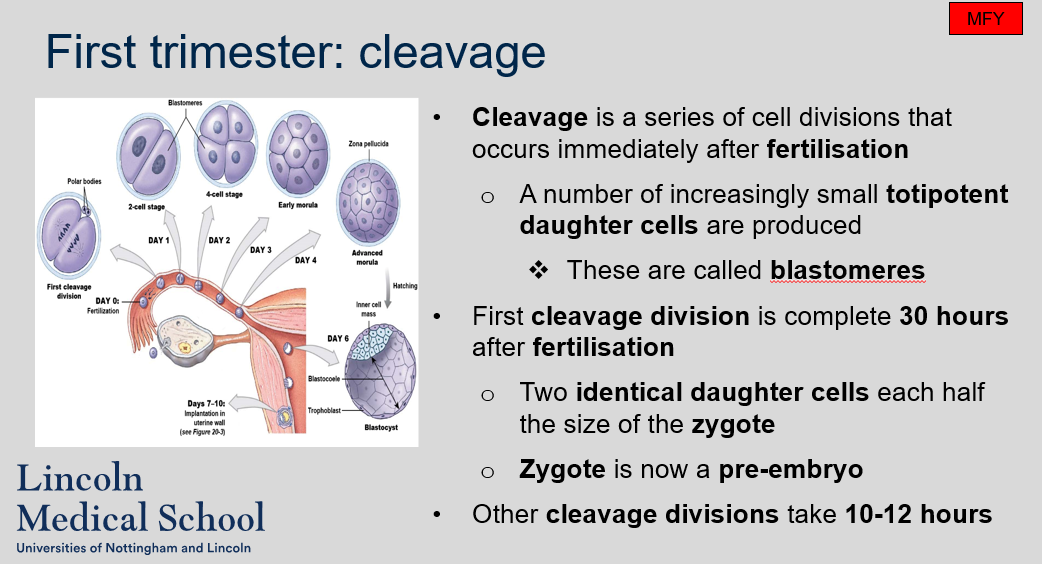
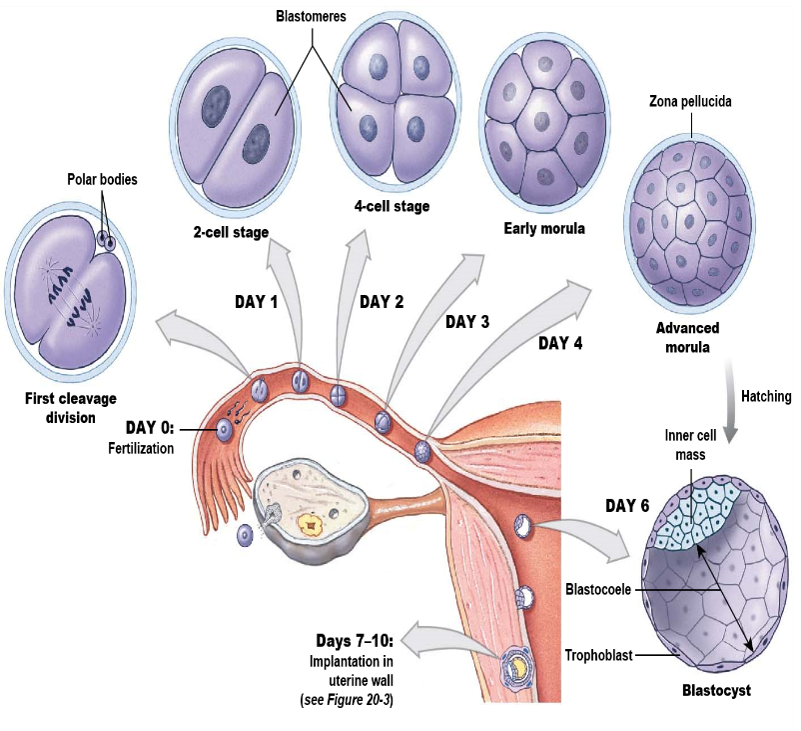
First trimester: cleavage
What is cleavage in the context of the first trimester of pregnancy?
Cleavage is a series of cell divisions that occur immediately after fertilization in the first trimester of pregnancy. During cleavage, the zygote divides into a number of increasingly small totipotent daughter cells called blastomeres. The first cleavage division is complete 30 hours after fertilization and produces two identical daughter cells, each half the size of the zygote, which is now a pre-embryo. Other cleavage divisions take 10-12 hours.
First trimester: cleavage
Can you label, describe and explain what this diagram is/shows?
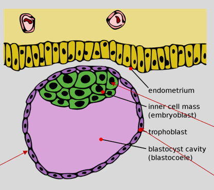
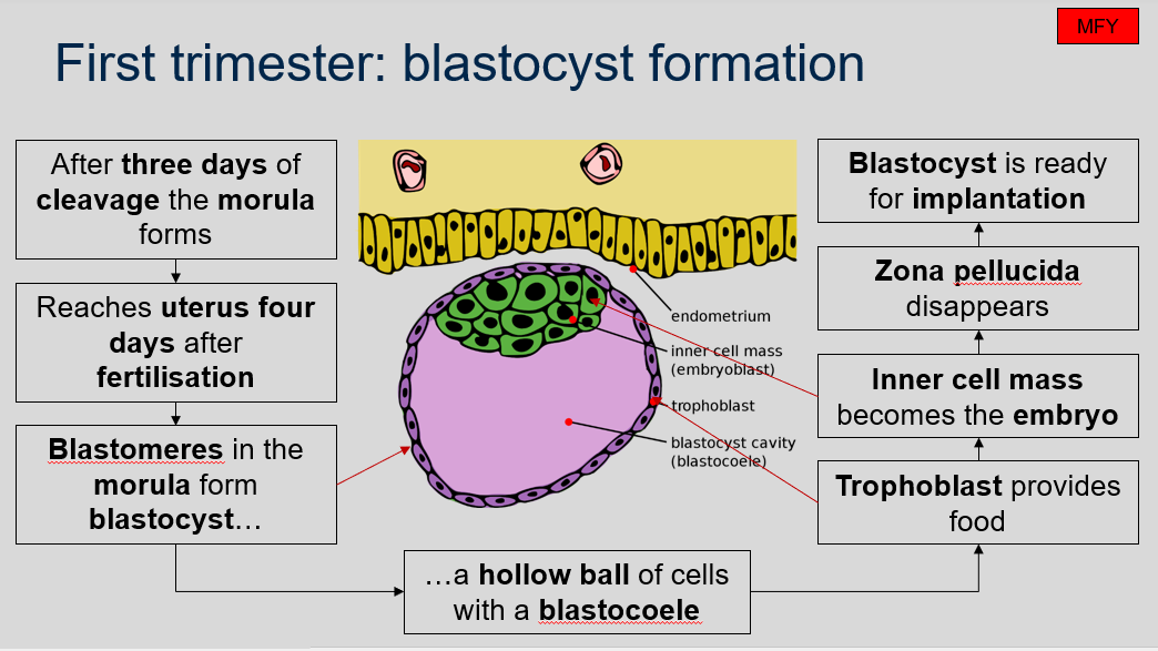
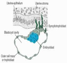
First trimester: blastocyst formation
What is blastocyst formation in the first trimester of pregnancy?
After three days of cleavage, the morula forms and reaches the uterus four days after fertilization. The blastomeres in the morula form a blastocyst, which is a hollow ball of cells with a blastocoel. The trophoblast, which is the outer layer of cells, provides nutrients to the developing embryo. The inner cell mass becomes the embryo itself. The zona pellucida, which is a glycoprotein layer surrounding the pre-embryo, disappears, and the blastocyst is now ready for implantation into the endometrium.
First trimester: blastocyst formation
Can you label, describe and explain what this diagram is/shows?
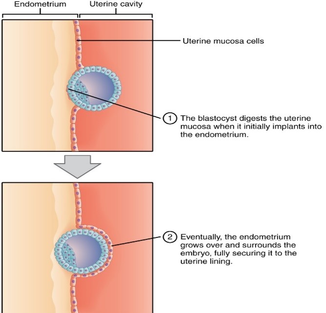
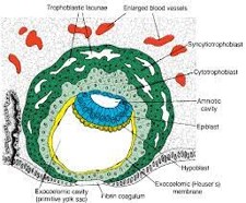
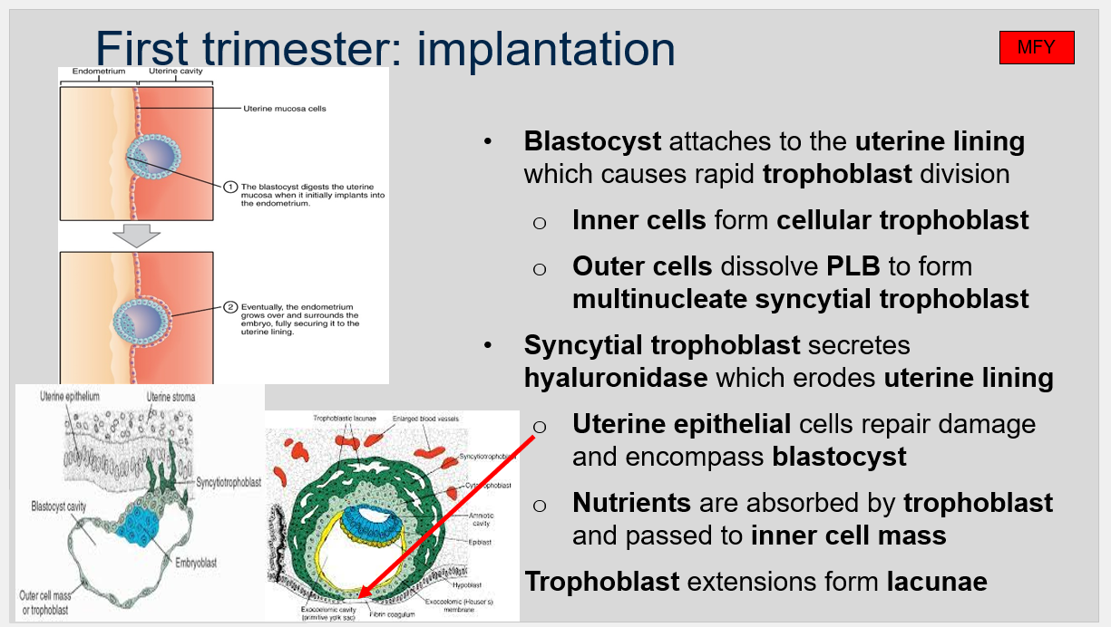
First trimester: implantation
What happens during implantation in the first trimester of pregnancy?
During implantation in the first trimester of pregnancy, the blastocyst attaches to the uterine lining, which causes rapid trophoblast division. The inner cells form the cellular trophoblast, while the outer cells dissolve the PLB (pellucida-like bodies) to form multinucleate syncytial trophoblast. The syncytial trophoblast secretes hyaluronidase, which erodes the uterine lining. The uterine epithelial cells repair the damage and encompass the blastocyst. Nutrients are absorbed by trophoblast and passed to the inner cell mass. Trophoblast extensions also form lacunae, which are spaces filled with maternal blood that will eventually form the placenta.
First trimester: implantation
Can you label, describe and explain what this diagram is/shows?
First trimester: implantation
Can you label, describe and explain what this diagram is/shows?
First trimester: implantation
Can you label, describe and explain what this diagram is/shows?
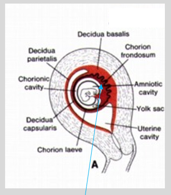
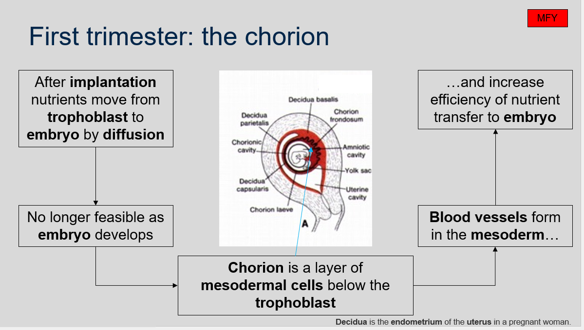
First trimester: the chorion
How do nutrients move from trophoblast to embryo after implantation?
What is the chorion?
What happens in the mesoderm of the chorion?
What is decidua?
Nutrients initially move from trophoblast to embryo by diffusion. However, as the embryo develops, this method becomes less feasible.
The chorion is a layer of mesodermal cells that develop below the trophoblast after implantation.
Blood vessels form in the mesoderm of the chorion, which increases the efficiency of nutrient transfer to the embryo.
Decidua is the endometrium of the uterus in a pregnant woman.
First trimester: the chorion
Can you label, describe and explain what this diagram is/shows?
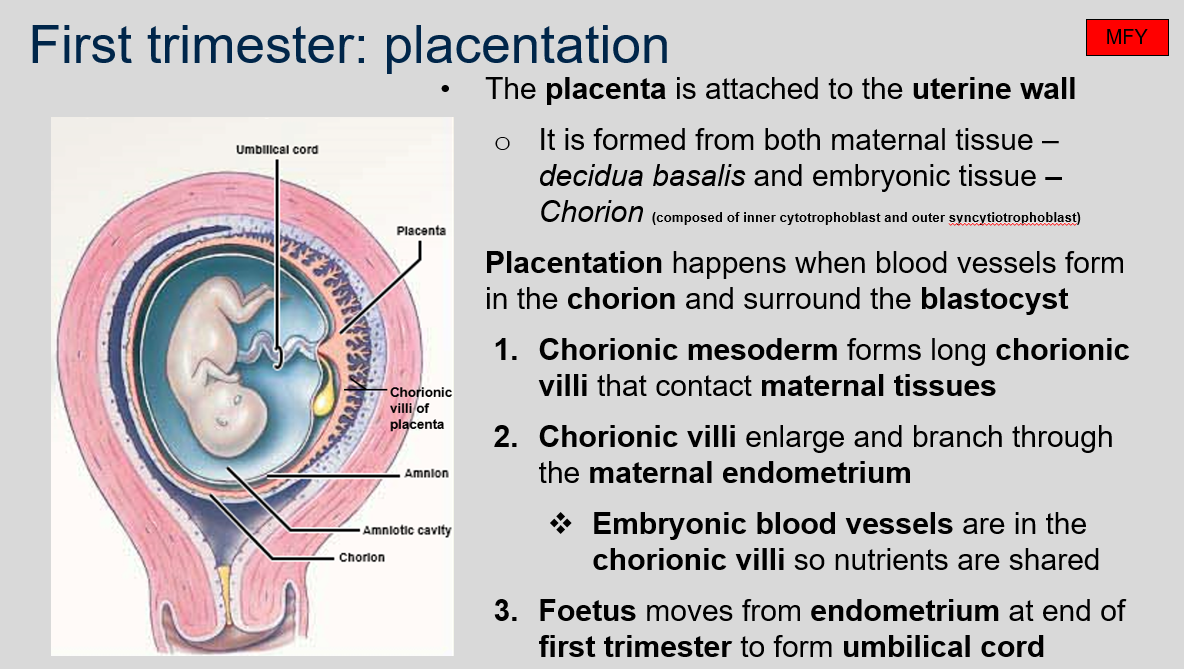
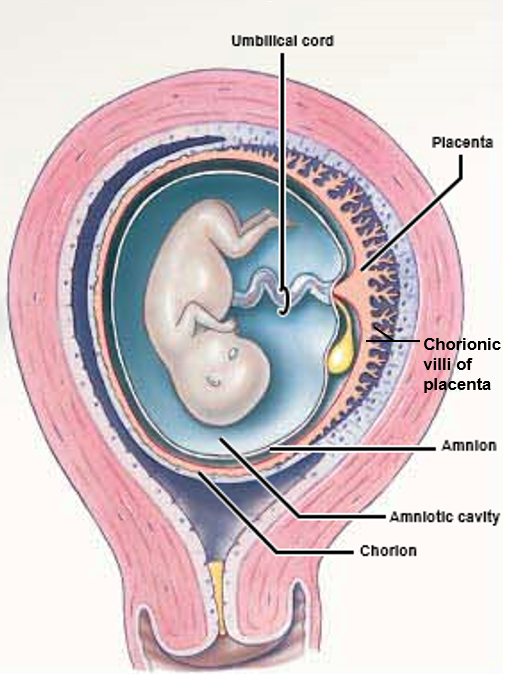
First trimester: placentation
What is the placenta and how is it formed in the first trimester?
The placenta is a vital organ that connects the developing fetus to the uterine wall to allow for the exchange of nutrients, oxygen, and waste products. It is formed in the first trimester of pregnancy from both maternal tissue (decidua basalis) and embryonic tissue (chorion), which is composed of inner cytotrophoblast and outer syncytiotrophoblast. Placentation occurs when blood vessels form in the chorion and surround the blastocyst. The chorionic mesoderm then forms long chorionic villi that contact maternal tissues and enlarge and branch through the maternal endometrium. Embryonic blood vessels are in the chorionic villi so nutrients are shared. Finally, the fetus moves from the endometrium at the end of the first trimester to form the umbilical cord.
First trimester: placentation
Can you label, describe and explain what this diagram is/shows?
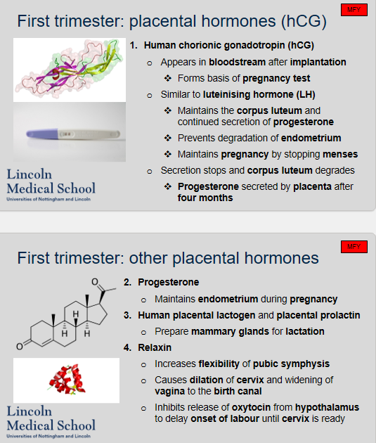
First trimester: placental hormones (hCG)
When does hCG appear in the bloodstream?
What is the basis of pregnancy test?
What hormone is hCG similar to?
What is the function of hCG in maintaining pregnancy?
When does the secretion of progesterone switch from the corpus luteum to the placenta?
What is the role of progesterone during pregnancy?
What is the role of human placental lactogen and placental prolactin during pregnancy?
What is the function of relaxin during pregnancy?
hCG appears in the bloodstream after implantation.
hCG is the basis of pregnancy test.
hCG is similar to luteinising hormone (LH).
hCG maintains the corpus luteum and continued secretion of progesterone, prevents degradation of endometrium, and maintains pregnancy by stopping menses.
The secretion of progesterone switches from the corpus luteum to the placenta after four months.
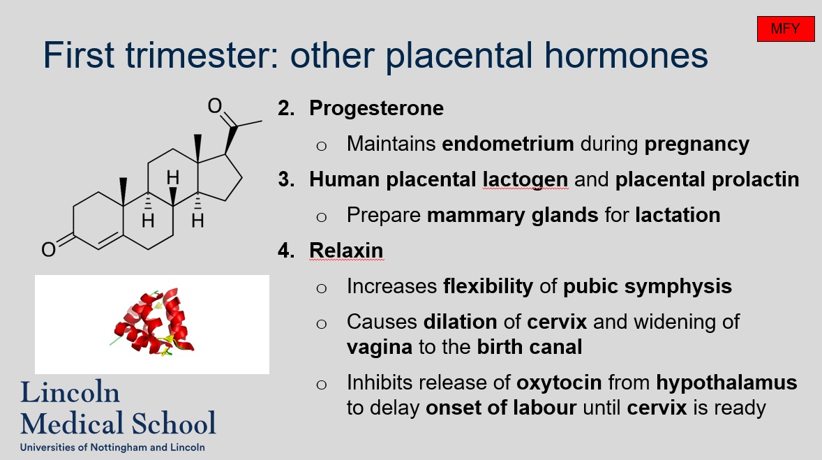
Progesterone plays an important role in maintaining the endometrium during pregnancy.
Human placental lactogen (hPL) and placental prolactin (PPL) are hormones produced by the placenta during pregnancy. Their role is to prepare the mammary glands for lactation, which is the process of producing and secreting breast milk.
Relaxin is a hormone secreted by the placenta during pregnancy. Its main function is to increase the flexibility of the pubic symphysis and relax the ligaments in the pelvic region to prepare for childbirth. It also helps to dilate the cervix and widen the vagina, making it easier for the baby to pass through the birth canal. Additionally, relaxin inhibits the release of oxytocin from the hypothalamus, which can delay the onset of labor until the cervix is ready for delivery.
First trimester: placental hormones (hCG)
Can you label, describe and explain what this diagram is/shows?
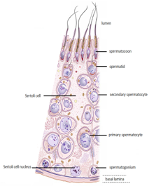
Male reproductive system
Can you label, describe and explain what this diagram is/shows?
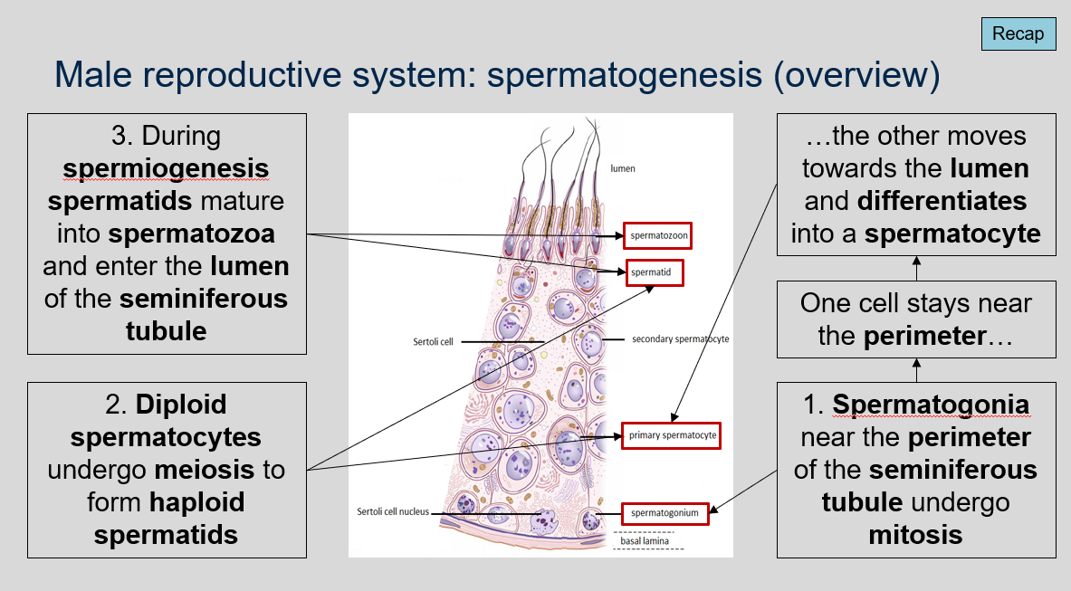
Male reproductive system: spermatogenesis (overview)
What is spermatogenesis and what are the main stages involved in it?
Spermatogenesis is the process of sperm cell development that occurs in the male reproductive system. The main stages involved in spermatogenesis are:
Spermatogonia mitosis: Spermatogonia, located near the perimeter of the seminiferous tubule, undergo mitosis. One cell stays near the perimeter, while the other moves towards the lumen and differentiates into a spermatocyte.
Meiosis: Diploid spermatocytes undergo meiosis to form haploid spermatids.
Spermiogenesis: During this stage, spermatids mature into spermatozoa and enter the lumen of the seminiferous tubule.