Leukocytes in Peripheral Blood IMAGE PRACTICE
1/61
Earn XP
Description and Tags
To those who want/need extra practice identifying leukocytes.
Name | Mastery | Learn | Test | Matching | Spaced | Call with Kai |
|---|
No study sessions yet.
62 Terms
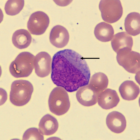
What is this leukocyte?
BAND NEUTROPHIL
The nucleus is band-shaped or contains rudimentary lobes that are connected by a thick band.
The cytoplasm is abundant, pink, and contains many small violet-pink neutrophilic or secondary granules.
Works in the body’s defense against bacteria. Their major function is as tissue phagocytes.
What is this leukocyte?
BAND NEUTROPHIL #2
The nucleus is band-shaped or contains rudimentary lobes that are connected by a thick band.
The cytoplasm is abundant, pink, and contains many small violet-pink neutrophilic or secondary granules.
Works in the body’s defense against bacteria. Their major function is as tissue phagocytes.
What is this leukocyte?
BAND NEUTROPHIL #3
The nucleus is band-shaped or contains rudimentary lobes that are connected by a thick band.
The cytoplasm is abundant, pink, and contains many small violet-pink neutrophilic or secondary granules.
Works in the body’s defense against bacteria. Their major function is as tissue phagocytes.
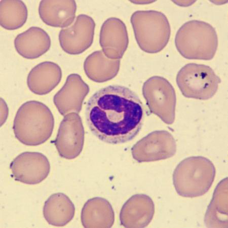
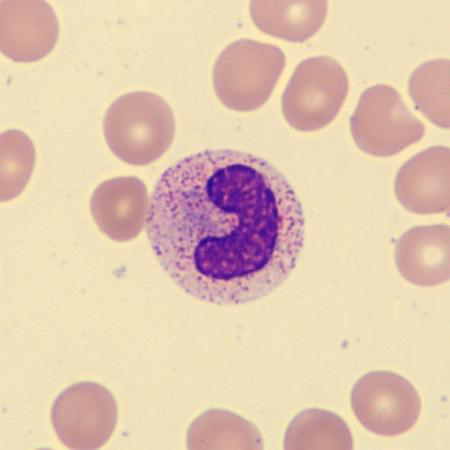
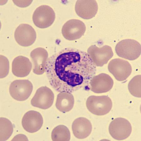
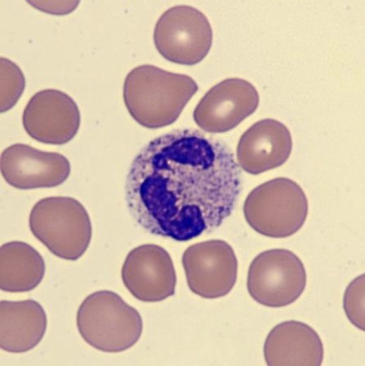
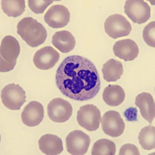
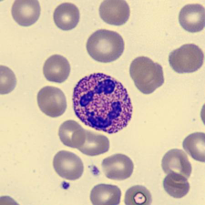
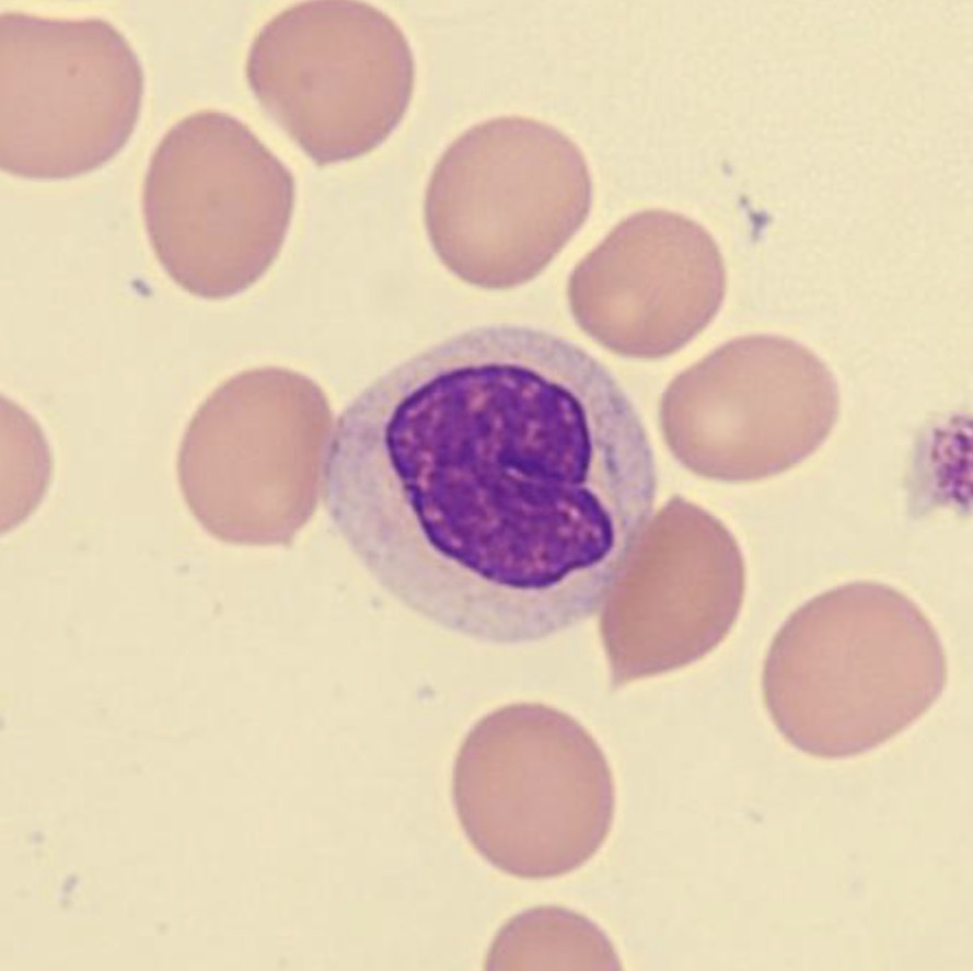
What is this leukocyte?
SEGMENTED NEUTROPHIL (PMN)
10–14 µm in diameter
The nucleus is segmented, usually 3–4 segments connected by a thin thread of chromatin that is coarse, stains violet, and is arranged in clumps.
Small nuclear appendages may be seen.
The cytoplasm is abundant, pink, with many small secondary granules.
Works in the body’s defense against bacteria. Their major function is tissue phagocytes.
What is this leukocyte?
SEGMENTED NEUTROPHIL (PMN) #2
10–14 µm in diameter
The nucleus is segmented, usually 3–4 segments connected by a thin thread of chromatin that is coarse, stains violet, and is arranged in clumps.
Small nuclear appendages may be seen.
The cytoplasm is abundant, pink, with many small secondary granules.
Works in the body’s defense against bacteria. Their major function is tissue phagocytes.

What is this leukocyte?
SEGMENTED NEUTROPHIL (PMN) #3
10–14 µm in diameter
The nucleus is segmented, usually 3–4 segments connected by a thin thread of chromatin that is coarse, stains violet, and is arranged in clumps.
Small nuclear appendages may be seen.
The cytoplasm is abundant, pink, with many small secondary granules.
Works in the body’s defense against bacteria. Their major function is tissue phagocytes.
What is this leukocyte?
SEGMENTED NEUTROPHIL (PMN) #4
10–14 µm in diameter
The nucleus is segmented, usually 3–4 segments connected by a thin thread of chromatin that is coarse, stains violet, and is arranged in clumps.
Small nuclear appendages may be seen.
The cytoplasm is abundant, pink, with many small secondary granules.
Works in the body’s defense against bacteria. Their major function is tissue phagocytes.
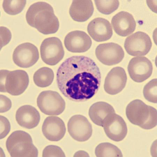
What is this leukocyte?
EOSINOPHIL
12–17 µm in diameter.
The nucleus is segmented, usually only two segments, with coarsely clumped, violet-staining chromatin.
The cytoplasm is abundant, containing many eosinophilic (orange or pink) secondary granules that are larger than neutrophilic granules and more uniform in size.
Works in the body’s defense against tissue parasites and allergic reaction.

What is this leukocyte?
EOSINOPHIL #2
12–17 µm in diameter.
The nucleus is segmented, usually only two segments, with coarsely clumped, violet-staining chromatin.
The cytoplasm is abundant, containing many eosinophilic (orange or pink) secondary granules that are larger than neutrophilic granules and more uniform in size.
Works in the body’s defense against tissue parasites and allergic reaction.
What is this leukocyte?
EOSINOPHIL #3
12–17 µm in diameter.
The nucleus is segmented, usually only two segments, with coarsely clumped, violet-staining chromatin.
The cytoplasm is abundant, containing many eosinophilic (orange or pink) secondary granules that are larger than neutrophilic granules and more uniform in size.
Works in the body’s defense against tissue parasites and allergic reaction.
What is this leukocyte?
EOSINOPHIL #4
12–17 µm in diameter.
The nucleus is segmented, usually only two segments, with coarsely clumped, violet-staining chromatin.
The cytoplasm is abundant, containing many eosinophilic (orange or pink) secondary granules that are larger than neutrophilic granules and more uniform in size.
Works in the body’s defense against tissue parasites and allergic reaction.
What is this leukocyte?
BASOPHIL
10–16 µm in diameter.
The nucleus is segmented but is often obscured by basophilic granules.
The cytoplasm is pale blue, containing purple-black secondary granules that can vary in number, size, and shape. The granules are water-soluble and may dissolve on staining, leaving clear areas in the cytoplasm.
Works in the body’s defense against allergic and inflammatory agents.
What is this leukocyte?
BASOPHIL #2
10–16 µm in diameter.
The nucleus is segmented but is often obscured by basophilic granules.
The cytoplasm is pale blue, containing purple-black secondary granules that can vary in number, size, and shape. The granules are water-soluble and may dissolve on staining, leaving clear areas in the cytoplasm.
Works in the body’s defense against allergic and inflammatory agents.

What is this leukocyte?
BASOPHIL #3
10–16 µm in diameter.
The nucleus is segmented but is often obscured by basophilic granules.
The cytoplasm is pale blue, containing purple-black secondary granules that can vary in number, size, and shape. The granules are water-soluble and may dissolve on staining, leaving clear areas in the cytoplasm.
Works in the body’s defense against allergic and inflammatory agents.
What is this leukocyte?
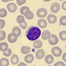
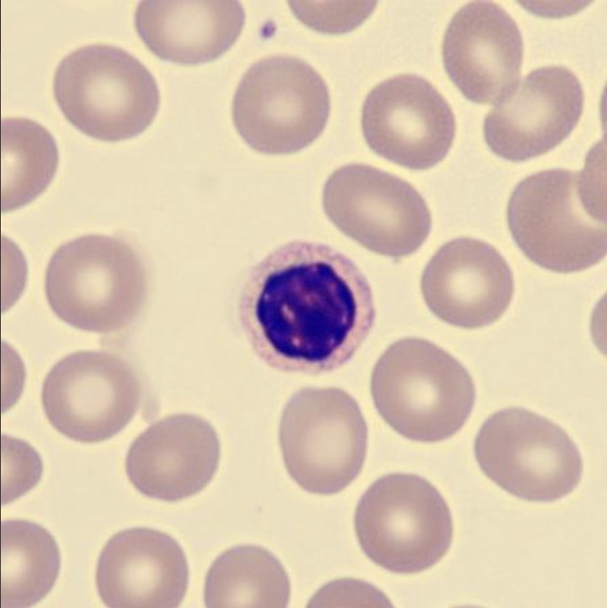
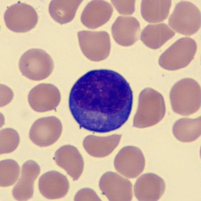
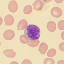
SMALL LYMPH
• 8–12 µm in diameter with high N:C ratio.
• The nucleus is round with coarse, dense chromatin.
• The cytoplasm is narrow, weakly basophilic, homogenous, and clear without inclusions.
• Works in the body’s defense against viral infections.
What is this leukocyte?
SMALL LYMPH #2
• 8–12 µm in diameter with high N:C ratio.
• The nucleus is round with coarse, dense chromatin.
• The cytoplasm is narrow, weakly basophilic, homogenous, and clear without inclusions.
• Works in the body’s defense against viral infections.

What is this leukocyte?
SMALL LYMPH #3
• 8–12 µm in diameter with high N:C ratio.
• The nucleus is round with coarse, dense chromatin.
• The cytoplasm is narrow, weakly basophilic, homogenous, and clear without inclusions.
• Works in the body’s defense against viral infections.
What is this leukocyte?
SMALL LYMPH #4
• 8–12 µm in diameter with high N:C ratio.
• The nucleus is round with coarse, dense chromatin.
• The cytoplasm is narrow, weakly basophilic, homogenous, and clear without inclusions.
• Works in the body’s defense against viral infections.
What is this leukocyte?
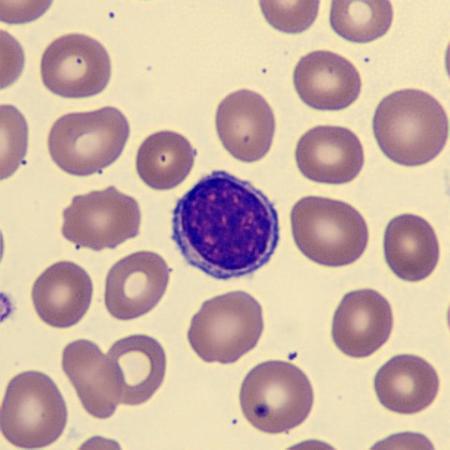
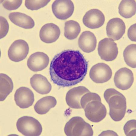
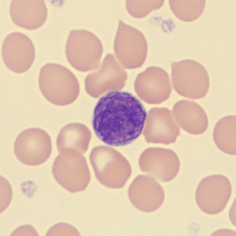
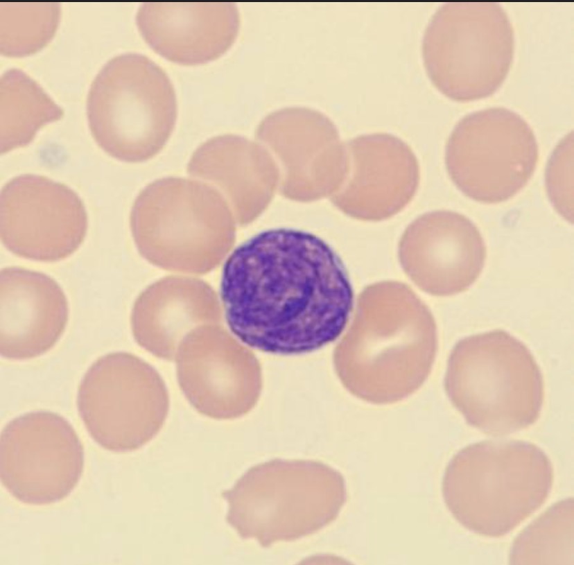
LARGE LYMPH
12–16 µm in diameter with an irregular outline.
The nuclear can appear irregular and the chromatin is not as coarse as in small lymphocytes.
The cytoplasm is abundant and tends to be light blue.
Works in the body’s defense against viral infections.

What is this leukocyte?
LARGE LYMPH #2
12–16 µm in diameter with an irregular outline.
The nuclear can appear irregular and the chromatin is not as coarse as in small lymphocytes.
The cytoplasm is abundant and tends to be light blue.
Works in the body’s defense against viral infections.
What is this leukocyte?
LARGE LYMPH #3
12–16 µm in diameter with an irregular outline.
The nuclear can appear irregular and the chromatin is not as coarse as in small lymphocytes.
The cytoplasm is abundant and tends to be light blue.
Works in the body’s defense against viral infections.
What is this leukocyte?
LARGE LYMPH #4
12–16 µm in diameter with an irregular outline.
The nuclear can appear irregular and the chromatin is not as coarse as in small lymphocytes.
The cytoplasm is abundant and tends to be light blue.
Works in the body’s defense against viral infections.
What is this leukocyte?
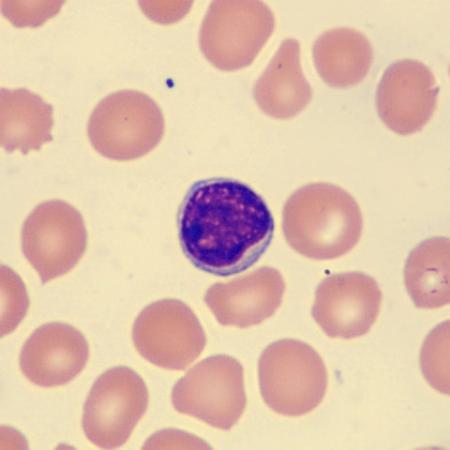
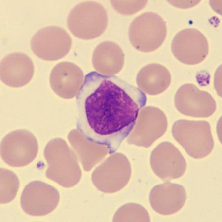
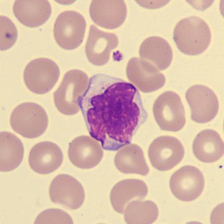
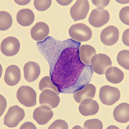
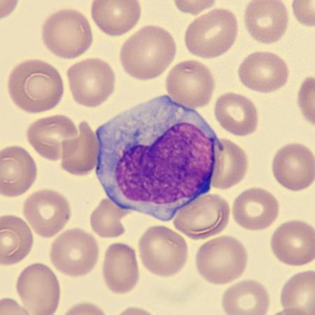
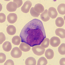
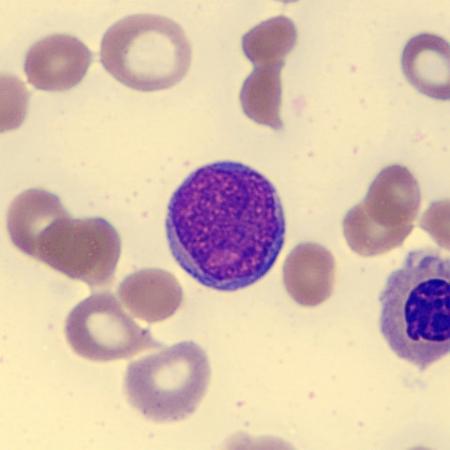
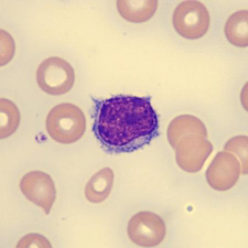
MONOCYTE
15–22 µm in diameter. Largest cell in peripheral blood.
The nucleus is irregular in outline, often kidney-shaped, and the chromatin is arranged in fine strands with sharply defined margins.
The cytoplasm is light blue-grey and contains numerous fine dust-like granules.
Vacuolation may be present.
Works in the body’s defense against bacterial and fungal infections.
Mainly differentiates into long-lived tissues macrophages.
What is this leukocyte?
MONOCYTE #2
15–22 µm in diameter. Largest cell in peripheral blood.
The nucleus is irregular in outline, often kidney-shaped, and the chromatin is arranged in fine strands with sharply defined margins.
The cytoplasm is light blue-grey and contains numerous fine dust-like granules.
Vacuolation may be present.
Works in the body’s defense against bacterial and fungal infections.
Mainly differentiates into long-lived tissues macrophages.

What is this leukocyte?
MONOCYTE #3
15–22 µm in diameter. Largest cell in peripheral blood.
The nucleus is irregular in outline, often kidney-shaped, and the chromatin is arranged in fine strands with sharply defined margins.
The cytoplasm is light blue-grey and contains numerous fine dust-like granules.
Vacuolation may be present.
Works in the body’s defense against bacterial and fungal infections.
Mainly differentiates into long-lived tissues macrophages.

What is this leukocyte?
MONOCYTE #4
15–22 µm in diameter. Largest cell in peripheral blood.
The nucleus is irregular in outline, often kidney-shaped, and the chromatin is arranged in fine strands with sharply defined margins.
The cytoplasm is light blue-grey and contains numerous fine dust-like granules.
Vacuolation may be present.
Works in the body’s defense against bacterial and fungal infections.
Mainly differentiates into long-lived tissues macrophages.
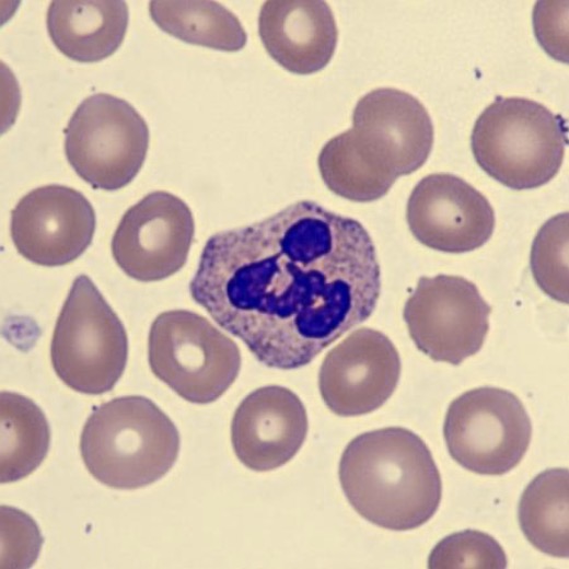
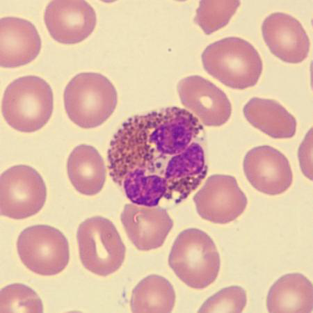
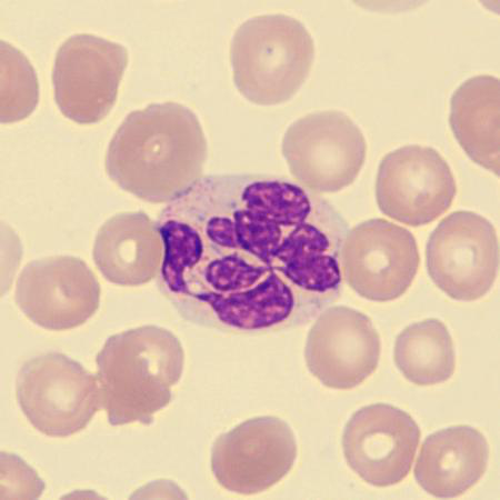
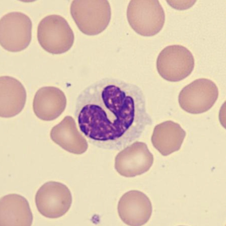
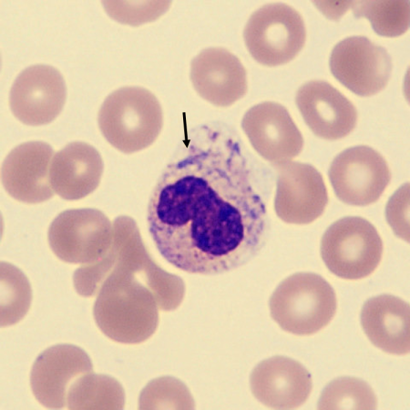
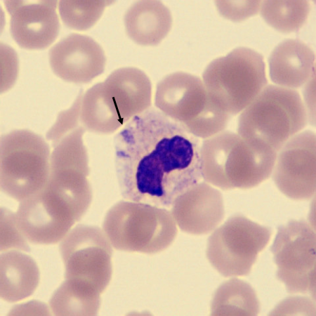
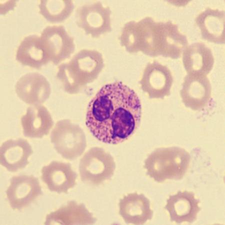
What is this neutrophil showing?
HYPERSEGMENTATION
• Hypersegmented neutrophils have an increased number of distinct nuclear lobes with increased numbers of neutrophils having five or more nuclear segments.
• Hypersegmented neutrophils have a correlation with Megaloblastic anemias.

What is this neutrophil showing?
HYPERSEGMENTATION #2
• Hypersegmented neutrophils have an increased number of distinct nuclear lobes with increased numbers of neutrophils having five or more nuclear segments.
• Hypersegmented neutrophils have a correlation with Megaloblastic anemias.
What is this neutrophil showing?
HYPERSEGMENTATION #3
• Hypersegmented neutrophils have an increased number of distinct nuclear lobes with increased numbers of neutrophils having five or more nuclear segments.
• Hypersegmented neutrophils have a correlation with Megaloblastic anemias.
What is this neutrophil showing?
HYPERSEGMENTATION #4
• Hypersegmented neutrophils have an increased number of distinct nuclear lobes with increased numbers of neutrophils having five or more nuclear segments.
• Hypersegmented neutrophils have a correlation with Megaloblastic anemias.
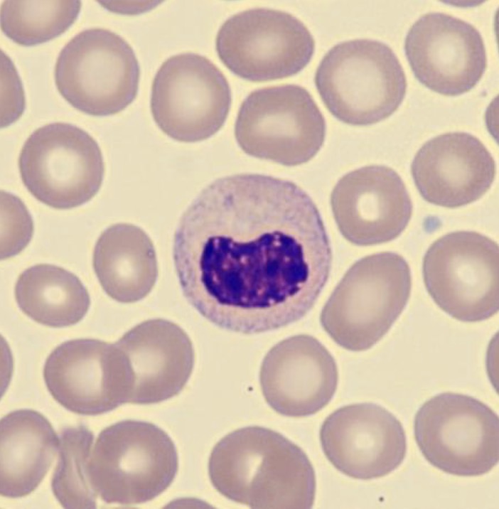
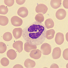
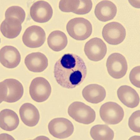
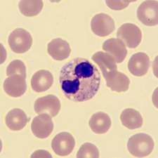
What is this neutrophil showing?
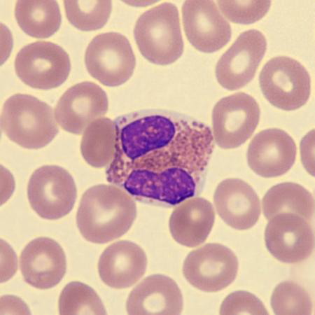
HYPOSEGMENTATION
• Hyposegmented are mature neutrophils and can be differentiated by their smaller nucleus and lower nuclear-to-cytoplasmic (N:C) ratio, along with condensed nuclear chromatin.
• Hyposegmented neutrophils are marked by the failure of normal nuclear lobe development during terminal differentiation and have coarse, clumped nuclear chromatin.
What is this neutrophil showing?
HYPOSEGMENTATION #2
• Hyposegmented are mature neutrophils and can be differentiated by their smaller nucleus and lower nuclear-to-cytoplasmic (N:C) ratio, along with condensed nuclear chromatin.
• Hyposegmented neutrophils are marked by the failure of normal nuclear lobe development during terminal differentiation and have coarse, clumped nuclear chromatin.
What is this neutrophil showing?
HYPOSEGMENTATION #3
• Hyposegmented are mature neutrophils and can be differentiated by their smaller nucleus and lower nuclear-to-cytoplasmic (N:C) ratio, along with condensed nuclear chromatin.
• Hyposegmented neutrophils are marked by the failure of normal nuclear lobe development during terminal differentiation and have coarse, clumped nuclear chromatin.
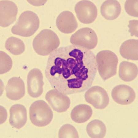
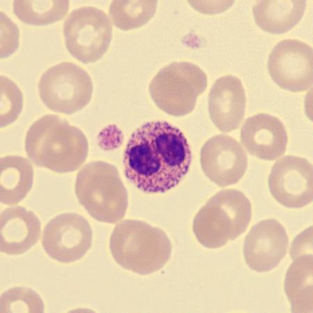
What is this neutrophil showing?
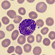
TOXIC GRANULATION
• Hypergranulated neutrophils contain coarse, dark-purple primary (azurophilic) granules that appear as a reactive response to infection or inflammation.
• This is a non-specific change caused by abnormal maturation of primary granules, leading to retention of their azurophilic staining properties.

What is this neutrophil showing?
TOXIC GRANULATION #2
• Hypergranulated neutrophils contain coarse, dark-purple primary (azurophilic) granules that appear as a reactive response to infection or inflammation.
• This is a non-specific change caused by abnormal maturation of primary granules, leading to retention of their azurophilic staining properties.

What is this neutrophil showing?
TOXIC GRANULATION #3
• Hypergranulated neutrophils contain coarse, dark-purple primary (azurophilic) granules that appear as a reactive response to infection or inflammation.
• This is a non-specific change caused by abnormal maturation of primary granules, leading to retention of their azurophilic staining properties.

What is this neutrophil showing?
TOXIC GRANULATION #4
• Hypergranulated neutrophils contain coarse, dark-purple primary (azurophilic) granules that appear as a reactive response to infection or inflammation.
• This is a non-specific change caused by abnormal maturation of primary granules, leading to retention of their azurophilic staining properties.
What is this neutrophil showing?
HYPOGRANULATION
• Hypogranulated neutrophils show reduced or absent cytoplasmic granules, giving mature neutrophils a smooth blue-grey appearance on the smear.
What is this neutrophil showing?
HYPOGRANULATION #2
• Hypogranulated neutrophils show reduced or absent cytoplasmic granules, giving mature neutrophils a smooth blue-grey appearance on the smear.
What is this neutrophil showing?
HYPOGRANULATION #3
• Hypogranulated neutrophils show reduced or absent cytoplasmic granules, giving mature neutrophils a smooth blue-grey appearance on the smear.
What is this leukocyte showing?
REACTIVITY
• Reactive lymphocyte is used to describe lymphocytes with a benign etiology.
• Abnormalities include increased cell size, immaturity of the nucleus including a visible nucleolus and lack of chromatin condensation.
• The nuclear outline can be irregular or present with lobulation.
• The cytoplasm appears basophilic, can contain vacuolation and have an irregular cell outline.
• Reactive lymphocytes have a tendency to surround adjacent cells and can be seen in viral infections.
What is this leukocyte showing?
REACTIVITY #2
• Reactive lymphocyte is used to describe lymphocytes with a benign etiology.
• Abnormalities include increased cell size, immaturity of the nucleus including a visible nucleolus and lack of chromatin condensation.
• The nuclear outline can be irregular or present with lobulation.
• The cytoplasm appears basophilic, can contain vacuolation and have an irregular cell outline.
• Reactive lymphocytes have a tendency to surround adjacent cells and can be seen in viral infections.

What is this leukocyte showing?
REACTIVITY #3
• Reactive lymphocyte is used to describe lymphocytes with a benign etiology.
• Abnormalities include increased cell size, immaturity of the nucleus including a visible nucleolus and lack of chromatin condensation.
• The nuclear outline can be irregular or present with lobulation.
• The cytoplasm appears basophilic, can contain vacuolation and have an irregular cell outline.
• Reactive lymphocytes have a tendency to surround adjacent cells and can be seen in viral infections.
What is this leukocyte showing?
REACTIVITY #4
• Reactive lymphocyte is used to describe lymphocytes with a benign etiology.
• Abnormalities include increased cell size, immaturity of the nucleus including a visible nucleolus and lack of chromatin condensation.
• The nuclear outline can be irregular or present with lobulation.
• The cytoplasm appears basophilic, can contain vacuolation and have an irregular cell outline.
• Reactive lymphocytes have a tendency to surround adjacent cells and can be seen in viral infections.
What is this leukocyte?
ABNORMAL LYMPH
• Abnormal lymphocyte with an accompanying description of the cells is used to describe lymphocytes with a suspected malignant or clonal etiology.
• Examples of abnormal lymphocytes are Hairy cells, Sézary cells, lymphoma cells, and prolymphocytes.
What is this leukocyte?
ABNORMAL LYMPH #2
• Abnormal lymphocyte with an accompanying description of the cells is used to describe lymphocytes with a suspected malignant or clonal etiology.
• Examples of abnormal lymphocytes are Hairy cells, Sézary cells, lymphoma cells, and prolymphocytes.
What is this?
AUER RODS
• Auer rods are sharply defined red rod or needle-like cytoplasmic inclusion formed by abnormal primary granule development.
• There may be several in a cell and may be arranged in bundles.
• Auer rods are mainly found in leukemic myeloblasts or abnormal promyelocytes.
• Auer rods can be found in Acute Myelocytic Leukemia (AML).
What is this?
AUER RODS #2
• Auer rods are sharply defined red rod or needle-like cytoplasmic inclusion formed by abnormal primary granule development.
• There may be several in a cell and may be arranged in bundles.
• Auer rods are mainly found in leukemic myeloblasts or abnormal promyelocytes.
• Auer rods can be found in Acute Myelocytic Leukemia (AML).
What is this?
AUER RODS #3
• Auer rods are sharply defined red rod or needle-like cytoplasmic inclusion formed by abnormal primary granule development.
• There may be several in a cell and may be arranged in bundles.
• Auer rods are mainly found in leukemic myeloblasts or abnormal promyelocytes.
• Auer rods can be found in Acute Myelocytic Leukemia (AML).
What is this leukocyte?
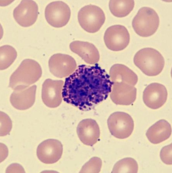
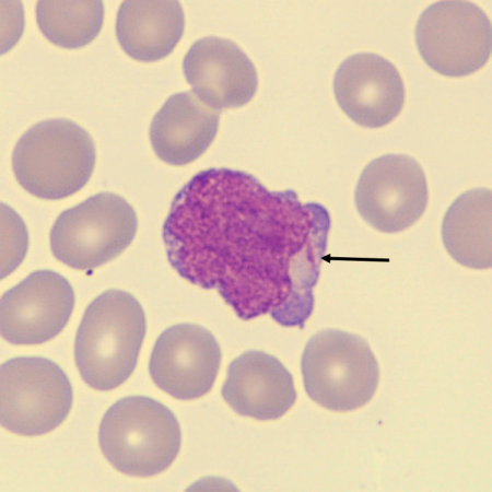
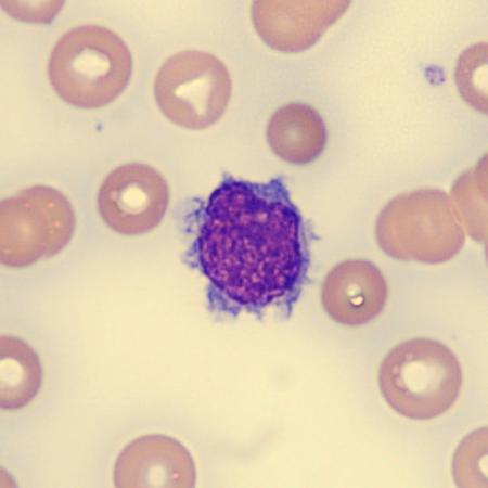
HAIRY CELL
• Hairy cells are larger than normal lymphocytes and have abundant pale blue-grey cytoplasm with fine hair-like projections.
• The nucleus varies in shape and may be round, oval, bean-shaped or bilobed.
• Hairy cell leukemia is a chronic B cell lineage leukemia with morphologically distinctive neoplastic cells.
What is this leukocyte?
HAIRY CELL #2
• Hairy cells are larger than normal lymphocytes and have abundant pale blue-grey cytoplasm with fine hair-like projections.
• The nucleus varies in shape and may be round, oval, bean-shaped or bilobed.
• Hairy cell leukemia is a chronic B cell lineage leukemia with morphologically distinctive neoplastic cells.
What is this leukocyte?
HAIRY CELL #3
• Hairy cells are larger than normal lymphocytes and have abundant pale blue-grey cytoplasm with fine hair-like projections.
• The nucleus varies in shape and may be round, oval, bean-shaped or bilobed.
• Hairy cell leukemia is a chronic B cell lineage leukemia with morphologically distinctive neoplastic cells.
What is this leukocyte?
SEZARY CELL
• Sezary cells can vary in size.
• The nuclear morphology, classically described as cerebriform, is the characteristic cytological feature of sezary cells.
• The nucleus has deep narrow clefts with superimposed and folded lobes giving it a very convoluted appearance.
• Sezary syndrome is a mature T-cell lymphoma with neoplastic T lymphocytes in the peripheral blood.
What is this leukocyte?
SEZARY CELL #2
• Sezary cells can vary in size.
• The nuclear morphology, classically described as cerebriform, is the characteristic cytological feature of sezary cells.
• The nucleus has deep narrow clefts with superimposed and folded lobes giving it a very convoluted appearance.
• Sezary syndrome is a mature T-cell lymphoma with neoplastic T lymphocytes in the peripheral blood.
What is this leukocyte?
SEZARY CELL #3
• Sezary cells can vary in size.
• The nuclear morphology, classically described as cerebriform, is the characteristic cytological feature of sezary cells.
• The nucleus has deep narrow clefts with superimposed and folded lobes giving it a very convoluted appearance.
• Sezary syndrome is a mature T-cell lymphoma with neoplastic T lymphocytes in the peripheral blood.
What characteristics is this neutrophil showing?
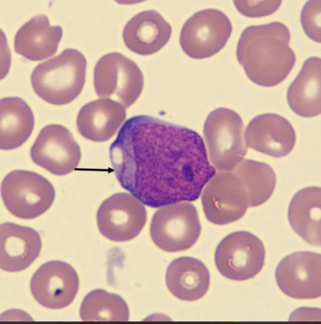
DÖHLE BODIES
• Döhle bodies are pale light blue or grey, single or multiple, cytoplasmic inclusions found near the periphery of the neutrophil.
• Döhle bodies are a non-specific reactive change but may also indicate May-Hegglin anomaly if associated with thrombocytopenia and giant platelets.
What characteristics is this neutrophil showing?
DÖHLE BODIES #2
• Döhle bodies are pale light blue or grey, single or multiple, cytoplasmic inclusions found near the periphery of the neutrophil.
• Döhle bodies are a non-specific reactive change but may also indicate May-Hegglin anomaly if associated with thrombocytopenia and giant platelets.
What characteristics is this neutrophil showing?
DÖHLE BODIES #3
• Döhle bodies are pale light blue or grey, single or multiple, cytoplasmic inclusions found near the periphery of the neutrophil.
• Döhle bodies are a non-specific reactive change but may also indicate May-Hegglin anomaly if associated with thrombocytopenia and giant platelets.
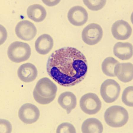
What characteristics is this neutrophil showing?
PEGLER HUËT ABNORMALITY
• Pelger Huët is an inherited or acquired (pseudo-Pelger-Huët) anomaly.
• It is characterized by decreased nuclear segmentation and a coarse chromatin clumping pattern of the Neutrophils.
What characteristics is this neutrophil showing?
PEGLER HUËT ABNORMALITY #2
• Pelger Huët is an inherited or acquired (pseudo-Pelger-Huët) anomaly.
• It is characterized by decreased nuclear segmentation and a coarse chromatin clumping pattern of the Neutrophils.
What characteristics is this neutrophil showing?
PEGLER HUËT ABNORMALITY #3
• Pelger Huët is an inherited or acquired (pseudo-Pelger-Huët) anomaly.
• It is characterized by decreased nuclear segmentation and a coarse chromatin clumping pattern of the Neutrophils.
What characteristics is this neutrophil showing?
PEGLER HUËT ABNORMALITY #4
• Pelger Huët is an inherited or acquired (pseudo-Pelger-Huët) anomaly.
• It is characterized by decreased nuclear segmentation and a coarse chromatin clumping pattern of the Neutrophils.