Structures and Functions of the Thigh
1/46
There's no tags or description
Looks like no tags are added yet.
Name | Mastery | Learn | Test | Matching | Spaced |
|---|
No study sessions yet.
47 Terms
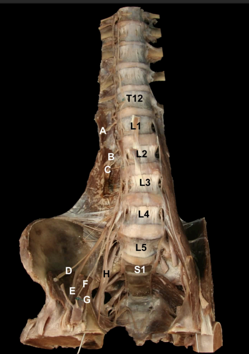
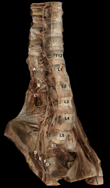
Identify the Nerves and their corresponding nerve levels indicated on this Anterior view of the Pelvis: Lumbar Plexus
A. Subcostal n (T12)
B: Iliophypogastric n (L1)
C: Ilioinguinal n, (L2)
D: Lateral Cutaneous n. of the Thigh (L2-L3)
E: Femoral n (L2-L4)
F: Genitofemoral n. (L1-L2)
G: Obturator n. (L2-L4)
H: Lumbosacral Trunk (L4-L5)
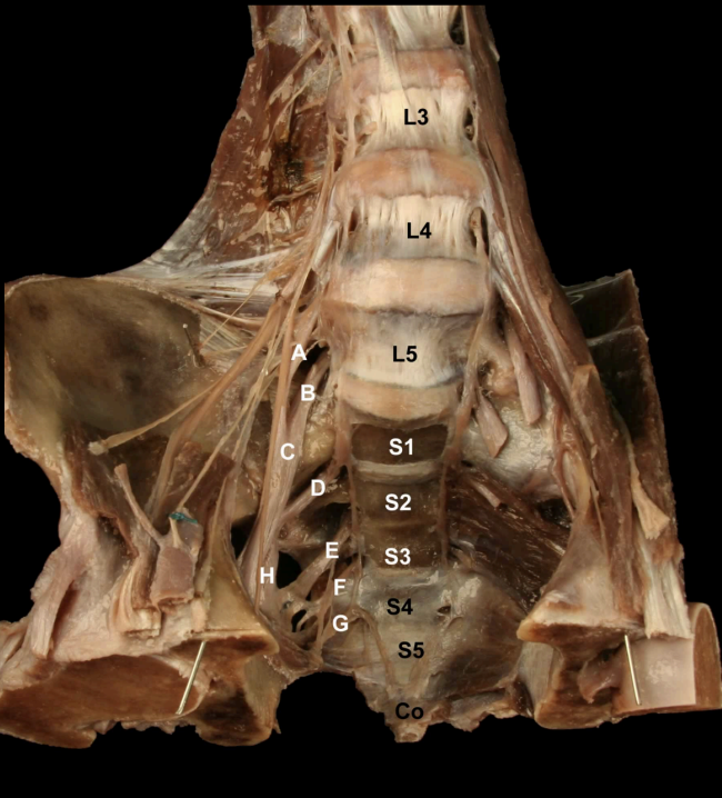
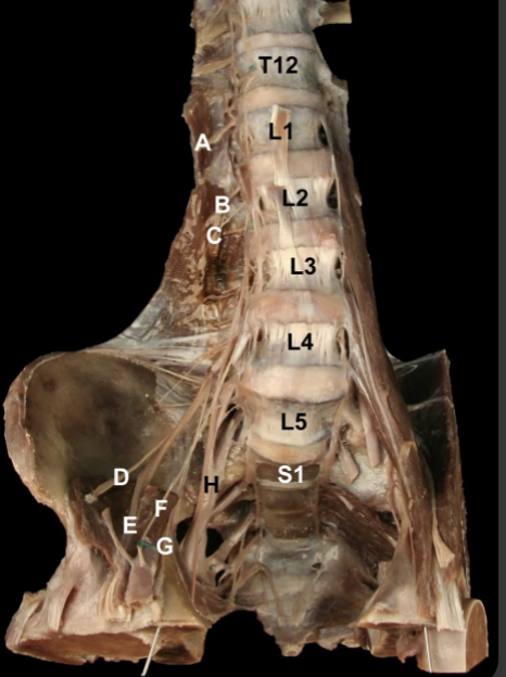
Identify the Nerves and their corresponding nerve levels indicated on this Anterior view of the Pelvis: Sacral Plexus
A. L4 Nerve Root
B: L5 Nerve Root
C: Lumbosacral Trunk (L4-L5)
D: S1 Nerve Root
E: S2 Nerve Root
F: S3 Nerve Root
G: S4 Nerve Root
H: Sciatic n. (L4-S3)
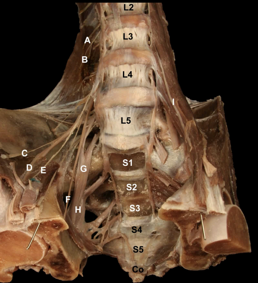
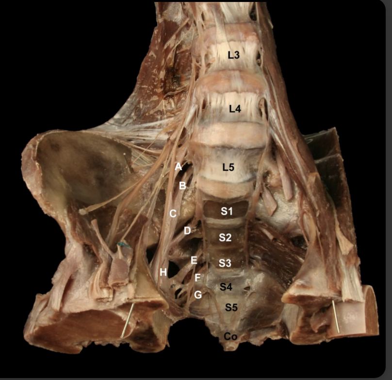
Try and Identify the nerves and nerve roots of he Lumbarsacral Plexus
A: Iliohypogastric n (L1)
B: Ilioinguinal n. (L1)
C: Lateral Cutaneous n. of the Thigh (L2-L3)
D: Femoral n. (L1-L4).
E: Geniofemoral n. (L1-L2)
F: Obturator n. (L2-L4)
G: Lumbosacral trunk (L4-L5)
H: Sciatic n. (L4-S3)
I: Genitofemoral n. (L1-L2).
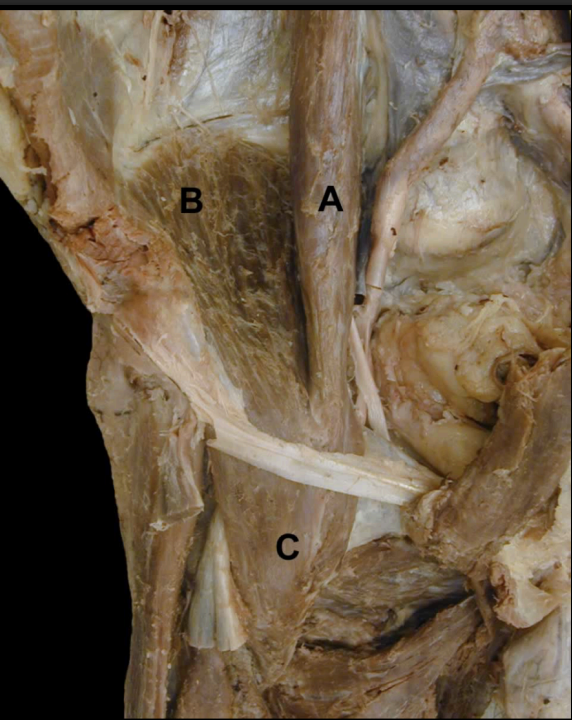
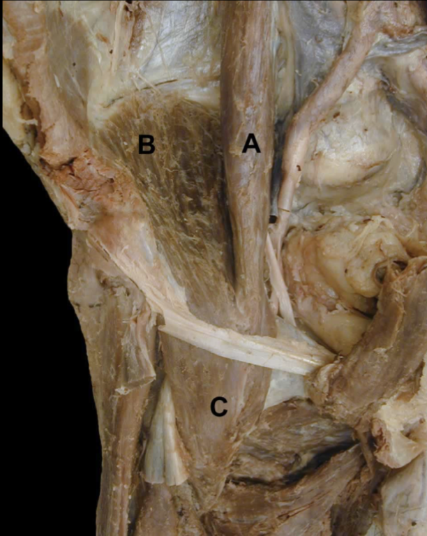
Identify the indicated muscles that make up the Iliopsoas Muscle
A: Psoas Major m
B: Iliacus m.
=
C: Iliopsoas m.
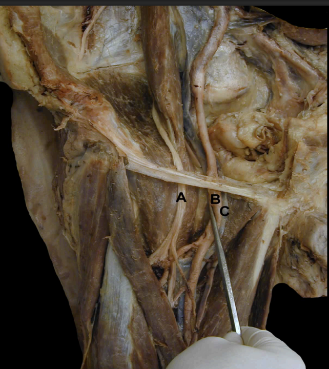
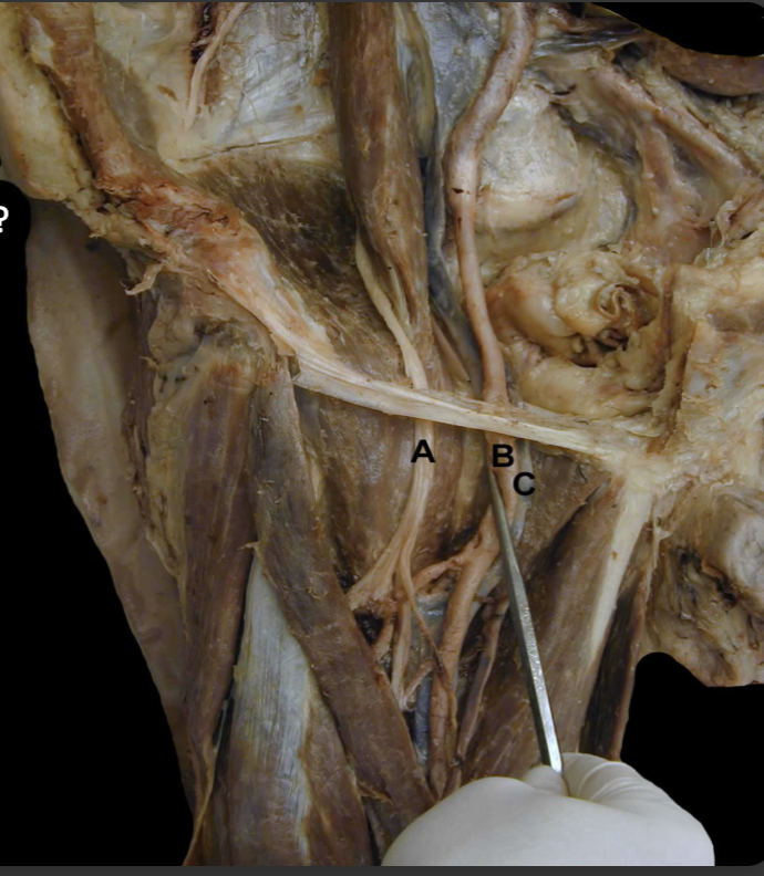
Identify the structures indicated of this Anterior View of the Thigh’s Femoral Triangle
A.: N: Femoral N.
B: A: Femoral A.
C: V: Femoral V.
D: L: Lymphatics (deep: not shown).
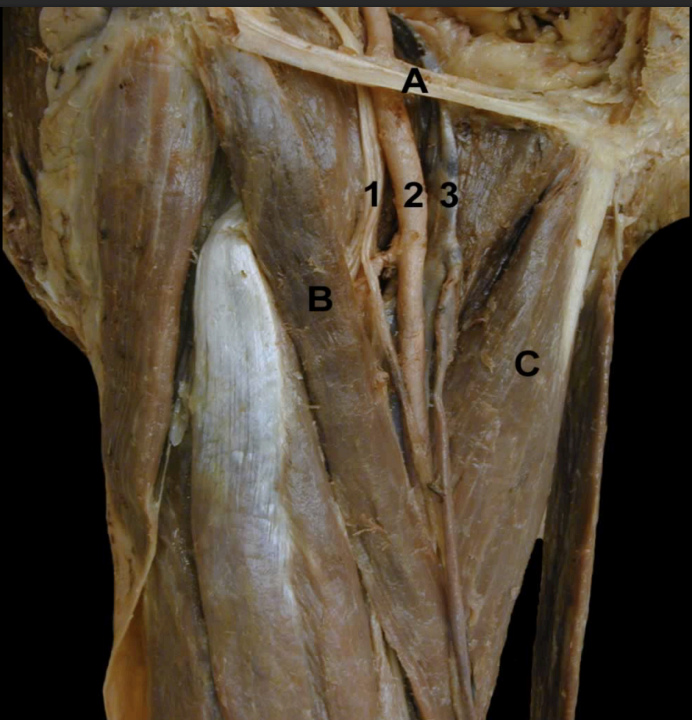
Identify the indicated structures on this Anterior View of the Thighs Femoral Triangle Boarders
A: Inguinal Lig.
B: Sartorius m.
C: Adductor longus m.
Contents (lateral to medial)
N: Femoral n.
A: Femoral a.
V: Femoral v.
L: Lymphatics
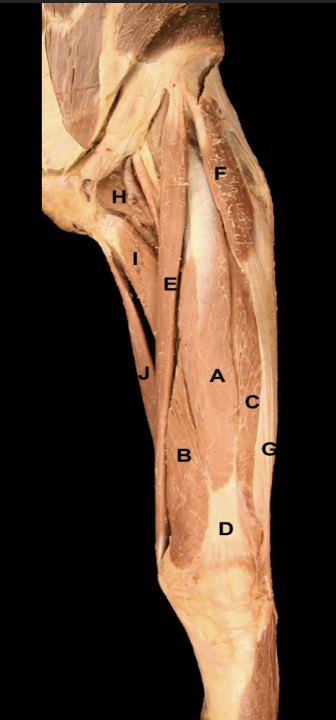
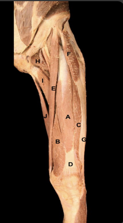
Identify the muscles on this Anterior View of the thigh
A: Rectus Femoris m
B: Vastus medialis m.
C: Vastus Lateralis m.
D: Quadricepes tendon m.
E: Sartorius m.
F: Tensor Fascia Lata m.
G: Iliotibial Tract
H: Pectineus m.
I: Adductor Longus m.
J: Gracilis m.
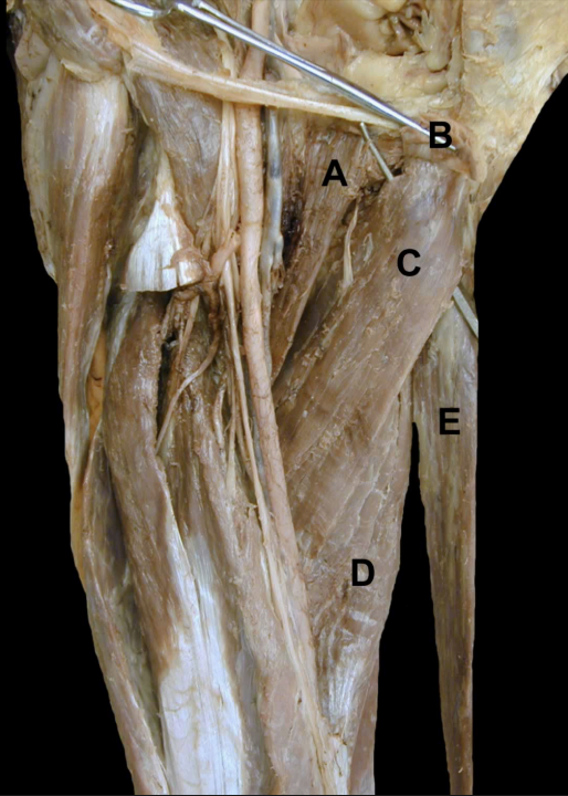
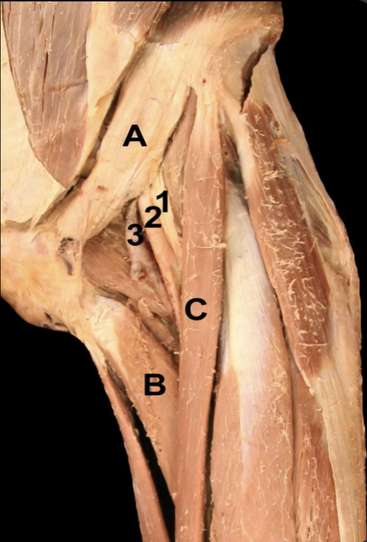
Identify the structures on this Anterior View of the thigh’s Adductors
A: Pectineus
B: Adductor longus m. (cut)
C: Adductor Brevis m.
D: Adductor Magnus m.
E: Gracilis m.
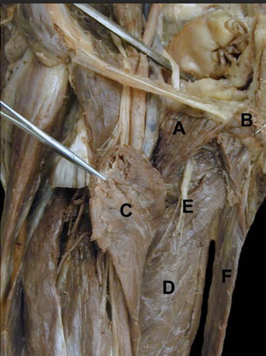
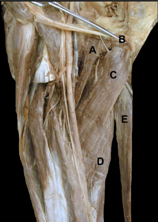
Identify the structures on this Anterior View of the thigh’s Adductors
A: Pectineus
B: Adductor longus m. (cut)
C: Adductor Brevis m. (cut)
D: Adductor Magnus m.
E: Obturator n.
F: Gracilis m.
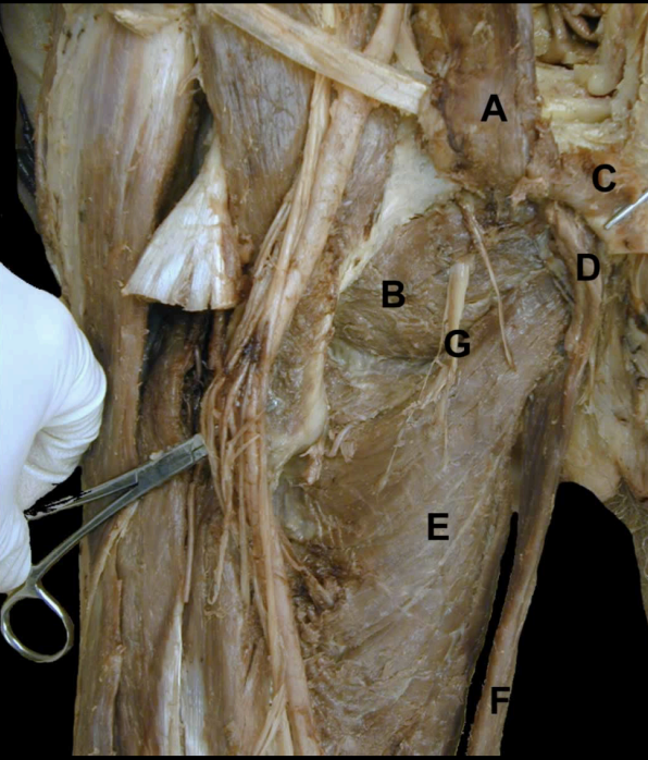
Identify the structures on this Anterior View of the thigh’s Adductors
A: Pectineus m.
B. Obtruator externus.
C: Adductor longus m. (cut)
D: Adductor Brevis m. (cut)
E: Adductor Magnus m.
F: Gracilis m.
G: Obturator n.
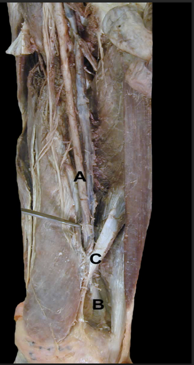
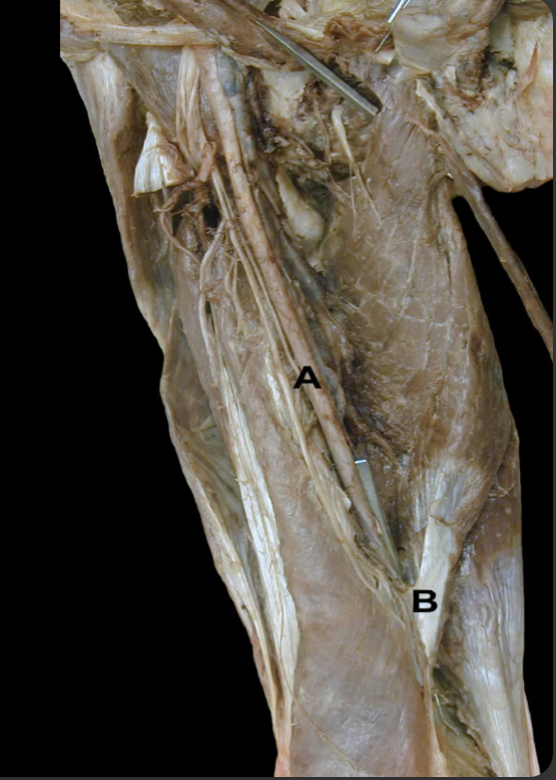
Identify the structures from this Medial View of the thigh’s Femoral Artery
A: Femoral a.
B: Popliteal a.
C: Adductor Hiatus
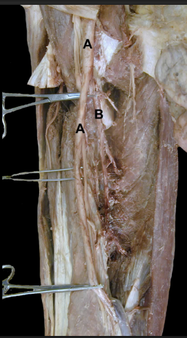
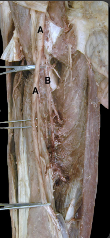
Identify the structures from this Anterior View of the thigh’s Femoral Artery
A: Femoral a.
B: Deep Femoral a.
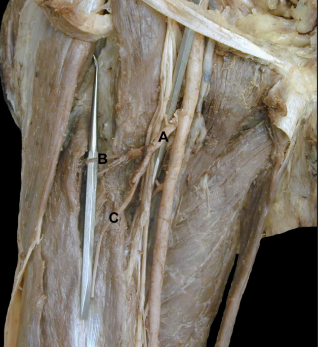
Identify the structures from this Anterior View of the thigh’s Lateral Femoral Circumflex Artery & Branches
A: Lateral Femoral Circumflex a.
B: Transverse Branch
C: Descending Branch
(Ascending branch not shown).
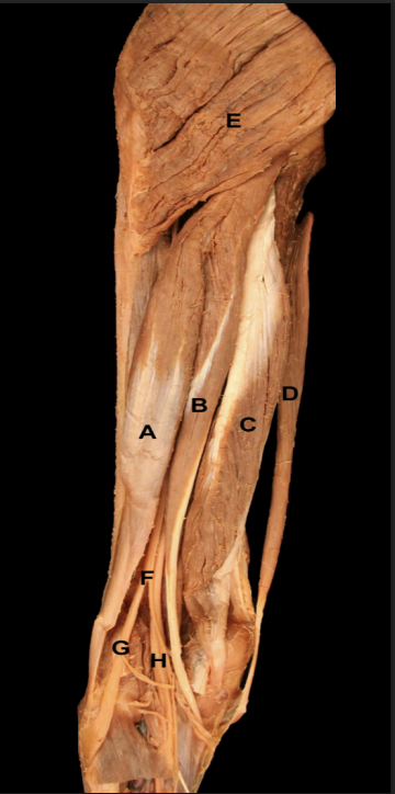
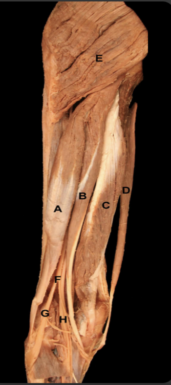
Identify the muscles of the Posterior Region of the thigh
A: Biceps femoris m.
B: Semitendinosus m.
C: Semimembranosus m.
D: Gracilis m.
E: Gluteus maximus m.
Neurovasculature:
F: Sciatic n.
G: Common fibular n.
H. Tibial n.
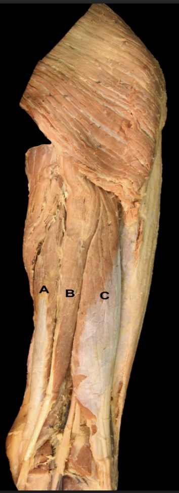
Identify the muscles of the Posterior Thigh’s Hamstrings
A. Semimembranosus m.
B. Semitendinosus m.
C. Biceps femoris m. (longhead)
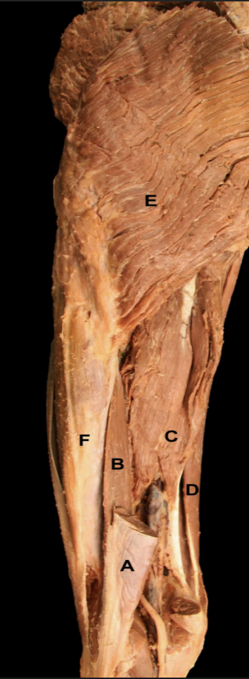
Identify the muscles of the Posterior Thigh
A: Biceps femoris m. (long head)
B: Biceps femoris m. (short head)
C: Semitendinosus m.
D: Semimembranosus m.
E: Gluteus maximus m.
F: Iliotibial Tract
A lesion of “D” will cause what impairment
Loss of sensation on the lateral aspect of the thigh
A lesion to “G'“ results in weakness of what movement of the lower leg?
Adduction (obturator nerve)
“H” splits into what 2 major nerves of the lower extremity
Common fibular n. & tibial n.
Name 2 muscles that share an action with “C”
Sartorius (hip flexion); Rectus femoris (hip flexion)
Quadratus femoris (external rotation); other external rotators
What are the borders of the Femoral Triangle?
Superior: Inguinal ligament
Lateral: Sartorius mm
Medial: Adductor Longus mm.
What are all the actions of “C”? Think about the force vectors at each joint
Hip flexion; Hip ER; hip abduction
Flexion at the knee
Which muscle lies just deep to “A”? What is the distal attachment of this muscle?
Quadriceps femoris vastus intermedius; Attaches on the Tibial tuberosity via the patellar ligament.
What muscles share the same insertion as “E”?
Gracilis insertion: Pes anserinus shared with the sartorius and semitendinosus
What causes “B”?
Adductor Hiatus: Created by a gap in the adductor magnus
“A” turns into what artery after passing through the adductor hiatus
The femoral artery is termed the popliteal artery after passing through adductor hiatus
Which muscle pictured are considered “hamstrings”?
A. Biceps femoris loghead
B. Semitendinosus
C: Semimembranosus
What makes a “True” Hamstring
Has an attachment on the ischeal tuberosity
Acts on both the hip and the knee joint
Innervated by the tibial division of the sciatic nerve
Posterior Compartment: Function, origin/attachment, & nerve innervation of the - Biceps Femoris (Long Head)
True hamstring
Function: Knee Flexion & hip extension
Origin/Attachment: Ischial tuberosity → fibular head on lower leg.
Nerve: Tibial division of sciatic nerve (L5-S2)
Posterior Compartment: Function, origin/attachment, & nerve innervation of the - Biceps Femoris (Short Head)
False hamstring
Function: Knee flexion
Origin/Attachment: Posterior Femur → Fibular head
Nerve: Fibular division of sciatic nerve (L5-S2)
Posterior Compartment: Function, origin/attachment, & nerve innervation of the - Semitendinosus
True Hamstring
Function: Knee flexion, hip extension, internal rotation
Origin/Attachment: Ischial tuberosity → “pes anserinus” anterior/medial tibia
Nerve: Tibial division sciatic nerve (L5-S2)
Posterior Compartment: Function, origin/attachment, & nerve innervation of the - Semimembranosus
True Hamstring
Function: Hip extension, knee flexion, & internal rotation
Origin/Attachment: Ischial tuberosity → post/medial tibia
Nerve: Tibial division sciatic nerve (L5-S2)
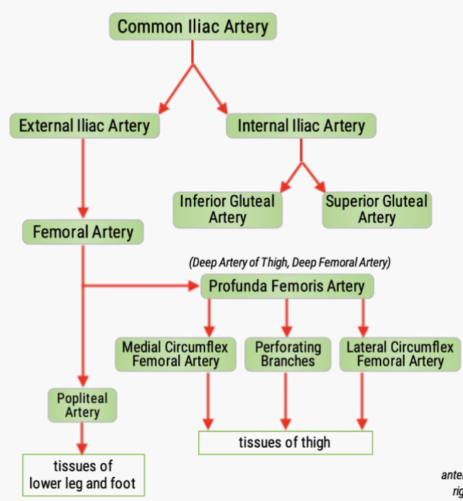
Flow of blood through the lower limbs
Anterior Compatment: Function, origin/attachment, & nerve innervation of the - Psoas Major
Function: Hip external rotation, hip flexion, flex trunk
Origin/Attachment: Lumbar spine (T12-L5) → Lesser trochanter
Nerve: Ventral Rami (L1-L3)
Anterior Compartment: Function, origin/attachment, & nerve innervation of the - Iliacus
Function: Hip flexion/Hip extension
Origin/Attachment: Iliac Fossa → Lesser trochanter
Nerve: Femoral n. (L2-L3)
Anterior Compartment: Function, origin/attachment, & nerve innervation of the - Sartorius
Function: Knee flexion, hip flexion, hip external rotation
Origin/Attachment: ASIS → Pes Anserineus
Nerve: Femoral Nerve (L2-L3)
Anterior Compartment (Quad Muscle): Function, origin/attachment, & nerve innervation of the - Rectus Femoris
Function: Hip flexion, knee extension
Origin/Attachment: AIIS → Tibial tuberosity
Nerve: Femoral n. (L2-L4)
Anterior Compartment (Quad Muscle): Function, origin/attachment, & nerve innervation of the - Vastus Intermedius
Function: Knee extension
Origin/Attachment: Anterior femur → Tibial Tuberosity
Nerve: Femoral n. (L2-L4)
Anterior Compartment (Quad Muscle): Function, origin/attachment, & nerve innervation of the - Vastus Lateralis
Function: Knee Extension
Origin/Attachment: P. Lateral femur → tibial tuberosity
Nerve: Femoral n. (L2-L4)
Medial Compartment of Thigh: Function, origin/attachment, & nerve innervation of the - Pectineus
Function: Hip Adduction
Origin/Attachment: Pubic Bone → Posterior/medial femur
Nerve: Femoral n. (L2-L3)
Medial Compartment of Thigh: Function, origin/attachment, & nerve innervation of the - Adductor Longus
Function: Hip Adduction
Origin/Attachment: Pubic Bone → Posterior/medial femur
Nerve: Obturator n. (L2-L4)
Medial Compartment of Thigh: Function, origin/attachment, & nerve innervation of the - Adductor Brevis
Function: Hip Adduction
Origin/Attachment: Body of pubis & inf. pubic ramus → Posterior/medial femur
Nerve: Obturator n. (L2-L3)
Medial Compartment of Thigh: Function, origin/attachment, & nerve innervation of the - Gracilis
Function: Hip Adduction
Origin/Attachment: Pubic Bone → Pes anerious
Nerve: Obturator n. (L2-L3)
Medial Compartment of Thigh: Function, origin/attachment, & nerve innervation of the - Adductor Magnus
Function: Hip Adduction
Origin/Attachment: Pubic Bone → Pes anerious
Nerve: Obturator n. (L2-L3)