Anatomy - Thorax Osteology and Arthrology
1/123
There's no tags or description
Looks like no tags are added yet.
Name | Mastery | Learn | Test | Matching | Spaced | Call with Kai |
|---|
No analytics yet
Send a link to your students to track their progress
124 Terms
The trunk is comprised of how many vertebrae?
33
What is the function of the thorax?
- To facilitate breathing
- Protection of vital organs
- Conduit for structures that pass through
What are the contents of the posterior components of the thorax?
12 thoracic vertebrae and their intervening intervertebral discs
What are the contents of the lateral thorax?
- ribs (12 on each side)
- three layers of flat muscles
What is the function of the flat muscles of the lateral thorax?
- Span the intercostal spaces between adjacent ribs
- Move the ribs
- Provide support for the intercostal space
What are the contents of the anterior thorax?
- Manubrium of sternum
- Body of sternum
- Xiphoid process
Each thoracic vertebrae articulates with
A rib
The function of the sternum is to
Protect mediastinal viscera, specifically the heart
The sternum is comprised of
- Manubrium
- Body
- Xiphoid process
The manubrium of the sternum lies at the level of
T3-T5
The body of the sternum lies at the level of
T5-T9
The xiphoid process of the sternum lies at the level of
T9-T10
The clavicular notch is an articulation point between the
Clavicle and manubrium
The surfaces of the clavicular notch are what shape?
Concave
The 1st costal notch is an articulation point between the
1st rib and manubrium
At the 1st costal notch, what type of joint is formed?
A synchondrosis joint with the costal cartilage of the 1st rib
The sternal angle of the manubrium is
A junction between the manubrium and the sternal body
The sternal body articulates with
Ribs 2-7
In children, the xiphoid process is
Cartilaginous and ossifies with age
The xiphoid process landmark for
- Superior limit of the liver
- Diaphragm
- Inferior border of the heart
A true rib directly articulates
To the sternum via costal cartilage
A false rib is
A rib that indirectly attaches to the sternum by attaching to the costal cartilage above
The true ribs are ribs _ to _
1-7
The false ribs are ribs _ to _
8-12
The floating ribs have
No attachment to the sternum
A typical rib consists of
- Head
- Neck
- Tubercle
- Body
- Angle
- Costal groove
The head of a typical rib contains
2 facets that articulate with corresponding vertebra and the superior vertebra
The neck of a typical rib connects the
Head to the body
The tubercle of a typical rib contains
A facet to articulate with the transverse process of the corresponding vertebra
The angle of a typical rib is an area where
The rib turns more anterolateral
The costal groove of a typical rib is
A concave internal surface of the body of the rib
The atypical ribs includes ribs
1st ,2nd ,10th-12th
What are the distinct landmarks of the first rib?
- single facet on head of rib to connect to T1
- Grooves for subclavian artery/vein
- Scalene tubercle
What are the distinct landmarks of the 2nd rib?
- Two facets on the head of the rib to connect to T1 and T2
- Tuberosity for serratus anterior
What are the distinct landmarks for ribs 10-12?
Single facet on head of rib to connect to their corresponding vertebrae (only connects to one vertebra)
What are the functions of the costal cartilages?
- Prolongs ribs anteriorly to connect to sternum
- Allows for elasticity of thoracic wall
The costochondral joint is formed between the
End of ribs and lateral end of costal cartilage
The sternocostal joint of the 1st rib is formed between the
1st costal cartilage and manubrium
The sternocostal joint of the 2nd - 7th ribs is formed between the
2nd-7th costal cartilages and the sternum
The sternocostal joints are reinforced by the
Anterior and posterior sternocostal radiate ligaments
During inhalation the upper ribs (1-7) and sternum move in the
Sagittal plane in a superior-anterior direction, and the sternum moves like a pump handle
During inhalation, the lower ribs move in
The frontal plane in a superior-lateral direction, and the ribs move like a bucket handle
What are the boundaries of the superior thoracic aperature
- T1 vertebra
- Left and right 1st ribs
- Manubrium
What are the contents of the superior thoracic aperture?
- Trachea
- Esophagus
- Blood vessels of the head and upper limb
- Sympathetic nerves
- Phrenic nerve (to diaphragm)
- Vagus nerve (to organs)
The are the boundaries of the inferior thoracic aperture?
- T12 vertebra
- Left and right 11th and 12th ribs
- Costal cartilages of ribs 7-10
- Xiphisternal joint
Inferior thoracic aperture
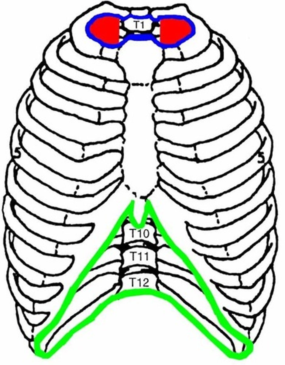
Superior thoracic aperture
What is the function of the spine?
- Protects spinal cord
- Supports weight of body above the pelvis
- Provides rigid base for head to function, as well as flexible axis for body to move
- Plays an important role in posture and locomotion
Of the 33 vertebrae, significant motion primarily occurs between the
Superior 25 vertebrae
The sacrum is formed by the
Fusing of the 5 sacral vertebrae
The coccyx is formed by the
Fusing of the 4 coccygeal vertebrae at around age 30
In lordosis the spine is
Concave posteriorly
In kyphosis, the spine is
Concave anteriorly
Cervical lordosis develops during
Infancy, when the child learns to extend head and look up while prone
Cervical kyphosis develops during
Fetal period when fetus is in flexed position
Lumbar lordosis is developed
Around 1 year of age, when child learns to stand
What is the function of the intervertebral discs?
- Permits movements between adjacent vertebrae
- Absorb shock during weight bearing activities
- Contributes to full length of vertebral column
The intervertebral disc is comprised of the
- Anulus fibrosis
- Nucleus pulposus
The anulus fibrosis is a
Concentric ring of fibrocartilage around the perimeter of the IV discs
The anulus fibrosis inserts onto the
Outer rim of vertebral bodies
The anulus fibrosis is thinner on the _ side compared to the _
Posterior; anterior
The nucleus pulposis allows for
Flexibility and load bearing of disc
The nucleus pulposis is the _ of the IV disc and is made up of
Core; 80% water
The intervertebral joint is an articulation between the
Vertebral bodies and intervertebral discs
A typical vertebrae consists of
- A vertebral body
- A vertebral arch
- 7 processes
The vertebra body is an _ structure that gives
Anterior; support to the spinal column
Why does the vertebra body get larger in size as you descend the column?
There is an increasing demand of support and to withstand more weight/pressure
The vertebral arch is comprised of the
Pedicle and lamina
The pedicle of the vertebral arch is
A cylindrical process that projects posterior from the body to meet with the laminae
The lamina of the vertebral arch is
A flat process that projects posteromedial to meet at the midline
The vertebral foramen is made up of
2 pedicles, 2 laminae, and the posterior surface of the vertebral body
The vertebral notches are
Indentations on the superior and inferior surfaces of each pedicle
The intervertebral foramen are formed from
The inferior notch of one vertebra, the superior notch of the vertebra beneath it, and the IV disc between the two vertebra
The superior and inferior articular processes of the vertebra project
Superiorly and inferiorly from the junction of the pedicle and lamina
The zygapophyseal joint is formed from the
Articular facet of the inferior articular process and the articular facet of the superior articular process of the vertebra beneath it
Due to the orientation of the zygapophyseal joints, the amount of each movement
Varies depending on each vertebra
The zygapophyseal joint is what type of joint?
Synovial; planar
The zygapophyseal joint is innervated by
PPRs of the corresponding segmental level
What are the motions of the zygapophyseal joint?
- Flexion/extension
- Lateral flexion
- Extension
Since each spinal division has different orientations of the facet joint, what is is possible
Different ranges of each movement
Cervical facet joints are at an angle of _ and allow for
45 degrees in the AP plane; all motions
Thoracic facet joints are _ and allow for
Vertical in the frontal plane; rotation and lateral flexion with some flexion and extension
What limits lateral flexion of the spine?
The ribs
The lumbar facet joints are _ and allow for
Vertical in the sagittal plane; flexion and extension with minimal rotation and lateral flexion
The 10th rib primarily articulates with the _ but does not always articulate with the _
10th vertebrae; 9th vertebrae
The anterior longitudinal ligament is fibrous band that extends from the _ to the _
Anterior tubercle of C1 to the sacrum; Anterior aspects of vertebral bodies and IV discs
The anterior longitudinal ligament limits
Spinal extension
The posterior longitudinal ligament is a fibrous band that extends from _ and attaches to _
The posterior aspect of C2 to the sacrum; The posterior aspects of the IV discs and loosely to posterior aspect of vertebral bodies
The posterior longitudinal ligament limits
Spinal flexion
Aside from being a spinal flexion limiter, what other purpose does the posterior longitudinal ligament serve
Prevention of posterior disc herniation
Ligamentum flavum connect
Adjacent lamina
What does the ligamentum flavum resist
Separation of the laminae during spinal flexion
The interspinous ligament connects
The bodies of adjacent SPs
The interspinous ligament limits _ and helps to maintain _
Spinal flexion; space between the SPs
The supraspinous ligament connects
The tips of SPs from C7 to the sacrum
At the cervical spine, the supraspinous ligament will become the
Nuchal ligament and attach to the occiput
The function of the supraspinous ligament is to
Limit spinal flexion and maintain space between SPs
The intertransverse ligament connects
Adjacent transverse processes
The function of the intertransverse ligament is to
Limit spinal lateral flexion and maintain space between the TPs
C7 (vertebra prominens) is at the level of which deep structures?
Apex of the lungs of thyroid isthmus