Module 1: from the Ground Up – Laying the Foundation
Module 1: from the ground up - laying the foundation
Section 01: Importance of anatomy
- Anatomy is the study of the structure
- word is derived from Greek and means “to cut apart.”
- parts of an organism are “cut apart” to ascertain their positions, relations, structure, and function
- Four areas of anatomy include:
- histology (microscopic features)
- gross anatomy (macroscopic features)
- neuroanatomy
- embryology
Unity of Form and Function
- form and function extricable linked
- organization of a bodily structure is instrumental as it is how a specific function is carried out → applied to all levels of organization:
- small molecules and cells to organ systems
- when a structure has proper form, there is proper function; when the form is disrupted, dysfunction can occur
Section 02: Organization of the human body and anatomical nomenclature
- microscopic (small) and macroscopic (large) structures of the human body can be organized by fundamental levels
- ==chemical leve==l → a molecule is a group of atoms bonded together
- ==cellular level== → cells are the smallest living structure and are formed from atoms and molecules
- tissues are similar cells that perform specialized functions
- ==Organ level== → organs are two or more tissues that work together to perform complex functions
- ==Organ system level== → the organ system level consists of related organs that work together to coordinate activities and achieve a common function
- ==Organismal level== → all body systems function interdependently in a single living human
- the body can also be organized into two main regions:
- ==Axial==: the axial region forms the main vertical axis of the body and includes the head, neck, and trunk o ==Appendicular==: the appendicular region consists of the limbs or appendages that attach to the axis
- Difference between the two: “axial region includes body structures along the midline, such as the head, spine, and trunk. This is different form the appendicular region that makes up our limbs, or structures further away from the body's midline.”
Organ Systems
- individual organs are organized into organ systems based on structure and function at macroscopic (large) levels
makes up our body covering and includes our skin and is associated with structures such as our hair and nails
consists of the bones and joints of the body
the system that contains the muscles and works together with the skeletal system for movement and support
includes the brain, spinal cord, and nerves that run throughout the body
: contains glands that produce and secrete hormones
- works with the nervous system to function in the integration and coordination of the body to act as a unit
- ^^Hormones: ^^molecules released from one cell into the blood that travels throughout the body to affect other cells
the system that starts at the mouth with a long tube and ends a the anus
allows you to breathe and includes the nose, air passageways, and lungs
consists of the lymphatic vessels, cells, and structure that can initiate an immune response
- ^^Lymphatic vessels: ^^thin-walled vessel structures like blood vessels that carry lymph (interstitial fluid of the body)
the system that includes the kidneys, ureters, bladder and urethra
- the respiratory, cardiovascular, lymphatic, and urinary systems function together in the processing and transportation of nutrients, oxygen, and waste products
system provides the means for sexual maturation and procreation
Body Cavities
- organ systems are enclosed with distinct spaces called body cavities → these spaces are important because they contain and protect our vital organs
- organ systems are held with distinct areas called body cavities
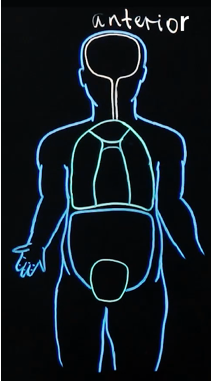
- Two views of the human body (anterior and lateral):
Anterior: front-on perspective
Lateral: side-on perspective 
Body Cavities
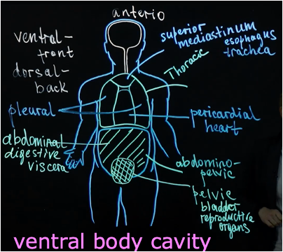
Ventral: at the front
- can be divided into two big groups:
- Thoracic: above the diaphragm → can be subdivided into three groups:
1. Superior mediastinum: contains the esophagus and trachea (sternum bone protects these structures and the aorta) 2. Pericardial: contains the heart 3. Pleural: contains the lungs
- Abdominopelvic: below the diaphragm
- The abdominal body cavity contains digestive viscera and the pelvic which includes the urinary bladder and reproductive organs.
Dorsal: at the back
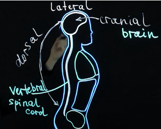
- can be divided into two cavities:
- Cranial: contains the brain and vertebral that contains the spinal cord
- Dorsal lateral: contains the brain and spinal cord (central nervous system
Language of anatomy
- Characteristics of anatomical positions include:
- standing upright
- feet parallel and on the floor
- head level and looking forward
- arms at side of the body
- palm facing forward and thumbs pointing away from the body
Directional terms
- provide precise descriptions of the location of structure relative to other structures in anatomical position → these terms are often paired with an associated term that means the opposite
%%Superior (cranial)/ Inferior (caudal)%% 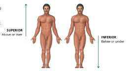
- superior: above or over
- inferior: below or under
%%Anterior (ventral)/Posterior dorsalal)%%
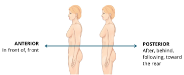
- anterior: in front of, front
- posterior: after, behind, following, toward the rear
%%Medial/Lateral%% 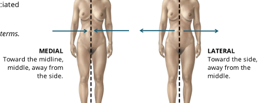
- medial: toward the midline, middle, away from the side
- lateral: forward
%%Proximal/Distal%% 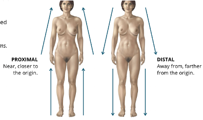
- Proximal: near, closer to the origin
- Distal: away from, farther from the origin
%%Superficial/Deep%% 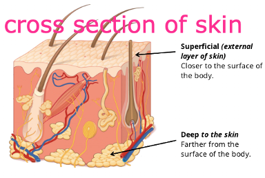
- Superficial (external layer of skin): closer to the surface of the body
- Deep (to the skin): farther away from the body
Parietal/Visceral
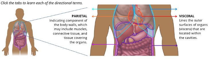
- Parietal: indicating component of the body walls, which may include muscles, connective tissue, and tissue covering the organs
- Visceral: lines the outer surfaces of organs (viscera) that are located within the cavities
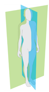
Planes and sections of the body
- planes or sections can be used to describe the location or direction of structures within the body in anatomical position
- different sections allow for their unique narrative of the internal environment
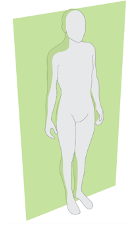
@@Saggital/Vertical plane@@

- A sagittal plane is a vertical plane that divides the body into left and right parts.
- When a sagittal plane passes through the midline of the body, it is referred to as the midsagittal plane.
@@Coronal/Frontal Plane@@
- A vertical plane that divides the body into superior and inferior parts (above the mid-body and below the midbody parts)
Horizontal/Transverse Plane
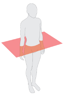
- the Transverse plane that horizontally divides the body into superior and inferior parts (above the mid-body and below the midbody parts)
Oblique plane
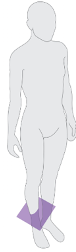
- plane that passes through the body at an angle
Longitudinal plane
- any plane that is perpendicular to the horizontal plane
- Saggital and coronal planes are examples of longitudinal planes
Section 03: Basic Tissue - Epithelium
- four basic types of tissue in the human body that are building blocks for every organ
- epithelium tissue
- connective tissue
- nervous tissue
- muscular tissue
Epithelium
- issued composed of closely apposed (side by side) cells with very little or no intervening intercellular substance
- ^^Apposed^^: in close position to juxtapose dot something else
- there are tow types of epithelium:
- ^^covering epithelium^^: cells that cover the external and internal surfaces
- ^^glandular epithelium: ^^cells that produce and secrete product, such as hormons
Characteristics of Epithlium
- epithelium cells can be observed in various shapes and layering throughout the body → regardless of shape, amount of layering, or location, epithelial tissue shares similar characteristics
Cellularity
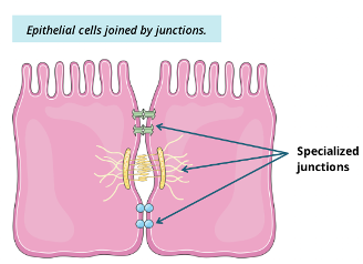
- adjacent cells care joined by 4 specialized junctions:
- tight junctions
- adhering junctions
- desmosomes
- gap junction
Polarity
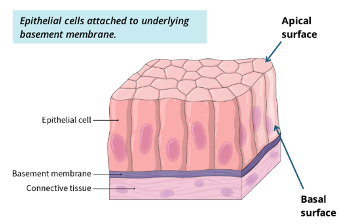
- an apithlial cell has an exposed (apical) surface that faces the exterior of the bouyd or internal space, and the basal surface where it is attached to the underlying tissue
Attatchment 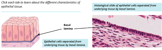
- Epithelial cells rest on and are attached to the basal lamina (basement membrane)
Avascularity
- epithelial tissues have no direct blood supply, The cells receives nutrients form the blood vessels in the underlying tissue
Regeneration
- epithelial tissues are renewed continuously
Functions of Epithelium
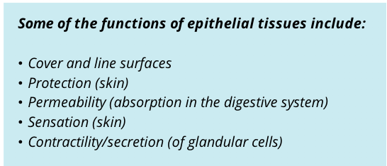
- Epithelial tissues have many functions, but not one single epithelium performs them all
Support and protection
- covers and lines external and internal surfaces of the body, protecting the underlying tissue from injury, pathogens, and dehydration (e.g.skin)
Permeability
- epithelium allows for substances to be absorbed into the body (e.g. epithelium lining the digestive system absorbs nutrients from food)
Sensation
- some epithelial tissues contain specialized cells that are able to detect sensory stimuli (e.g. skin senses, touch senses, taste)
Secretion
- some epithelial cells are specialized to secrete specific substances (e.g skin secretes lubricating oil, enzymes, and hormones are secreted by the digestive system)
Classifying Epithelium
- the body contains different kinds of epithelia → may be classified based upon its cell organization or its shape
Cell organization
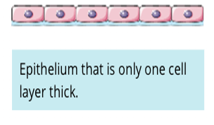
==Simple==: only one cell layer thick
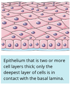
==Stratified==: two or more cells layer thick, only the deepest layer of cells is in contact with basal lamina (basement membrane)
Cell Shape
- may be classified by the shape of the cell
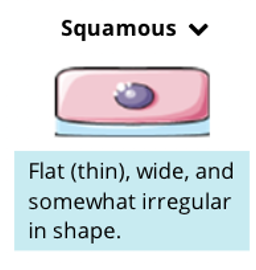
==Squamous==: thin, wide, and irregular shape
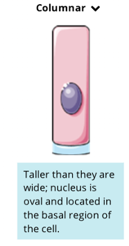
Columnar: tall with an oval nucleus located near the basal region of the cell
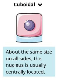
Cuboidal: approximately the same size of all sides with a centered nucleus
Naming Epithelial Tissue
- epithelium tissues are named by combining their two classificaitons
Cell organization + Cell shape → (first name) + (last name)
**Simple Epithelium ***only one cell layer thick
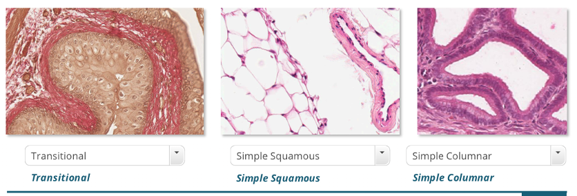
]]Simple Squamous]]
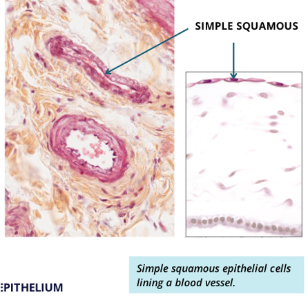
- single layer of flattened cells
- can be found lining blood vessels
- blood vessels are used to transport materials to and from cells, they are energy efficient because the thin barrier allows for rapid exchange
- single layer of flattened provides a thin barrier allowing material to travel a short distance between the two
]]Simple Cuboidal]]
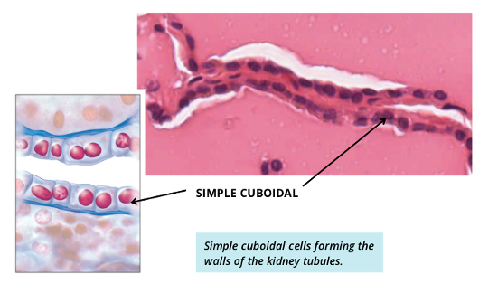
- single layer of cube cells
- can be found lining some glands
]]SImple Columnar]]
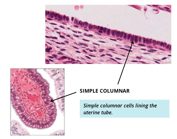
- single layer of column-shaped cells
- columnar epithelium can be found lining the gastrointestinal (GI) tract
- similar to simple squamous in the blood vessels, the single layer of cells allows for rapid secretion or absorption of material
Stratified Epithelium * two or more cell layers thick, only the deepest layer is in contact with the basal lamina
]]Stratified Squamous]]
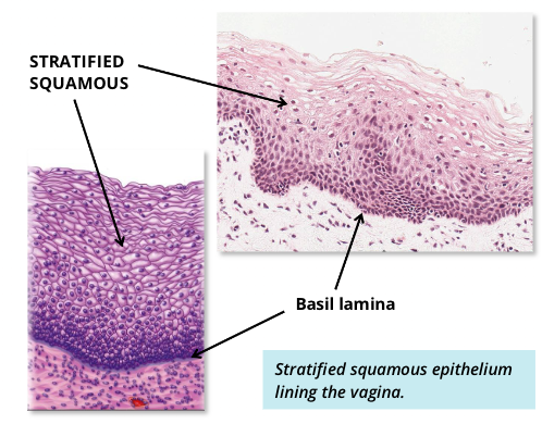
- multiple layers of flat-shaped cells
- the basal cells may be more cuboidal in shape, but the apical (surface level of superficial cells) display a squamous flattened shape
- stratified squamous epithelium makes up the most superficial layer of skin as multiple thin layers of small cells. this allows the skin to protect deeper structures from abrasion and damage
]]Stratified Cuboidal]]
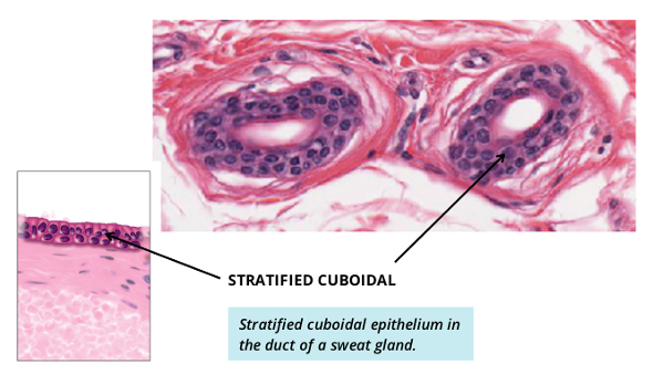
- multiple layers of cube-shaped cells
- can be located in some ducts in the glands
- functions of these cells include secretion, protection, and strengthening the walls of the ducts of glands
]]Stratified Columnar]]

- multiple layers of column-shaped cells
- relatively rare in the human body
- cells can be found in the male urethra, these cells function as a protection and produce secretion
Other Types of Epithelium
- two types of epithelium do not fit the organizational framework
]]Transitional]]
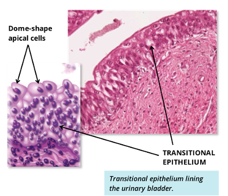
- multiple layers of epithelium cells that allows for stretching
- can vary in shape depending on whether the tissue si stretched or relaxed
- characterized by the presence of domed-shaped surface cels which is a reflection of the relaxed state; when stretched, these surface cells flatten out
- epithelium cells in the urinary bladder can change shape as urine accumulates in the bladder
- transitional epithelium also lines the ureters and proximal end of the urethra (near the bladder)
]]Pseudostratified (ciliated columnar)]]
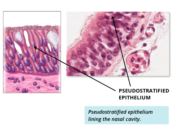
- comprise only a single layer or cells and its cell nuclei is positioned ina manner suggestive of stratified epithlium
- cilia or hair-like projections on the surface cells help in moving mucous
- this type of epithelium can be found throughout most of the respiratory tract, a system where mucous must be transported
Section 04: Basic Tissue - Connective Tissue
- conenctive tissue is most widespread and abundant type of tissue in the human body
- the most diverse of the four tissue types with a wide variety of functions
- ranges in consistency from gel-like softness to areolar connective tissue to the hardest of bone
Functions of the Connective Tissue
- the functions of connective tissue are primarily to support, anchor, and connect various parts of the body
%%Support and protection: %%the bones of the skull protect the brain. the kidneys are surrounded by fat padding and protect it
%%Provides s structural framework for the body: %%cartilage supports body structures such as the windpipe (trachea), ears, and nose. Bones of the skeleton provide the framework for skeletal muscles
%%Medium for exchange of nutrients and metabolic waste: %%blood serves as a medium that carries gases, nutrients, wastes, and blood cells to different parts of the body
%%Storage and repair: %%bone stores minerals such as calcium; fat serves as a major energy reservoir for the body
%%Defence: %%connective tissue performs this function in several ways such as acting as a physical barrier, through white blood cells (macrophages, neutrophils), and antibody production (plasma cells)
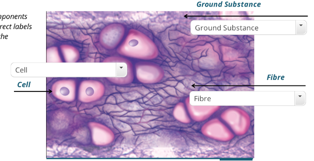
Components of Connective Tissue
- connective tissue exists in multiple forms; all types have there basic structural elements: cells, fibres, and intercellular substance (ground substance and the different proteins present in it)
Cells of Connective Tissue
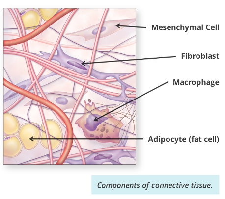
- many kinds of cells can be found in connective tissues (CT)
- large and low-diversity cells types can be found in CTs
- some cells in CTs are fixed (they are permanent residents in the connective tissue (fibroblasts))
- others are wandering (they are transient migrants who have entered the CT from the blood in response to specific stimuli (macrophages which are white blood cells))
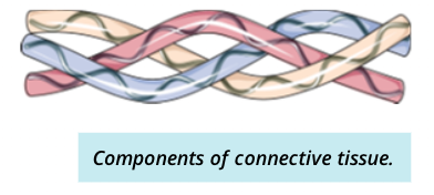
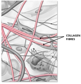
Fibres of Connective TIssue
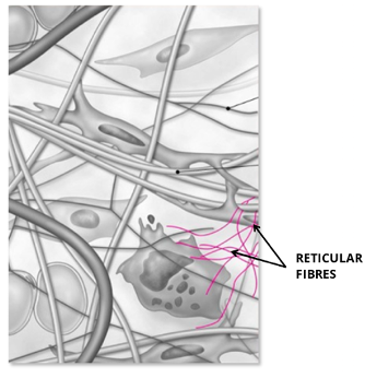
- there are three types of fibres serete dby fibroblasts: collagen fibres, reticular fibres, and elastic fibres
- each type of fibres is formed by proteins made of long peptide chains
- different components and proportions of fibre types lead to diverse function of various CT
- different components and proportions of fibre types lead to diverse function of various CT
@@Elasstic Fibres: @@thin and branched, elastic fibres appear wavy or curly and they have rubber-like material that can stretch (gives tissues flexibility) 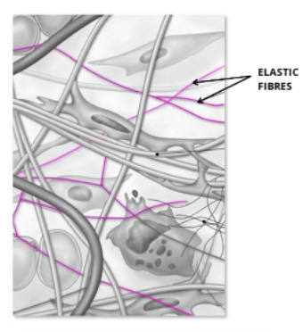
@@Collagen Fibres: @@most common type of fibre, these are flexible fibres with high tensile strength.
- the microscopic structure of collagen fibre appears similar to a rope (provides tissues with tensile strength and responsible for resisting large forces)
@@Recticular Fibres: @@thin fibres that form a branching interwoven network with no common alignment
@@Dysfunctional collagen fibres sysmtoms (EDS): @@
- Laxity or looseness in tissue
- loose skin with high elasticity or strethc
- fragile skin that cannot resist large forces
- flexible joints leading to joint pain and eventually arthritis
Type of Connective Tissue
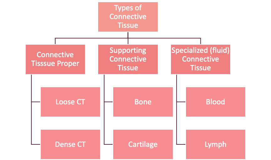
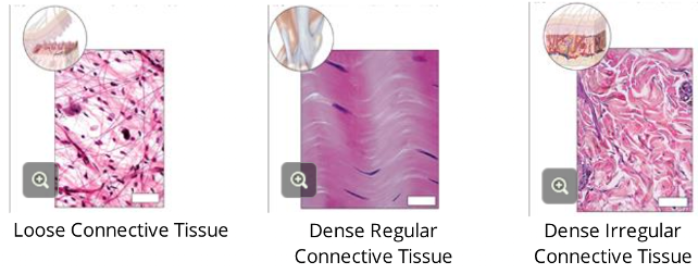
==Loose CT:== 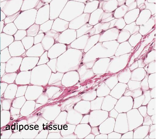
- has more ground substance with a few CT fibres
- contains elastin fibres for flexibility and collagen fibres to anchor the tissue
- adipose tissue is a specialized type of loose connective tissue where fat cells (adipocytes) account for most of the ovluem of this tissue
- this tissue acts as padding, insulates against heat loss through the skin, and serves as a packing filler around and between structures
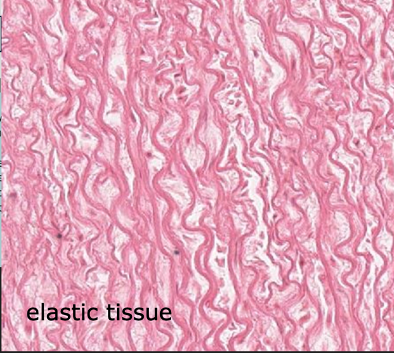
==Dense CT:==
- has less ground substance with more CT fibres
- elastic tissue is a specialized type of dense regular connective tissue composed of bundles of thick parallel elastic fibres between which we find some collagen and fibroblasts → due to a fibrous nature, this tissue has the ability to stretch and recoil
- Dense CT can be found in ligaments, tendons, and surrounding some blood vessels → places that need a lot of resistance
==Bone:==
- important structural tissue that is the framework of the body
- bone is rgit because its ground substance is mineral
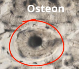
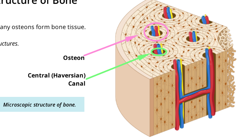
- main structure eunit in bone is called the osteon
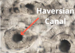
- ^^osteons^^: are forms of concentric rings of compact bone, with a central Haversian canal which houses blood vessels and nerves
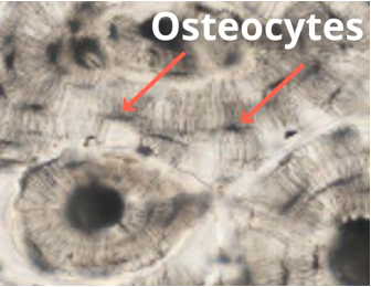
- Osteocytes (bone cells): are visible trapped within the rings of mineralized bone matrix
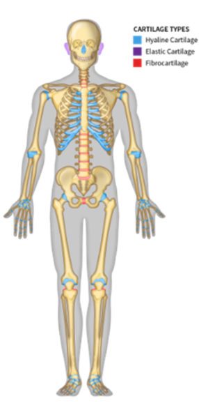
==Cartilage:==
- a structural component of the body
- further classified into hyaline cartilage, fibrocartilage, and elastic cartilage
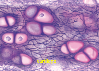
- mainly chondrocytes, found in spaces called lacunae
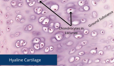
- derived from “hyalos” in Greek → means glass
- appears galssy under a microscope
- wear and tear resistant type of tissue → strong but also allows flexibility (found in movable joints)
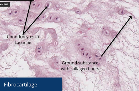
- named after the many fibres within it
- the chondrocytes (cartilage cells) are found within a dense arrangement of collagen fibers
- strongest type of cartilage →found in places that act as shock absorbers
- discs between our vertebra (spin) and the meniscus, fiberious cartilage between knee joint
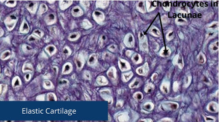
- chondrocytes are also found within a network of fibres, but they ar elastic type of fibres
- found in more flexible places like the external ear
:

- fluid within blood vessels and the heart
- ground substance in blood is fluid (plasma presented in blood)
- the cells in blood include red blood cells (erythrocytes); **white blood cells (**leukocytes; platelets
- contains various cells and proteins and preforms various essential functions within the body such as:
- connecting different parts fo the body, providing metabolic support, also contains the three main components: cells, ground substance, and fibres
:
- interstitual fluid (fluid that bathes cells) is collected into thin-walled lymphatic vessels and transported to the cardiovascular system
Supporting Connective Tissue:
- support connective tissue provides a strong durable framework to protect and support soft body tissues → cartilage and bone are two types of supporting connective tissue
Subsection 01: Cartilage
- important structural component fo the the body, composed of firm tissue but is softer and more flexible than bone
- unique conentive tissue found in many areas of the body, including:
- the joints between moveable bones
- between the vertebrae in the spine
- ears and nose
- bronchial tubes or airway
Components of Cartilage
^^Cells: ^^cellins in aprtilage are primarily chondorcytes, which are locate throughout the ground substance in small spaces called lacune that contain one of more cells
^^Fibres: ^^this can include various collagen or elastic fibres scattered throughout the cartilage
^^Ground substance: ^^firm gel that makes cartilage solid → cells called chondrocytes are located throughout the intercelluar substance
Additional components of cartilage
^^Perichondrium^^: dense irregular connective tissue that envelops cartilage to provide nutrients to the cartilage → not all types of cartilage have perichondrium
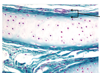
^^Lacuna: ^^small spaces in the cartilage that house one or more chondrocytes (the major cells of the cartilage) 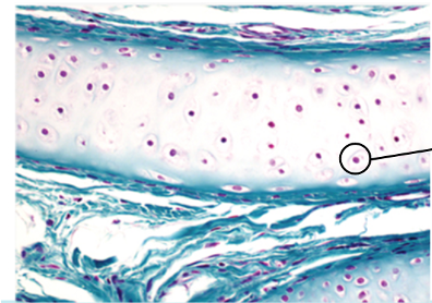
Why do somehave perichondrium?
- cartilage is avascular, therefore unable to get nutrients from direct blood supply
- the perichondrium is present to help provide nutrients to the cartilage and remove waste products
| Types of Cartilage | Anatomical Characteristics | Lcoation |
|---|---|---|
| {{{{ | wear resistant tissue that is designed to bear and distribute weightstrong, rubbery, flexible tissue- most common type of cartilage | joint surface of moveable joints, walls of nose, trachea bronchi (upper respiratory tract), and ribs |
| {{Fibrocartilage{{ | tough and inflexible form of cartilage- durable and resistant to compression | intervertebral discs and symphysis pubis |
| {{Elastic{{ | more flexible than hyaline cartilage | external ear, Eustachian tube (connects ear to nose)< and epiglottis (barrier to trachea during swallowing food or drinks) |
Subsectoin 02: Bone
- another important structural component of the body, its functions include:
- support
- locomotion
- protection
- blood cell production
- mineral metabolism
Composition of Bone
- bone compositioncan be divided into two broad categories ():
- cells, fibres, ground substance
- minerals, salts, inorganic components (mainly calcium phosphate) provide for the rigidity of bone
Structural unit of bone
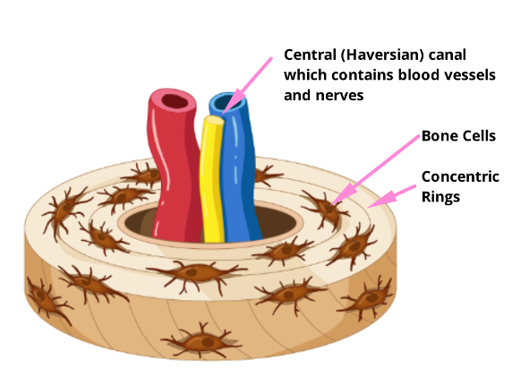
- bone has structured pattern of repeating cylindrical structures (osteons)
- eat osteon is made up of concentric rings with a hallow central canal (Haversian canal)
- the cellular components of bone are locate between concentric rings
Microscopic Structure of Bone
Connective Tissue Summary:
- cartilage and bone demonstrate how differences in cell density and organization produce a variety of tissues with