Oxygen Effect/ LET and RBE
1/60
There's no tags or description
Looks like no tags are added yet.
Name | Mastery | Learn | Test | Matching | Spaced | Call with Kai |
|---|
No analytics yet
Send a link to your students to track their progress
61 Terms
Reoxygenation
Cells are more sensitize to radiation in the presence of oxygen
Out of all pharmologic radiosensitizers,
oxygen has been determined to be the most effective and practical
Oxygen enchancement ratio (OER)
Ratio of the dose needed to kill a certain number of hypoxic cells to the dose needed to kill the same number of oxygenated cells
OER equation
D hypoxic/ D oxygen
Once a free radical has reacted with DNA it is called a
DNA radical
Oxygen fixation hypothesis
In the absence of O2, certain protein components like sulfhydryl groups bind to the DNA radical to help facilitate repair
However, if O2 is in the cellular environment, it will bind to the DNA radical instead to form RO2, which makes the damage permanent (fixes/freezes repair) by blocking the action of other helpful repair molecules
DNA radicals are more highly-reactive and susceptible to binding of
O2
DNA that has been damaged via the direct method of interaction is
NOT susceptible to oxygen fixation
DNA that has been damaged via the indirect method of interaction is
HIGHLY SUSCEPTIBLE to oxygen fixation
(predominantly low LET)
In order for a cell to be affected by the block that oxygen creates to DNA damage repair,
Repair must be possible in the first place
(not doable with single event killing)
Areas of single-event killing on cell survival curve are
Less affected or completely unaffected to the presence of oxygen
Areas of cell survival curve dominated by sublethal damage repair (shoulder) or double event killing (quadratic component) are most affected by
Oxygen
Oxygen needs to be delivered before the cell
starts to repair itself
Oxygen must be present
1) at the exact moment of irradiation
2) within microseconds (10-6s) of the irradiation
OER range for low LET
2.5 (low doses) -3.5 (high doses)
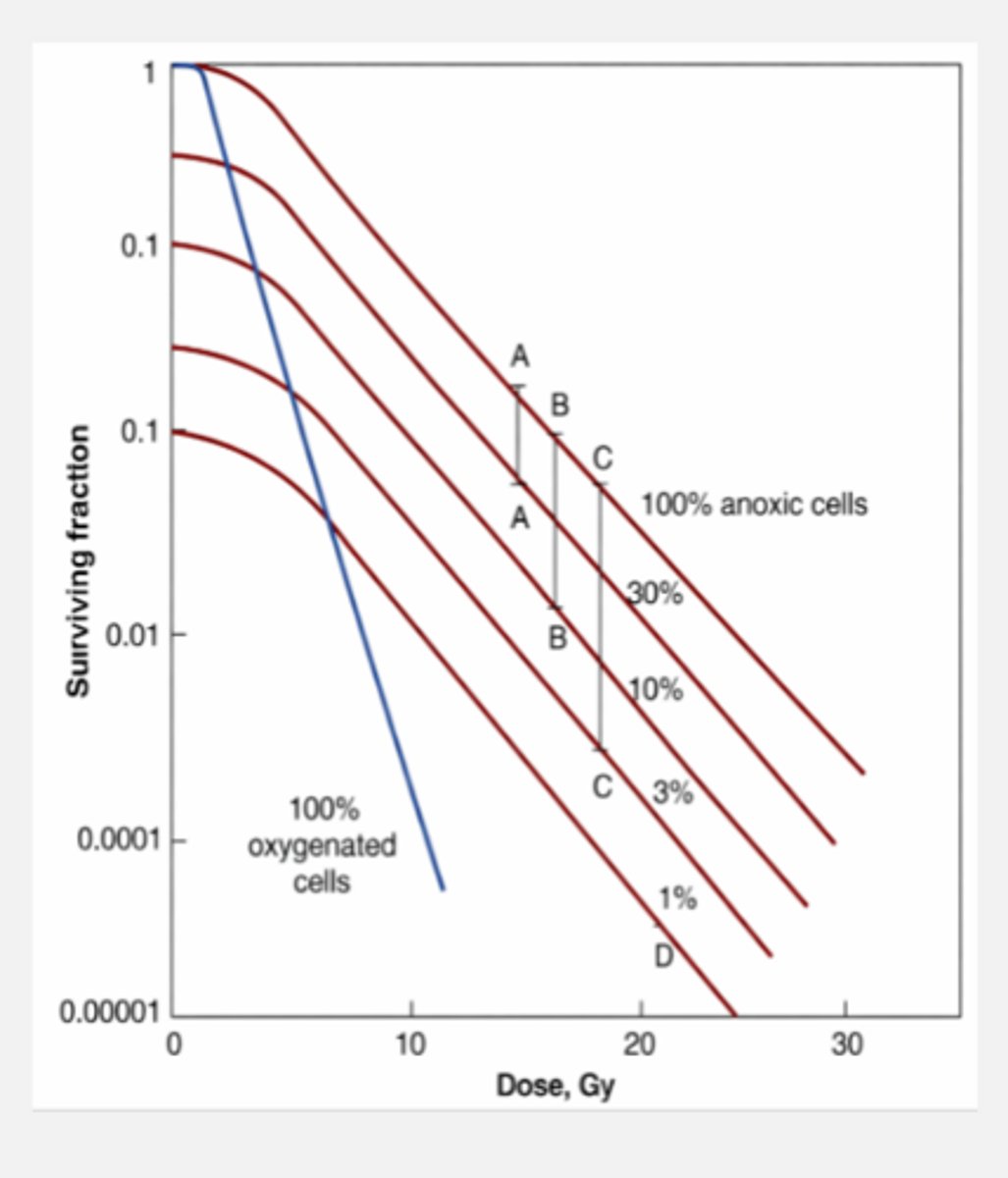
OER on graph
Oxygenated cell populations are significantly more radiosensitive
Hypoxic cell populations are more radioresistant
The green box on high-dose assay curve represented by low-dose assay graph
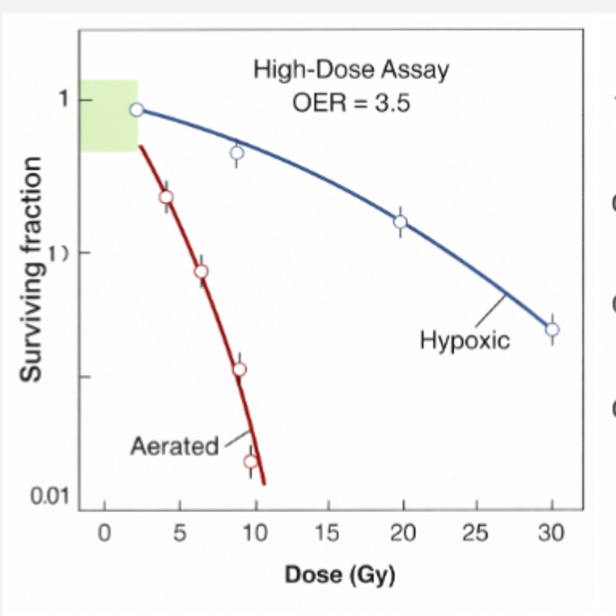
Why does the area of the curve in the green box have a lower OER?
This is the lowest-dose region of the curve, where cell survival is incredibly good
If cells are going to die here, it is most likely due to single event killing, which is unaffected by oxygen fixation
Higher dose needed typically under
hypoxic conditions
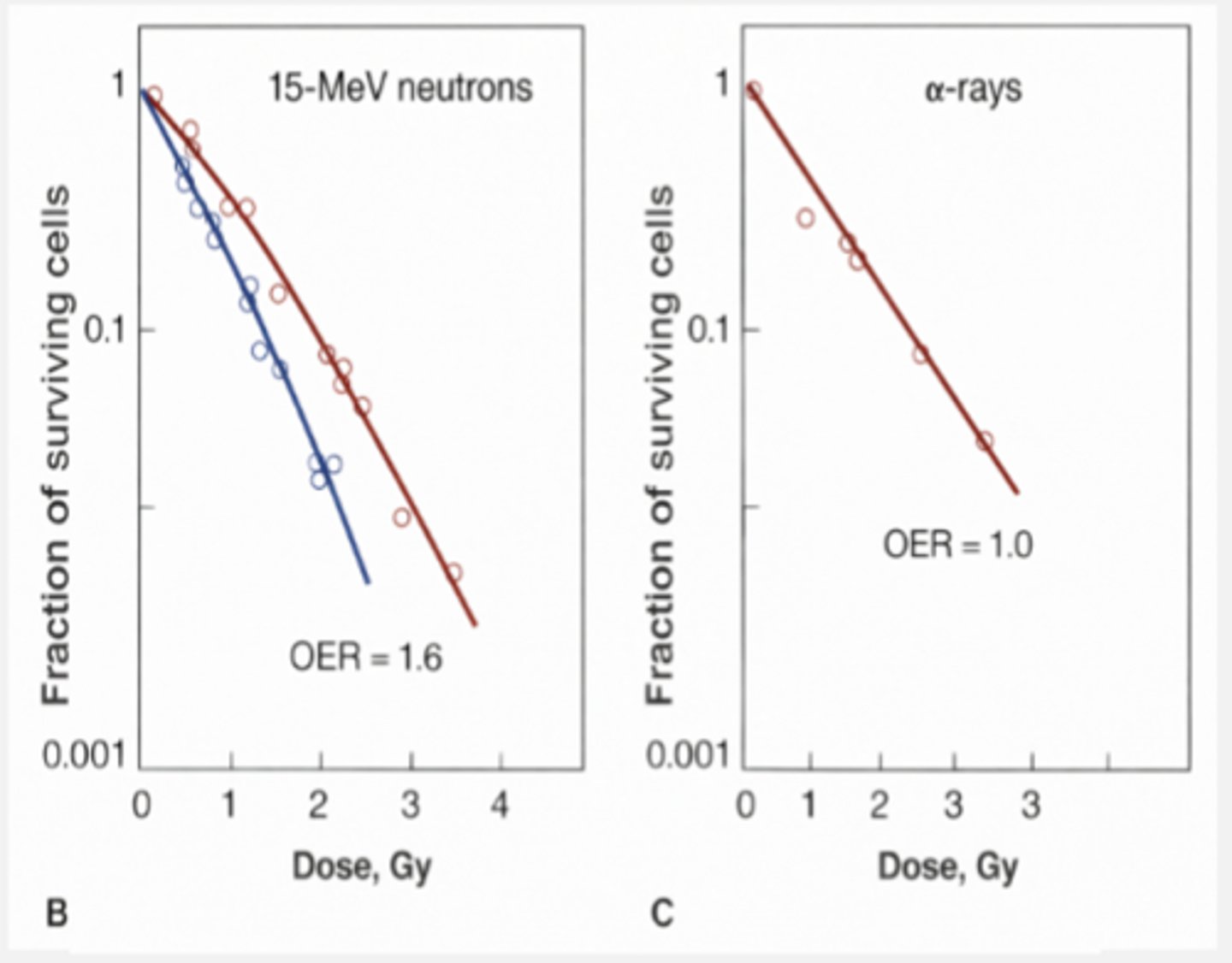
OER value for alpha particles
Nonexistent or 1
OER for neutrons
1.6
OER is diminished or nonexistent for
1) Radiation that damages DNA via direct interactions (high LET)
2) Radiation that is dominated by single-event killing (high LET)
Oxygen concentration needed to observe an increase in radiosensitivity of a cell population
5% needed for a dramatic increase in cell killing
Beyond 5% cell killing
remains the same even if the concentration of oxygen is increased
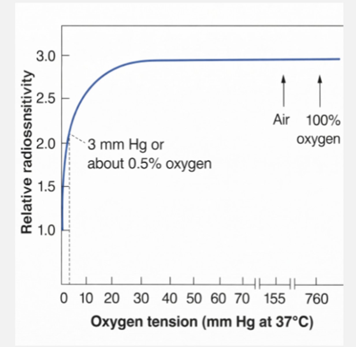
Very small amounts of oxygen produce big effects, but once all DNA damage has been made permanent by oxygen,
increasing the oxygen concentration does not result in more cell killing
Different tissues in the body exist at different levels of
hypoxia
Oxygen status effects radiosensitivity of those tissues
Tumors composed of varying levels of
hypoxic cells
42 types of human tumors contain up to
50% hypoxic cells, making it harder to treat
average of 14% hypoxic cells
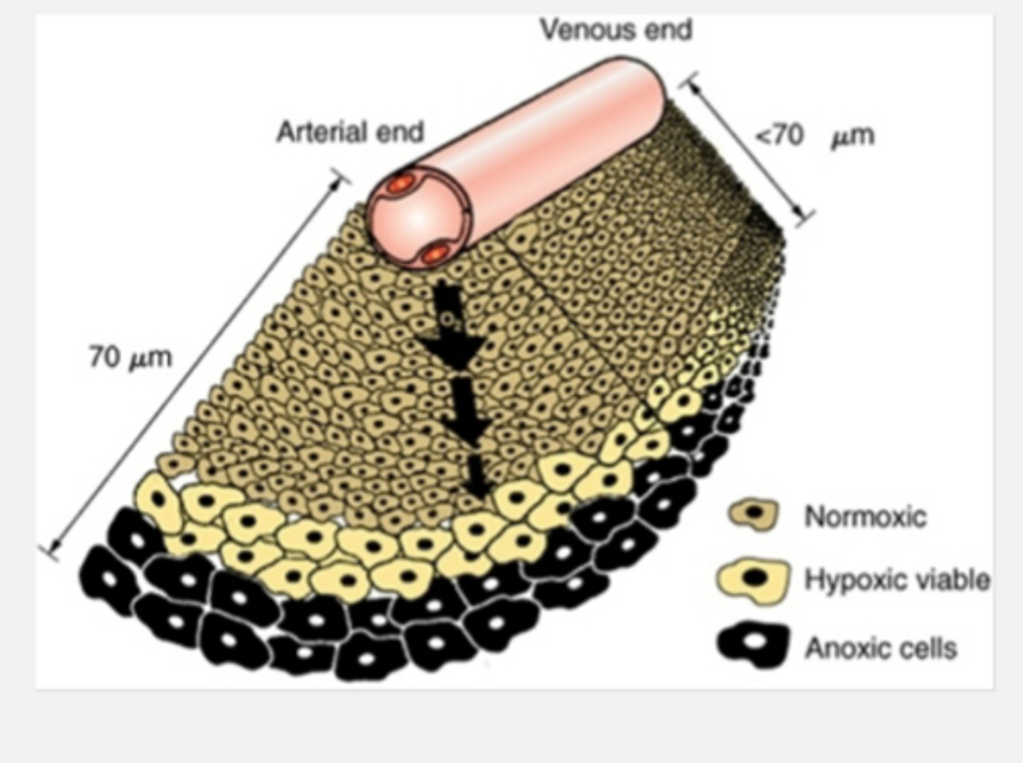
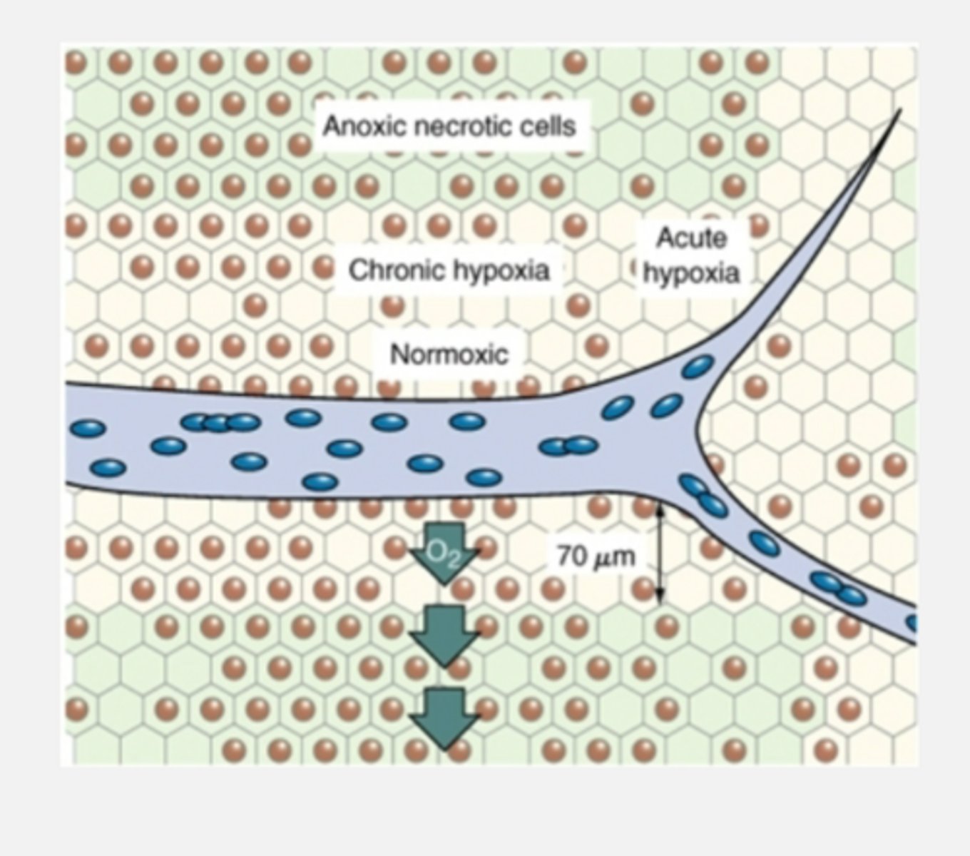
Chronic hypoxia
Centers of large tumors tend to be chronically hypoxic/anoxic due to distance from capillary beds
Cells between oxygenated and anoxic layer may be hypoxic enough to be radioresistant but oxygenated enough to promote tumor growth
Acute hypoxia
Tumors can become acutely hypoxic due to blockage of a blood vessel
TRANSIENT OR INTERMITTENT
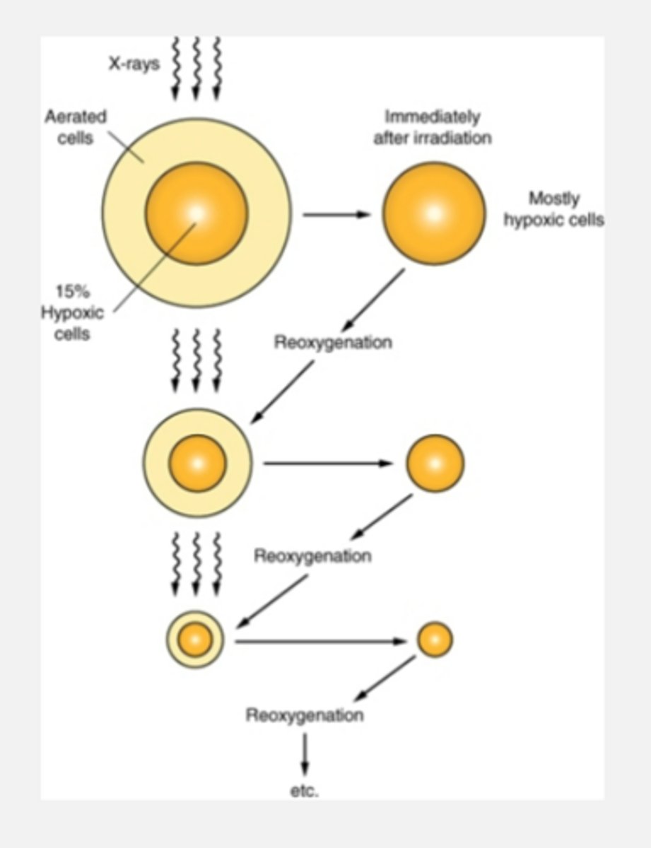
Fractionation to increase radiosensitivity of chronically hypoxic tumors
Delivering a dose of radiation kills a layer of oxygenated cells, allowing the hypoxic cells underneath to receive oxygen again (reoxygenation)
Note: fractionation ALLOWS necrotic cells to reoxygenate (making it more effective to kill tumors cells)
Layers of cells in tumor
Oxygenated (outside)
Hypoxic (middle)
Anoxic (inner)
Three types of cells tumors consist of
Oxygenated
Acutely hypoxic
Chronically hypoxic
Acutely hypoxic cells reoxygenate in a matter of
hours
Chronically hypoxic cells reoxygenate over
Days
Distance between survival curves represent number of
hypoxic cells remaining in tumor
Low LET components
Create free electrons in a medium
Electrons incredibly light and fast
High LET components
Create recoil protons in a medium, and also more direct ionization
Protons and alpha particles more heavy and lumbering
Linear energy transfer (LET)
Average energy deposited in a given material over a given distance
How is LET reported?
keV/um
keV/micrometers
Advantages of utilizing concept of LET
1) there are clear LET patterns that can be seen in cell survival curves
2) allows us to speak in broad terms about different types of radiation
general framework for describing the biological effects of radiation
Disadvantages of utilizing the concept of LET in radiobiology
1) different ways to calculate LET
-track average (breaking up sections in equal distance)
-energy average (amount of energy deposit)
Note: the two calculation methods can give significantly different results in certain situations
2) not the most accurate framework for radiobiology
Relative biological effectiveness (RBE)
Used to compare the different degrees of biological damage from different types of radiation
MUCH MORE COMPLETE PARAMETER THAN LET
Measure of killing efficiency
Takes radiation we are testing and compares to the gold standard
Gold standard in RBE evaluations
250 kV x-rays (D250)
RBE equation/ratio
The ratio of the dose of 250 kV x-rays to the dose of the test radiation that results in EQUIVALENT BIOLOGICAL EFFECT
D250/Dr
D250: gold standard (250 kV x-rays)
Dr: dose of test radiation
Two mandatory experimental conditions for calculating RBE
1) Must be using the SAME CELLS
2) Must be using the SAME BIOLOGICAL ENDPOINT
What changes in RBE?
DOSE
Example of RBE
10 Gy/ 6.6 Gy
=1.5
so test radiation is 1.5x more effective
LD50
Important biological endpoint for relative biological effectiveness (RBE) experiments
-cell survival/death is a common and useful biological endpoint used for RBE
The radiation dose required to kill HALF of the study population
Meaning of RBE as it relates to biological impact of different radiation types
MEASURES CELL KILLING EFFICIENCY
Example: RBE of 1.5 for neutrons means that the neutrons were 1.5 times more efficient at killing cells than 250 kV x-rays (if RBE less than 1, usually means it is worse)
RBE is influenced by
1) LET
-higher LET, more RBE
2) Dose
-higher dose, more RBE
3) number of fractions
-more fractionations, more RBE
4) dose rate
5) type of biological system
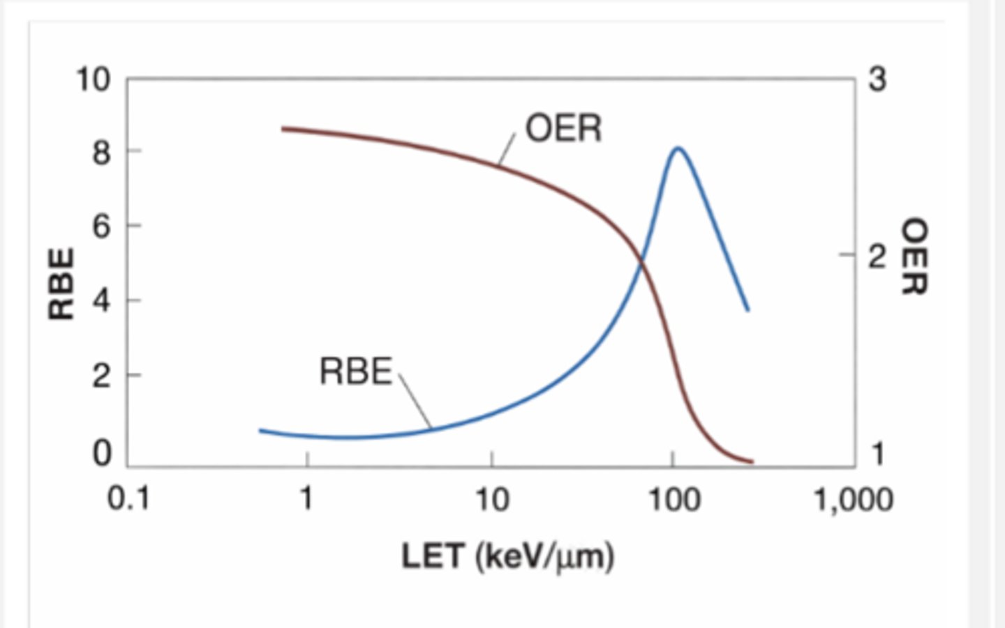
Once LET exceeds 10 keV/micrometer, RBE
increases dramatically
As LET approaches 100 keV/micrometer, RBE
decreases dramatically
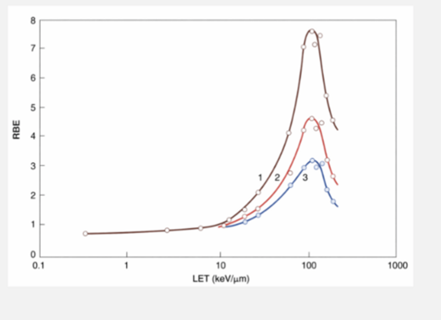
Peak of 100 keV/micrometer LET
Average distance between ionizing events is about the same width as the DNA double helix
Ionizations are perfectly spaced to create double strand breaks
-killing effiency is good
Before the peak
Probability of two DSB is low
After peak
Many DSB are produced, but extra energy is wasted
OER and RBE at 100 keV/micrometer
RBE rapidly increases
OER rapidly decreases
Radiation weighting factors (Wr)
Practical application of RBE
takes into account
-RBE
-type of effect produced (cancer and hereditary effects)
-considerations of lower doses and dose rates
Wr for low LET
1
Wr for high LET
5-20
Equivalent dose (Sv) equation
Absorbed dose (Gy) x Weighting factor
100 rem= 1 Sv
Equivalent dose: Dosimeters
-deposition of energy into crystals is true absorbed dose (Gy)
-once the dose is determined, that value is multiplied by a radiation weighting factor appropriate to the radiation type
-the application of a weighting factor of any kind transforms the unit Gy (absorbed dose) into the unit Sv (equivalent dose)