physio exam 2
0.0(0)
Studied by 4 peopleCard Sorting
1/111
Earn XP
Description and Tags
Last updated 5:53 PM on 3/27/23
Name | Mastery | Learn | Test | Matching | Spaced | Call with Kai |
|---|
No analytics yet
Send a link to your students to track their progress
112 Terms
1
New cards
optogenetics
* light and genetics
* new tool for correlating neuronal circuit activity with behavior
* controlling neuron activity by using light and genetic engineering
* mouse infected with virus = light sensitive channels that produce an AP without change in voltage
* when exposed to blue light = AP, channelrhodopsins
* new tool for correlating neuronal circuit activity with behavior
* controlling neuron activity by using light and genetic engineering
* mouse infected with virus = light sensitive channels that produce an AP without change in voltage
* when exposed to blue light = AP, channelrhodopsins
2
New cards
name of student who worked with him
Scott Varga
3
New cards
optogenetics in Drosophilia larva
* genetically modified to have light sensing channels
* exposed to blue light 470nm, curled up when exposed
* test locomotor symptoms to look for improvement
* exposed to blue light 470nm, curled up when exposed
* test locomotor symptoms to look for improvement
4
New cards
transmission of information within a single neuron: flow info, morpho features, cable properties of axons, myelin, passive propagation, Rm Rl
* ion channels > ionic currents > AP
* input @ dendrites, cell body - integration, AP generated at axon hillock, axon propagates AP to terminals
* morphological feature: long & thin processes
* cable properties of axons: decays with distance
1. cytoplasmic R to electrical signals
2. leaking through plasma membrane - insulation = bad
* myelin = mostly lipid, insulates electrical flow
* passive propagation: current
* higher membrane resistance = insulation better
* smaller diameter axon = high resistance, longitudinal resistance (Rl)
* membrane resistance = Rm = resistance to current flow out across membrane?
5
New cards
passive spread of electrical signals: lambda
* lambda = length constant, represents distance at which decaying voltage change is 37% of its value at the origin
* lambda = sqrt(Rm/Rl)
* high Rm, Low Rl = large lambda
* current decreases = expo decay
* Vx=Vo\* e^-x/lambda
* lambda = Vo @37%
* expo decrease in membrane current
* lambda = sqrt(Rm/Rl)
* high Rm, Low Rl = large lambda
* current decreases = expo decay
* Vx=Vo\* e^-x/lambda
* lambda = Vo @37%
* expo decrease in membrane current
6
New cards
speed of propagation depends on 3
1) temperature
2) greater lambda length constant
3) # channels
2) greater lambda length constant
3) # channels
7
New cards
how to increase lambda 2
1) increase axon size: Rl will decrease because less resistance
2) increase myelination: increases Rm
2) increase myelination: increases Rm
8
New cards
saltatory conductance
AP jumping propagation because of myelination, cannot stop in myelin, Na channels are only expressed at node of ranvier, so AP jumps around, AP propagation is much faster in mammals
9
New cards
_ axon = __ resistance = _ flow
_ lambda = __ propagation speed
_ lambda = __ propagation speed
bigger axon = less resistance = greater flow
10
New cards
what happens if axon too long with no nodes of ranvier
AP cannot travel to terminals, current decays too fast
nodes of ranvier VG Na and K channels help continue AP → more channels = increases speed of propagation
nodes of ranvier VG Na and K channels help continue AP → more channels = increases speed of propagation
11
New cards
propagation of a nerve impulse AP - 2
\
\
1) local circuits - AP propagation
2) unidirectional - excitability Na channels
\
* VG lets Na+ into cell, inside cell = negative, +/- charges attracted to each other = local current, ions are moving bidirectionally
* AP needs to move in one direction
* how AP move in one direction? Na inactivation ends local circuit/ back propagation
\
12
New cards
two functions of Na inactivation
1) overexcitation
2) unidirectional direction of AP
2) unidirectional direction of AP
13
New cards
velocity of nerve impulse conduction increases with
increasing axon diameter with and without myelination
14
New cards
transmission of information between neurons
stimulus + sensory neuron + CNS/ interneuon + motor neuron + ouput (muscle contraction)
15
New cards
echolocation + bats/moth
echolocation: noctural encounter between moth and bat
bat generates ultrasonic sound
object nearby (moth) reflects sound back to bat
moth is also able to detect ultrasonic sound
moth makes rapid unpredictable movements to confuse the bat and escape
bat generates ultrasonic sound
object nearby (moth) reflects sound back to bat
moth is also able to detect ultrasonic sound
moth makes rapid unpredictable movements to confuse the bat and escape
16
New cards
how is sensory information conveyed to the NS - 3 steps
* transduction: convert external stimulus to internal signal (electrical - voltage), interneurons cannot recognize direct external stimulus, must make it so they understand
* amplification: of signal, separate background noise from important signal, DOESN’T always occur
* transmission: signal to CNS where signal is integrated/ processed with other signals
* amplification: of signal, separate background noise from important signal, DOESN’T always occur
* transmission: signal to CNS where signal is integrated/ processed with other signals
17
New cards
transduction
* requires sensory receptor cell to convert external stimulus to electrical signal
* sensory organs: specialized for reception of particular kinds of stimuli (includes sensory receptor cells)
* sensory systems: sense organs and all of their associated central processing areas (organs, neurons, receptors, etc all)
\
sensory receptors (receipt, transduction, amplification) → sensory input → CNS (integration of information) → motor output → effectors (response to signals)
18
New cards
labeled lines principle
*The sensory modality or quality of sensation associated with a stimulus depends solely on which receptor cells are stimulated, rather than on how they are stimulated*.
sensory systems are separate but can work together
labeled lines: sensory neurons → CNS
touch and smell are directly connected
taste, light, and sound require secondary sensory neurons, no direct
sensory systems are separate but can work together
labeled lines: sensory neurons → CNS
touch and smell are directly connected
taste, light, and sound require secondary sensory neurons, no direct
19
New cards
classification of sensory receptor cells 4
1) sensory modality: subjective nature of sensory stimulus (good/bad/pretty/ugly/etc)
2) forms of stimulus energy: light, touch, sound, balance, pressure, etc
3) mechanisms of transduction
4) location of the source of the stimulus energy: exteroceptor = external stimuli, interoceptor = internal stimuli
2) forms of stimulus energy: light, touch, sound, balance, pressure, etc
3) mechanisms of transduction
4) location of the source of the stimulus energy: exteroceptor = external stimuli, interoceptor = internal stimuli
20
New cards
2 kinds of sensory transduction mechanisms
types of sensing
types of sensing
1. ionotropic transduction: direct, faster
* ligand gated channels
2. metabotropic transduction: slower, more amplified, indirect
* G-protein mediated signaling
\
mechanoreception + touch
vestibular organs + hearing
chemoreception and taste
olfaction
photoreception + visual sensory processing
signal → collection → transduction → processing → action
21
New cards
types of synapses
electrical by direct ion coupling
chemical with NT
chemical with NT
22
New cards
electrical synapses: structure, vert/invert, current measured?, coupling coeff, advant/disad, squid + cockroach?, retina + heart?
* gap junctions: 6 monomers of connexin = connexons = pore which allows direct conductance of ions
* connexin = vertebrates, innexin = invertebrates
* current measured between pre/post synaptic cells (can be hard to distinguish for gap junx)
* **coupling coefficient**: gap junctions efficacy can be different = V2/V1 (decays over time)
* advantages of gap junx: rapid bc direct ion conductance
* disadvantages: little flexibility bc can’t control flow of ions in synaptic transmission
* giant squid axon + cockroach: use electrical synapses for fast escapes
* vert. retina + heart = synchronized
23
New cards
chemical synapses + postsynaptic receptors
* presynaptic: vesicles and NT release
* postsynaptic receptors: 1) ionotropic receptors 2) metabotropic receptors
* slower, flexibility higher, lots of vesicles, synaptic cleft, vesicles contain NT = chem signals = ligands, ligands bind postsynaptic R’s = neurotransmitter receptors
* ionotropic receptors: ligand-gated, direct, fast,
* metabotropic receptors: GPCR, slow, amplification, indirect, secondary messengers
24
New cards
fast chemical synapses example
postsynaptic density
synaptic potentials
synaptic currents
functional basis
postsynaptic density
synaptic potentials
synaptic currents
functional basis
* fast chemical synapses: neuromusclar junction → acetylcholine
* end plate region: unique structure, NMJ
* Schwann cell sheath: myelin PNS
* post synaptic density: dense aggregates (receptors) at the membrane
* functional basis: nerve is stimulated, recording potential in presynaptic cell, muscle AP, epp is unique to neuromuscular junctions which is higher than the threshold level of muscle AP
synaptic potential: graded change in resting membrane potential in post-syn cell
* epps: end plate potentials, not used in brain, neuromuscular junctions only
* PSP: post synaptic potential
* EPSP: excitatory postsynaptic potential (ex: epp)
* IPSP: inhibitory, drive the membrane potential away from threshold
synaptic currents: ion flow through channels in response to NT release, produces depolarization in rising phase of EPSP
* EPSC/IPSC: excitatory/inhibitory postsynaptic currents, trigger PSP’s
25
New cards
sequence of events of synaptic transmission at NMJ
1. AP arrives: generated at axon hillock, propagated to presynaptic terminal, role is to depolarize terminal in order to open Ca channels
2. VG Ca channel’s open: influx Ca so NT is released
3. NT (Ach) released: cannot be available all the time, Ach is quickly taken back up to stop contraction
4. ACh receptors open: Ach binds to ionotropic receptors Na / K channels, Na in, K out
5. EPSP: depolarization membrane
6. muscle AP - contraction
7. ACh degradation of AChE: Ach is cut by enzyme to make choline
8. recycled choline: choline taken back up by presynaptic cell and reused by adding a acetyl group back on
26
New cards
neuronal synaptic potentials resemble those @NMJ
2 factors that affect hyperpolarization
2 factors that affect hyperpolarization
1. excitatory CNS synapse: glutamate in mammalial CNS
2. inhibitory CNS synapse: GABA
\
* excitatory NT R: Na+ K+ ions
* inhibitory NT R: Cl- ions = hyperpolarize
* excitatory CNS synapse SMALLER than in muscle
2 factors: loosing positive charges + gaining negative charges (Cl-)
27
New cards
pros of chemical synapses v electrical - 4
* amplify current flow
* can be excitatory or inhibitory unlike electrical which are almost always excitable
* one-way: either excites OR inhibits, electrical can do both
* more modifiable in their properties: plasticity of NS, electrical are not flexible
* can be excitatory or inhibitory unlike electrical which are almost always excitable
* one-way: either excites OR inhibits, electrical can do both
* more modifiable in their properties: plasticity of NS, electrical are not flexible
28
New cards
2 major differences between neuronal + neuromusclar EPSP
* CNS vertebrates use glutamate instead of acetylcholine
* NMJ has larger EPSP than neuronal
* ACh gives greater EPSP than glutamate, GABA gives small IPSP
* NMJ has larger EPSP than neuronal
* ACh gives greater EPSP than glutamate, GABA gives small IPSP
29
New cards
presynaptic release of neurotransmitters 4
1. neurotransmitters: Ach synthesized + stored in presynaptic terminal
2. voltage-gated and Ca2+ dependent
3. quantal + vesicular
4. synaptic vesicle recycling
30
New cards
neurotransmitters: 3 statues, 7 major types,
ways to terminate NT signaling 4
ways to terminate NT signaling 4
1. exogenous application of the NT → same physiological effects
2. must be released when presynaptic neuron is active
3. must be blocked by the same agents that block the transmission?
\
* 7 major types: Ach, GABA, Glutamate, Dopamine, Serotonin, Nor-epi, glycine
* ionotropic: GABA, Ach, glutamate
* metabotropic: nor-epi, serotonin, dopamine
* termination of NT signaling:
1. enzymatic degradation:
2. reuptake: choline taken up by presynaptic + acetyl = Ach
3. enzymatic modification: NE is methylated = NE stops working
4. NT receptor desensitization: signal is terminated until receptor is able to recover - takes time
31
New cards
voltage-gated and Ca2+ dependence
* synaptic response is depolarization dependent
* **Block Ca2+ channels? block AP?**
* PSP is graded: stronger stimulation = more NT released, intensity dependent
* AP is all or nothing: as long as current is greater than threshold level it will occur
* depolarization = increases calcium influx = NT dependent release
* NT release is depolarization and Ca2+ release dependent
* Na Channel blocker = TTX
* K channel blocker = TEA
32
New cards
quantal release of NT
* miniature EPSP: smaller version of as neuromuscular EPSP, small depolarizations with no stimulation in response to A quantum, proves spont. release quantum
* quanta = multi-molecular packet = vesicles
* quanta was named before vesicles were discovered
* a physiological concept to vesicles
* more stimulation = more quanta released
* evoked miniature EPSP’s are quantal
* quanta = multi-molecular packet = vesicles
* quanta was named before vesicles were discovered
* a physiological concept to vesicles
* more stimulation = more quanta released
* evoked miniature EPSP’s are quantal
33
New cards
vesicular release NT
vesicular explained via cellular/molecular
1. docking: place vesicle in correct place to release NT
* V-snare: protein on vesicle, synaptobrevin
* T-snare: syntaxin + SNAP 25
* vesicles move around randomly until V+T snare find each other
2. Ca2+ entry:
* AP depolarizes presynaptic terminal to open Ca channels
* **calcium sensor ?**
* synaptoagmin binds Ca2+, bound vesicle can fuse with membrane
3. fusion: releases NT
* NT release can be controlled by Ca
34
New cards
botox
cleaves synaptobrevin to stop overexcitation of cells, no docking, enzyme is stable
35
New cards
synaptic vesicle recycling 2
1. classical: requires ATP, clathrin-mediated: coated with clathrin, NT are fully released, slower, greater amplitude of PSP
1. kiss-and-run: don’t fully bind/fuse, fast, NT not fully released, no energy required, amplitude PSP is smaller
36
New cards
postsynaptic mechanisms: structure, function of NT receptors AChR’s
pentamer: 5 subunits: 2 alpha, beta, gamma, epsilon
subunits have 4 transmembrane domains M1-M4
M2 creates the channel
2Ach bind 2 alpha subunits
AChR gating by the alpha subunits → will only bind ACh
subunits have 4 transmembrane domains M1-M4
M2 creates the channel
2Ach bind 2 alpha subunits
AChR gating by the alpha subunits → will only bind ACh
37
New cards
reverse potential: defintion, direction of __, sign of__
when Vm = Erev, how does current go with Ik and Ina dominant??
emf equation
max PSP
when Vm = Erev, how does current go with Ik and Ina dominant??
emf equation
max PSP
* reversal potential: the value of Vm that would reverse EPSP/IPSP
* look at the direction of ionic current and the sign of the postsynaptic potential reverse
* Ik is dominant towards Vm = ENa and hyperpolarization, current out
* Ina is dominant towards Vm=Ek and Ach release, current in
* emfx = Vm - Ex
* max PSP: emf = Vm-E, -90-(60) = -150mV
* look at the direction of ionic current and the sign of the postsynaptic potential reverse
* Ik is dominant towards Vm = ENa and hyperpolarization, current out
* Ina is dominant towards Vm=Ek and Ach release, current in
* emfx = Vm - Ex
* max PSP: emf = Vm-E, -90-(60) = -150mV
38
New cards
Ach receptor 3 names
* channel (2Ach bind)
* ligand gated
* non-selective cations
* ligand gated
* non-selective cations
39
New cards
integration at synapses
* each neuron has more than one synaptic contant
* 1 neuron forms \~1000 synapses (1 soma?)
* bigger soma = less resistance
* more Na channels = fires more easily
* closer to axon hillock (further from soma) there are more Na channels where the synaptic potential can be greater than the threshold potential to generate an AP
* 1 neuron forms \~1000 synapses (1 soma?)
* bigger soma = less resistance
* more Na channels = fires more easily
* closer to axon hillock (further from soma) there are more Na channels where the synaptic potential can be greater than the threshold potential to generate an AP
40
New cards
spatial vs temporal summation
stimuli integration
stimuli integration
spatial: multiple stimuli, synapses A+B+C+D generate AP
temporal: same synapse but sends signal at different times to generate AP (meets threshold)
\
stimuli can be integrated: excitatory current + inhibitory current = difference, (bigger excite + small inhibitory = small excite)
temporal: same synapse but sends signal at different times to generate AP (meets threshold)
\
stimuli can be integrated: excitatory current + inhibitory current = difference, (bigger excite + small inhibitory = small excite)
41
New cards
synaptic plasticity
LTP: induction, maintenance, retrograde?
LTD
tetanus stimulation
LTP: induction, maintenance, retrograde?
LTD
tetanus stimulation
changes in synaptic strength (spatial and temporal is similar but more basic and less important)
long-term potentiation: long-lasting enhancement of synaptic transmission following intense stimulation
induction: NMDA receptor-dependent
maintenance: Ca2+ dependent second messenger pathways → increase GluR sensitivity
retrograde signal → NO release to presynaptic terminals to increases NT release
long term depression: suppressing synaptic strength
tetanus stimulation: constant high freq stimulation?
42
New cards
glutamate, Ach
glutamate CNS primary excitation NT
Ach primary excitatory in NMJ
* glutamate receptors
* ionotropic
* non-cation channels
Ach primary excitatory in NMJ
* glutamate receptors
* ionotropic
* non-cation channels
43
New cards
NMDA receptors vs nonNMDA-R
Ca role
Ca role
* activated by glutamate
* NMDA able to conduct Ca ions and Na and K ions
* non-NMDA: small PSP
* NMDA: larger PSP, not easy to open, requires LTP to open, conducts Na,K,Ca,
* presynaptic signal opens glutamate Na/K channels = stronger stimulation, NMDA can open
* glutamate is caged and needs to be open
* Ca concentration increases → steps → generates retrograde signal generator (NO) → presynaptic terminal to enhance NT release
* Ca2+ can bind PKC calmodulin and kinases
44
New cards
doogie mouse
overexpression of NMDA receptors, causes increased intelligence/cognition
45
New cards
muscarinic vs nicotinic AChR
muscarinic: inhibitory, metabotropic, opens K channels = hyperpolarization, lowers heart rate
\
nicotinic: excitatory, ionotropic in NMJ
\
Ach typically excitatory but depends on the type of receptors
\
nicotinic: excitatory, ionotropic in NMJ
\
Ach typically excitatory but depends on the type of receptors
46
New cards
NT receptors for slow metabotropic actions - G-protein mediates synaptic transmission
metabotropic: no channel pore
G-protein alpha subunit activates ion channel, it can activate or inhibit
G-protein alpha subunit activates ion channel, it can activate or inhibit
47
New cards
G proteins in postsynaptic cells act via intracellular effectors - 3
g proteins can act via non-cAMP signaling pathway - 3
g proteins can act via non-cAMP signaling pathway - 3
1. NT can mediate different synaptic effects by activating different kinds of receptors
2. G protein: membrane-delimited action
3. ion channels can be gated by signal other than voltage or ligands
\
1. IP3 and DAG
2. cGMP
3. direct modulation
48
New cards
3 things make synapse flexible
* flexibility of receptors and NT, ionotropic and metabotropic
* summation: temporal and spatial, signaling can be different
* synaptic plasticity
* summation: temporal and spatial, signaling can be different
* synaptic plasticity
49
New cards
why muscles organized
in order to generate enough power
50
New cards
types of muscles
intercalated disk
intercalated disk
striated: skeletal, cardiac
smooth
gap junction that has electrical synapse, used in heart muscles
smooth
gap junction that has electrical synapse, used in heart muscles
51
New cards
skeletal muscle organization
what is the building block of muscle fibers
what is the building block of muscle fibers
muscle fiber (cell)→ myofibrils → sarcomere → myofilaments → actin + myosin
\
building block = sarcomeres
\
building block = sarcomeres
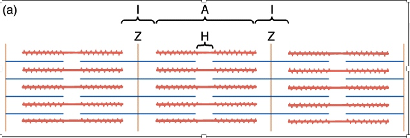
52
New cards
z disc to z disc
actin? myosin?
I band
H zone
M line
A band
actin? myosin?
I band
H zone
M line
A band
z disc to z disc = 1 sarcomere
actin binds z disc, structural support, can see striation, thin filament
myosin = thick filament
I band = z discs are, actin only
H zone = only myosin, no actin
M line = bisects the H zone, myosin, center of sarcomere
A band = length of myosin
actin binds z disc, structural support, can see striation, thin filament
myosin = thick filament
I band = z discs are, actin only
H zone = only myosin, no actin
M line = bisects the H zone, myosin, center of sarcomere
A band = length of myosin
53
New cards
myofilament structure
actin: stabilize heavy chain structure, aka G-actin (globular), polymerization of G-actin forms chains of 2 stranded helix of F-actin, has myosin binding sites
\
tropomyosin and troponin complex: enzymes that control/ regulate muscle contraction
\
myosin: thick heavy chains = tails and head, light chain = hinge, molecular motor, myosin ATPase hydrolyzes ATP forms cross-bridges, has actin and ATP binding sites
\
titan and nebulin provide structural support
\
tropomyosin and troponin complex: enzymes that control/ regulate muscle contraction
\
myosin: thick heavy chains = tails and head, light chain = hinge, molecular motor, myosin ATPase hydrolyzes ATP forms cross-bridges, has actin and ATP binding sites
\
titan and nebulin provide structural support
54
New cards
muscle contraction
myosin ATPase = molecular motor
sarcomere length shortens
rigor mortis = ATP never binds to site = stiff body after death
1. actin bound to site on myosin = ATP site is free to bind, = rigor
2. ATP binds to ATP-binding site, conformational change, myosin and actin detach
3. hydrolysis of ATP by myosin, energy stored in cross-bridge, muscles are at REST
4. actomyosin: actin and myosin bind together, requires Ca2+, by energy provided by ATP hydrolysis ?
5. power stroke: myosin head cocks + actin slides = shortens
6. ADP unbound, new ATP binds to myosin head and releases actin
55
New cards
excitation-contraction coupling
muscle contraction is coupled with excitation
ATP + Ca2 = tension
No Ca2+ ATP = tension decreasing
ATP is not responsible for tension (muscle contraction)
Ca initiation muscle contraction
Ca2+ in excess increases muscle tension
Calcium concentration dependent contraction
role of calcium = cross-bridge attachment enhanced
ATP increases, myosin hydrolyses ATP more = more cross bridge cycles (1 ATP = 1 cycle, 2 ATP = 2 cycle… more cycles – more muscle contractions)
\
3 components of troponin: prevent contraction
Troponin C: binds to Ca2+, conformational change, unblocks myosin binding site on actin, synaptotagmin?, binds myosin?,
Troponin I = binds to actin
Troponin T = binds to tropomyosin (which covers myosin binding sites on actin), pulls tropomyosin away exposing the site so myosin can bind and create crossbridges
When Ca2+ binds to troponin C, there is a conformational change, makes troponin I further away
Troponin T is away from the actin binding site, myosin can bind
56
New cards
the triad
transverse-tubule + 2 sacroplasmic reticulums
T-tubule: Ach → EPSP, generates muscle AP which travels through t-tubule, DHP-R
SR: Ca2+ stores, RyR
DHP-R: voltage sensitive Ca2+ channel, when AP travels down t-tubule, opens channel, blocks RyR
RyR: becomes unblocked when DHP-R is open, releases Ca2+ which travel through DHP-R, sensitive to ryanodine
T-tubule and SR are connected, AP opens Ca channels, DHP-R blocks RyR, when DHP-R active it shrinks and calcium can be released from RyR and binds TN-C, then myosin binding side of actin exposed → powerstroke
57
New cards
summary of excitation-contraction coupling
1. AP in motor neuron → ACh release
2. Ligand-gated ACh channels open
3. AP in muscle → depolarizing T-tubules
4. conformational DHPR → RyR opens
5. Ca2+ release → TN-C
6. Cross bridges → power stroke
7. muscle contraction
8. Ca taken back up into SR when not needed
58
New cards
mechanics of muscle contraction
antagonistic pairs, isometric vs isotonic contractions, concentric vs eccentric
3 “types” of muscle contractions
elastic component
antagonistic pairs, isometric vs isotonic contractions, concentric vs eccentric
3 “types” of muscle contractions
elastic component
antagonistic pairs: skeletal muscles working in pairs, one muscle shortens and the other lengthens
isotonic contraction: IT: muscle generates enough tension to overcome load, goes above the force required for the load, type examples
* Concentric contraction: concentration moving closer to the center of the body, shortening conctration
* Contractile component: group of sarcomeres, actively contracting
* Eccentric contraction: muscle lengthening, happens like when walking downhill?, generates power, like when holding dumbbell and tending arm out = lengthening, muscle lengthening can damage muscles if lengthens too far, small lengthening room,
* Elastic component: tendon – serial, parallel - connective tissues / cell membranes surrounding muscles, passively stretching
isometric contraction: IM: muscle length does not change, muscles are contracted but length does not change, like when you try to move a wall (it doesn’t), tension is generated – force is required to move load - Muscle doesn’t overcome the load = length stays the same
3 types muscle contractions: isometric, isotonic, eccentric
59
New cards
length-tension relation
Active tension like upside down U, passive tension increases as active tension decreases
Passive tension: pulling a rubber band, there is a point where tension begins
Length-tension relation: not solely determined by contractile component, we need elastic component
Sarcomere length: 3 phases increases, plateau, decreasing
maximum muscle contraction = no power, shortest
maximum muscle length: stretched too far, not enough actin myosin overlap
60
New cards
length-tension math
Resting Tension
* Change Length
* Find Tension
Pre-load weight
* Change Tension
* Find Length
Contracting
* Passive → Active
* Change is either T or L
Work = force x distance
* gram x mm
\
Resting tension: passive
Contracting: active
Work = F x d = tension x length = g\*mm = tension \* (length1 – length 2)
To find maximally shortened length: active curve, find where on the curve where x = y, then find parallel x? green + pink, have to look back to SHORTEST length, THEN substract the two numbers
61
New cards
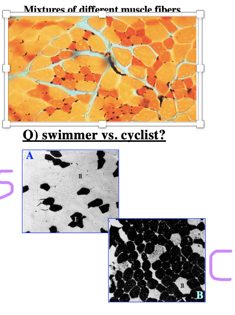
fiber types in vertebrate skeletal muscle
swimmer vs cyclist
swimmer vs cyclist
2 groups of muscles : tonic and twitch
Tonic fibers: no AP, yes membrane potential differences, don’t want eye to twitch,
Twitch/ phasic fibers: most musculoskeletal fibers, generate AP
1. Slow oxidative: SO, type 1: ATP steadily provided, contracts slowly, not easily tired – used for long term, O2 dependent require hemoglobin is why the slow oxidative is red, red colored cells, steak – red meat
2. Fast oxidative glycolytic, FOG type 2a: ATP provided by glycolysis, rapid repetitive movements, orange, in between rapid and long term, low fatigue
3. Fast glycolytic, FG type 2x: no oxygen needed, yellow/white, rapid contraction, chicken breast, fast fatigue
Black dots: capillary, surround red cells, do not surround yellow cells because they do not need oxygen,
\
Second pictures:
Darker color = red , white color = FG is bigger, why? In order to supply oxygen need to be smaller = greater SA, but FG doesn’t need O2
Swimmer v cyclist: swimmer – short term sprints - have less red cells, cyclists – endurance training – have more red cells
62
New cards
types of skeletal muscle fibers:
name, myoglobin, metabolism, strength, fatigue resistance, capillary blood supply
name, myoglobin, metabolism, strength, fatigue resistance, capillary blood supply

63
New cards
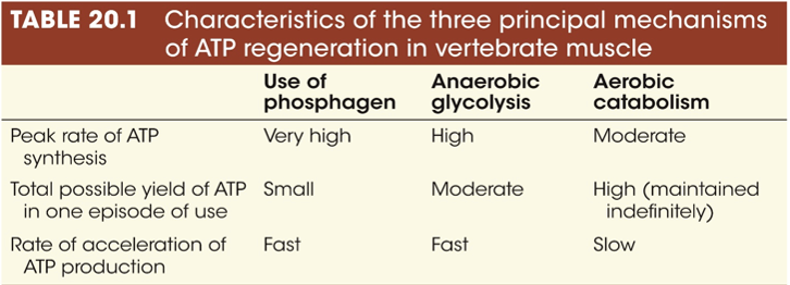
energetics of muscle contraction: ATP use and regen
ATP consumption:
* myosin ATPase: detachment from actin, power stroke
* calcium pumps: pumps Ca2+ back into SR
ATP regeneration during muscle activity:
* high energy phosphatates (creatine-p)
* anaerobic glycolysis
* aerobic catabolism - used in type 1 fibers
* myosin ATPase: detachment from actin, power stroke
* calcium pumps: pumps Ca2+ back into SR
ATP regeneration during muscle activity:
* high energy phosphatates (creatine-p)
* anaerobic glycolysis
* aerobic catabolism - used in type 1 fibers
64
New cards
speed of muscle contraction/relaxation
1. myosin heavy chain isoforms: the rate of ATP hydrolysis varies (SO = slow)
2. sequestration of Ca2+ by Ca2+ ATPAse: Ca2+ needs to be pumped back into SR
speed of contraction: FG > FOG > SO
different muscle types
65
New cards
color, diameter of fiber, levels of glycolysic enzyme, function in animal - SO FOG FG
SO: red, small diameter, low level of glycolytic enzymes, used in animal posture
FOG: red/orange, intermediate diameter, intermediate glycolytic enzyme, standing/ walking/ repetitive movements
FG: white, large diameter, high levels of glycoltic enzymes, jumping/ burst of fast running
FOG: red/orange, intermediate diameter, intermediate glycolytic enzyme, standing/ walking/ repetitive movements
FG: white, large diameter, high levels of glycoltic enzymes, jumping/ burst of fast running
66
New cards
muscle endurance vs resistance training
exercise: muscle in use
**endurance training**
1. endurance: FG → FOG fibers
2. angiogenesis: capillary density increases by VEGF (more VEGF expressed = more capillaries made) (VEGF increases w exercise)
3. density of mitochondria increases why? increased O2 levels, more ox phos needs to happen = more mito required
**resistance training**
1. hypertrophy: actin and myosin synthesis, myofibrils increase
2. FG → FOG fibers
**PGC-1a in endurance and resistance exercise**
* transcription coactivator binds to TF to regulates genes involved with energy metabolism, controls endurance and resistance training why? increase gene expression to increase synthesis of new components in training
* 2 different subtypes: PGC-1a1 and PGC-1a4
* PGC-1a1: endurance, increases VEGF, fatty acid oxidation, mitochondria, and myosin heavy chain 1 + 2
* PGC-1a4: resistance, increases IGF-1 and decreases myostatin
why? myostatin inhibits muscle mass increase, IGF-1 increases glycolysis, more ATP taken up, more glucose required to generate power
67
New cards
atrophy: muscle not in use
by denervation, tenotomy (cutting the tendon), immobilization, microgravity
few SO type 1 fibers
muscle atrophy with age = sarcopenia
EXCEPTION: black bears and aestivating frog hibernation… NO atropy why? how? they minimally maintain, oxidative stress reduction, SOD superoxide dismutase inhibits ROS
few SO type 1 fibers
muscle atrophy with age = sarcopenia
EXCEPTION: black bears and aestivating frog hibernation… NO atropy why? how? they minimally maintain, oxidative stress reduction, SOD superoxide dismutase inhibits ROS
68
New cards
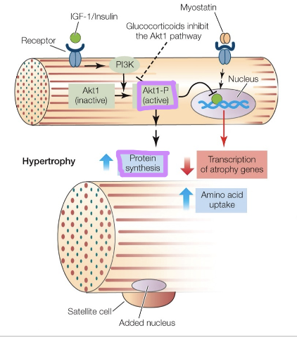
regulating muscle mass
homeostasis between hypertrophy and atrophy
PGC-1a, hypertrophy?
myostatin and PI3K-Akt1 pathway control hypertrophy and atrophy
\
myostatin: negative growth regulator, myostatin mutants = double muscled, causes atrophy = muscle degradation, suppresses protein synthesis, increases atrophy genes
\
PI3K-Akt1: hypertrophy, overall gene expression is increased, enhances muscle growth, Akt1-p inhibts myostatin binding
\
69
New cards
neuronal control of muscle contraction
vertebrates:
1. antagonistic pairs - flexor and extensor muscles
bicep and tricep not working at the same time even tho they both are activated by Ach, one is contracting the other is not, pairs are not stimulated at the same time, only exception = shivering
2. excitatory Ach
3. increases muscle tension, muscle fiber tension is set, 1 AP causes 1 tension/twitch, how does muscle tension increase? Motor units
* no gradation in a motor unit but high-frequency AP
* activating more motor units
* different types of muscle fibers
\
1 Motor unit = 1 motor neuron + all muscle fibers its attached to (\~2-3), we have several different motor units, motor units not overlapped, something is heavy? more motor units are used
Activating more motor units: continually stimulation from AP = tetanic stimulation, tension is at maximum
70
New cards
smooth muscle
no sarcomere, no t-tubule
more randomized, instead of z-disc = dense body
dense body: actin and myosin overlap and bind,
single unit SM: work through gap junction via electrical synapse, depolarize and contract together as one unit, myogenic = beginning in muscles (impulse comes from muscle), intestinal tract - moves as one
\
multi unit SM: no gap junction, neurogenic = beginning in neurons (impulse originates from neurons), much easier to regulate power of muscle contraction, iris muscle - carefully controlled
71
New cards
contraction of smooth muscles
Ca from internal and external sources
* hormones and NT released
* G-protein linked Ca channel - metabotropic
* VG Ca2+ channel - ionotropic
* Ca from SR - not well developed in smooth muscles
* Ca binds calmodulin, binds and activates myosin light chain kinase, kinase phosphorylates myosin LC, activates myosin ATPase = begins contraction, phosphatase removes PO4 from MLC
Ca2+ pumps into plasma and SR
no troponin
myosin-linked regulation
contracts and variable, diverse, not designed to generate large power like skeletal muscles
\

72
New cards
unusual features of SM contraction
latch state: vertebrates, ATP binding is very slow, cross-bridges are “latched” to actin and maintain tension without using ATP, termination: myosin LC dephosphorylation?
\
catch state: invertebrates, mollusks, muscles are stiff and resistant to stretch, produced by the formation of a rigid network of connections between myofilaments, ACh can activate, serotonin relaxes, after the catch state Ca2+ should be gone from cytoplasm?
\
catch state: invertebrates, mollusks, muscles are stiff and resistant to stretch, produced by the formation of a rigid network of connections between myofilaments, ACh can activate, serotonin relaxes, after the catch state Ca2+ should be gone from cytoplasm?
73
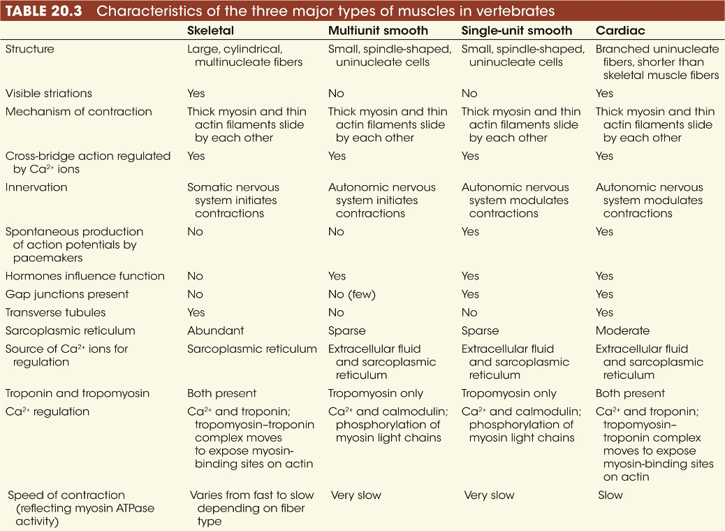
New cards
characteristics of 3 major types of muscles in vertebrates
74
New cards
cellular physiology
cellular signaling
membrane transport
neuronal communication
sensory system
motor structure and function
energetics of living cells
thermoregulation
membrane transport
neuronal communication
sensory system
motor structure and function
energetics of living cells
thermoregulation
75
New cards
systems physiology
lung + O2 → blood circulation → tissues
nutrients → blood circulation → kidney
thermoregulation: animal maintains its own body temp regardless of external temperature
nutrients → blood circulation → kidney
thermoregulation: animal maintains its own body temp regardless of external temperature
76
New cards
circulatory systems
function: carry blood/hemolymph into close contact with every cell in the body
well developed structure: heart, arteries, veins, capillaries
two CV systems: open and closed
well developed structure: heart, arteries, veins, capillaries
two CV systems: open and closed
77
New cards
two types of CV circulation
1. open CV:
* blood circulated slowly
* contained in space called hemocoel
* baths tissues freely in blood (not circulated through capillaries)
* 20-40% of body volume is hemolymph
* blood pumped through heart
* low pressure
* disadvantages: slow nutrient delivery, gas exchange, and metabolic rates
* advantages: no BP, less energy required, better regulation of body temperature
2. closed CV:
* blood is compartmentalized from other body fluids
* smaller volume
* rapid distribution
* divided heart allows different pressures in pulmonary and systemic loops
* disadvantages: requires lots of energy
* advantages: less volume \~5% body, rapid distribution
78
New cards
overview of mammalian circulatory system
propulsive organ = heart
blood vessels = arterial and venous systems, capillaries
distribution of blood throughout vascular tree
arteries \~16-18%
capillaries \~ 5-7%
veins \~ 50%
heart \~15%
pulmonary system \~12%
blood vessels = arterial and venous systems, capillaries
distribution of blood throughout vascular tree
arteries \~16-18%
capillaries \~ 5-7%
veins \~ 50%
heart \~15%
pulmonary system \~12%
79
New cards
physical and mechanical properties of cardiovascular system 4
flow: movement of blood is controlled primarily by the heart
pressure: hydrostatic/hydraulic pressure
resistance: law of bulk flow and ohms law
velocity: depends on part of circulatory system
pressure: hydrostatic/hydraulic pressure
resistance: law of bulk flow and ohms law
velocity: depends on part of circulatory system
80
New cards
flow rate
how much blood flowing at a given time
F = /\\P/R = law of bulk flow
most important
peripheral blood flow = not heart
central circulation = heart
cardiac output (Q) units volume pumped per unit time
Q = V x F
V = stroke volume = volume ejected in one contraction
F = frequency of heartbeats beats/min
F = /\\P/R = law of bulk flow
most important
peripheral blood flow = not heart
central circulation = heart
cardiac output (Q) units volume pumped per unit time
Q = V x F
V = stroke volume = volume ejected in one contraction
F = frequency of heartbeats beats/min
81
New cards
stroke volume
SV: the difference between EDV and ESV, blood left over in heart?
SV = EDV - ESV
EDV: end-diastolic volume, before contraction, heart is fully relaxed, highest amount of blood,
ESV: end-systolic volume, end of contraction, smallest volume, maximum blood ejected
high BP = hard to eject blood
SV varies with HR, pressures in atria ventricles, aorta etc
SV = EDV - ESV
EDV: end-diastolic volume, before contraction, heart is fully relaxed, highest amount of blood,
ESV: end-systolic volume, end of contraction, smallest volume, maximum blood ejected
high BP = hard to eject blood
SV varies with HR, pressures in atria ventricles, aorta etc
82
New cards
average volume and flow and cardiac output in humans
V = 70 mls F = 72 beats/min
cardiac output through aorta = 5040mls / min
flow remains equal when viewed collectively in all compartments
closed system = smaller volume = more efficient
cardiac output through aorta = 5040mls / min
flow remains equal when viewed collectively in all compartments
closed system = smaller volume = more efficient
83
New cards
pressure
mmHg torr Pa
blood pressure: hydrostatic + hydraulic
hydrostatic pressure: force exerted against wall, static
hydraulic pressure: pressure in fluid which is in motion, moving water
goes down pressure gradient high → low
blood pressure: hydrostatic + hydraulic
hydrostatic pressure: force exerted against wall, static
hydraulic pressure: pressure in fluid which is in motion, moving water
goes down pressure gradient high → low
84
New cards
flow is proportional to the ___
difference in pressure /\\P
pressure gradient is important
at high and low pressures, same resistance, flow can be the same
ex: P1: 100mmHg P2: 80mmHg AND P1: 50mmHg P2: 30mmHg, /\\P = 20mmHg for both
pressure gradient is important
at high and low pressures, same resistance, flow can be the same
ex: P1: 100mmHg P2: 80mmHg AND P1: 50mmHg P2: 30mmHg, /\\P = 20mmHg for both
85
New cards
resistance
relationship between pressure and flow
the law of bulk flow or ohms law
flow between two points is proportional to the pressure difference AND inversely related to the resistance
Flow = /\\P/R
higher resistance = flow rate decreases
the law of bulk flow or ohms law
flow between two points is proportional to the pressure difference AND inversely related to the resistance
Flow = /\\P/R
higher resistance = flow rate decreases
86
New cards
flow is inversely related to resistance
Q = /\\P/R I = /\\E/R
as resistance increases, flow decreases
things that increase R: 1) small tube radium 2) long tube length 3) high fluid viscocity
which is most important? radius
vasoconstriction and vasodilation: changing tube radius, practical way to regulate cardiovascular system
as resistance increases, flow decreases
things that increase R: 1) small tube radium 2) long tube length 3) high fluid viscocity
which is most important? radius
vasoconstriction and vasodilation: changing tube radius, practical way to regulate cardiovascular system
87
New cards
pressure decreases with increasing ______
place of highest pressure change
major site of resistance
largest blood vessel
smallest diameter
place of highest pressure change
major site of resistance
largest blood vessel
smallest diameter
distance from the heart
arterioles are major site of resistance, mainly due to sharp decrease in radius
arterioles have greatest drop in pressure, they have the largest drop in size between aorta and capillaries
vena cava = largest blood vessel
capillaries diameter is the smallest \~6-10um
arterioles are major site of resistance, mainly due to sharp decrease in radius
arterioles have greatest drop in pressure, they have the largest drop in size between aorta and capillaries
vena cava = largest blood vessel
capillaries diameter is the smallest \~6-10um
88
New cards
resistors in series vs parallel
series: Rtot = R1+R2+R3
parallel: 1/Rtot= 1/R1 + 1/R2 + 1/R3
if R’s are the same
series: 3R parallel: 1/3R
parallel resistors make it easier to reduce resistance and increase flow which makes CV work less
series resistors make flow slower
most vascular beds are arranged in parallel → minimizes resistance
parallel: 1/Rtot= 1/R1 + 1/R2 + 1/R3
if R’s are the same
series: 3R parallel: 1/3R
parallel resistors make it easier to reduce resistance and increase flow which makes CV work less
series resistors make flow slower
most vascular beds are arranged in parallel → minimizes resistance
89
New cards
entire circulatory system cardiac output
Q = mean blood pressure / total peripheral resistance
90
New cards
peripheral circulation
arterial system
venous system
capillary exchange
anatomy of arteries, veins, capill, very different because of different pressures
atrial system: deals with highest pressure, more thick = less compliant, smooth muscle
venous system: thin and compliant, like a balloon?, vena cava lowest blood pressure,
venous system
capillary exchange
anatomy of arteries, veins, capill, very different because of different pressures
atrial system: deals with highest pressure, more thick = less compliant, smooth muscle
venous system: thin and compliant, like a balloon?, vena cava lowest blood pressure,
91
New cards
arteries
4 functions
4 functions
get stiffer, less compliant, smaller with increasing distance from heart → PRESSURE RESERVOIR
4 functions:
1) passage of blood to capillary system + venial system
2) pressure reservoir: diastolic = lowest pressure, systolic = pressure high, the average pressure maintain by heart is lower than arterial why? heart is contracting much bigger, arterioes are not relaxing as much as the heart = higher pressyre
3) dampening of pressure, when pressure gets to capillaries there is no more oscillations
4) control of blood distribution: precapillary sphincters, more blood to stomach during digestion
\
at periphery, pulse pressure oscillations are much reduced and resistance in arteriolar beds controlled by vasoconstriction
diastolic = relaxing, systolic = contracting
92
New cards
peripheral circulation 3 parts
1) arterial system 2) venous system 3) capillary exchange
\
arterial system: highest pressure = pressure reservoir, more thick = less compliant, thick smooth muscle?
venous system: highest volume, thin + compliant, vena cava = lowest blood pressure
anatomy of arteries, veins, capillaries are very different because of different pressures
\
arterial system: highest pressure = pressure reservoir, more thick = less compliant, thick smooth muscle?
venous system: highest volume, thin + compliant, vena cava = lowest blood pressure
anatomy of arteries, veins, capillaries are very different because of different pressures
93
New cards
arteries
4 functions:
1. __passage of blood__ to capillary system + venous system
2. **pressure reservoir**:
3. __dampening of pressure:__ when pressure reaches capillaries it is no longer oscillating,
4. __control of blood distribution:__ precapillary sphincters
gets stiffer, less compliant, smaller with increasing distance from heart
at periphery, pulse pressure oscillations are much reduced and resistance in arteriolar beds controlled by vasoconstriction
diastolic: relaxing
systolic: contracting
94
New cards
why is average heart pressure lower than arterial pressure
heart contractions are much bigger (big oscillations), arterioles are not relaxing as much as heart (smaller oscillations at higher pressure)
95
New cards
compliance in venous system
compliance: volume change when there is a pressure change, bigger volume change = more compliant
change in volume (V) / change in pressure (Ptm: transmural)
* high compliance: small increases in pressure produce larger increases in volume
* lower compliance: stiff, takes more pressure to increase volume
\
11 mmHg \~10% of arterial pressures
\~25-30 fold more compliant than arteries
\
less elastic tissues = less recoil = greater compliance
act as volume reservoir, susceptible to venous pooling
blood vessel lining is very thin = easy to increase volume
\
veins can vasoconstrict
sympathetics adrenergic fibers can change their compliance → occurs after hemorrhage to compensate for reduced venous volume, also shifts with position (laying down to standing up)
96
New cards
capillary exchange: types, structure, functions, regulation of functions
3 types capillaries:
1. continuous
2. fenestrated
3. sinusoidal
structure:
* one cell layer of endothelial cells, no smooth muscles, 6-10 micrometer diameter
* precapillary sphincters: can allow or block blood flow
* autonomic input: controls precap-sph, can redirect blood flow to areas that need it
* microcirulatory bed: venule, arteriole, precap-sph, capillary, RBC, etc
filtration and diffusion:
starling forces: blood pressure and osmotic pressure
97
New cards
types of capillaries
found in body where
found in body where
Continuous: small cleft 4nm wide, AA and glucose cannot pass, ions can pass through and water, exchange is minimal, largely found in brain, muscles, lung
Fenestrated capillary: like window, glucose and small AA can pass, in between continuous + sinusoidal, kidney allow glucose to pass through,
Sinusoidal: really big to pass proteins, found in liver, paracellular gap
Fenestrated capillary: like window, glucose and small AA can pass, in between continuous + sinusoidal, kidney allow glucose to pass through,
Sinusoidal: really big to pass proteins, found in liver, paracellular gap
98
New cards
movement of substances across capillary walls - 3 mechs
diffusion: random molecular motion
* Ficks Law: Flux, Js = D*** A \* (C1-C2) / thickness
filtration/reabsorption: net movement due to fluid (blood) or osmotic pressure
* more related to circulatory system
* osmotic pressure + blood pressure contribute to direction of material movement
transcytosis:
* endocytosis, vesicular transport, exocytosis
* large proteins across epithelium
* unique to capillary
99
New cards
starling forces in capillaries
blood pressure > osmotic pressure = filtration
blood pressure < osmotic pressure = uptake
filtration more towards arterial end of cap
uptake more towards venous end of cap
**blood pressure = driving force for filtration**
**osmotic pressure = driving force for uptake**
blood hydraulic pressure decreases along cap
colloid osmotic pressure = oncotic pressure
osmotic + blood pressure regulate filtration and uptake
100
New cards
flux across capillaries
reflection coefficient
reflection coefficient
J = \[K(Pcap-Pint)\] - \[o(pi-cap - pi-int)\]
Pcap leaves cell? pi-cap enters cell?
Pcap = capillary blood (hydraulic) pressure
Pint = interstitial fluid pressure
pi = osmotic pressure
K = Coefficient of Hydraulic Conductivity
sigma, o = Reflection Coefficient
\
Sigma: highest - continuous (x\~1) > fenestration (0
Pcap leaves cell? pi-cap enters cell?
Pcap = capillary blood (hydraulic) pressure
Pint = interstitial fluid pressure
pi = osmotic pressure
K = Coefficient of Hydraulic Conductivity
sigma, o = Reflection Coefficient
\
Sigma: highest - continuous (x\~1) > fenestration (0