development of the Back
1/96
There's no tags or description
Looks like no tags are added yet.
Name | Mastery | Learn | Test | Matching | Spaced | Call with Kai |
|---|
No analytics yet
Send a link to your students to track their progress
97 Terms
skull
axial
scapula
appendicular
arm bones
appendicular
ribs
axial
coccyx
axial
pelvic girdle
appendicular
os coxae
appendicular (hips)
sternum
axial
verterbral column
axial
clavicle
appendicular
pectoral girdle
appendicular
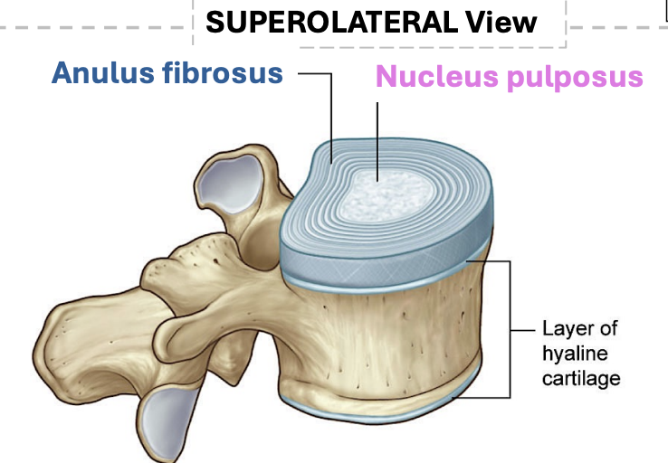
Intervertebral disc is made of what
cartilage
msucles aroudn the verterbral column are called
Epaxial

muscles associated with the body wall
Hypaxial
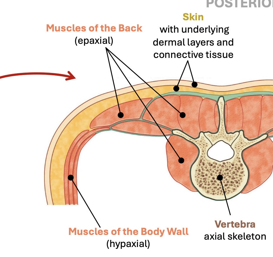
are limb muscles hypxaial or epaxial?
hypxaial
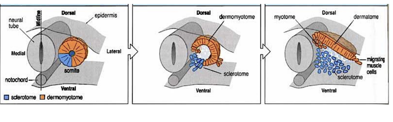
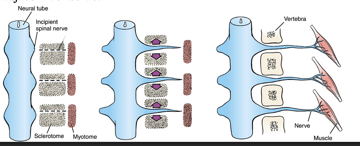
the paraxial mesoderm gives rise to what 3 things?
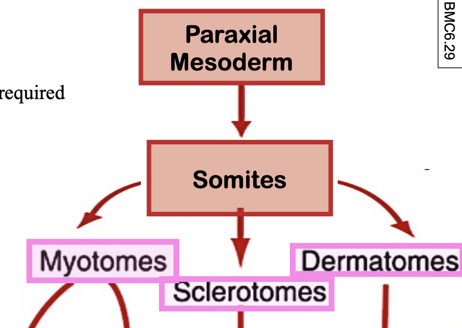
dermatomes give rise to
dermis of skin
when do somites start forming?
end of third week
part of the notochord persists to form a part of the intervertebral disc called the —
nucleus pulposus
a specialized region of mesoderm that marks the cranial end of the embryonic axis.
the prechordal plate
The prechordal plate acts as an—
anterior signalling centre
The prechordal plate helps pattern the — and —
developing forebrain
and overlying neural plate.
the notochord acts as a Signalling centre – it releases morphogens (e.g., —
Sonic hedgehog
what does Sonic hedgehog help do?
pattern surrounding tissues, including the neural plate and the paraxial mesoderm, helping establish somite formation
As development proceeds, the primitive streak regresses in what direction?
caudally
The primitive streak also functions as a signaling center, secreting — and —
FGFs and Wnt proteins.
what activates the segmentation clock?
When the streak has regressed sufficiently, cranial mesoderm cells encounter a sufficiently low concentration of “caudal morphogens”(??)
The first — pairs of somitomeres do not go on to form somites
seven
The first seven pairs instead contribute to the development of —
skeletal muscle of the head instead of the trunk
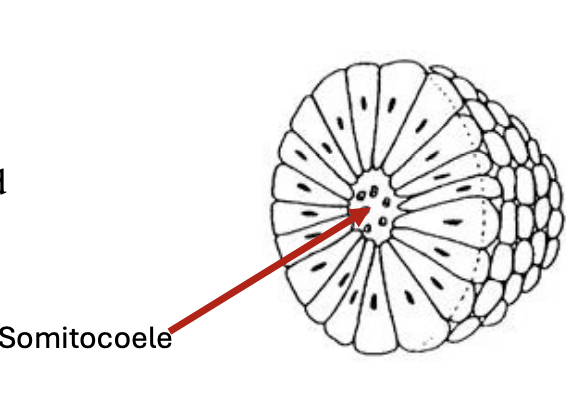
somitomeres have what kind of cells?
compact mesenechymal cells
what happens to the cells in the somitomeres?
undergo a mesenchymal-to epithelial transition
once they have undergone the MET, what happens now?
the epithelial-like cells organize around a central cavity called the somitocoele
First pair of somites appears ~embryonic day 20 in the—region.
occipital
Somite pairs appear sequentially at a rate of —
~3 pairs per day.
A total of ~ — somite pairs form in humans
~42-45
The final number is reduced to ~—somite pairs, with the last few pairs disappearing to correspond to the segmentation of the adult vertebral column
38
Somite period ends ~ day X
30
how many occipital somites?
5
how many cervical somites?
7
how many thoracic somites?
12
how many lumbar somites?
5
how many sacral somites?
5
how many coccygeal somites?
4
Somites are most prominent during week - and -
4 and 5.
what carnegie stage do we stop using somites?
14 (13 is the last one to include it)
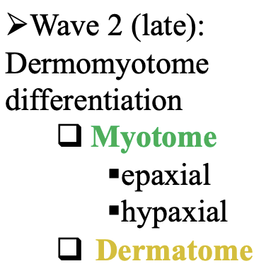
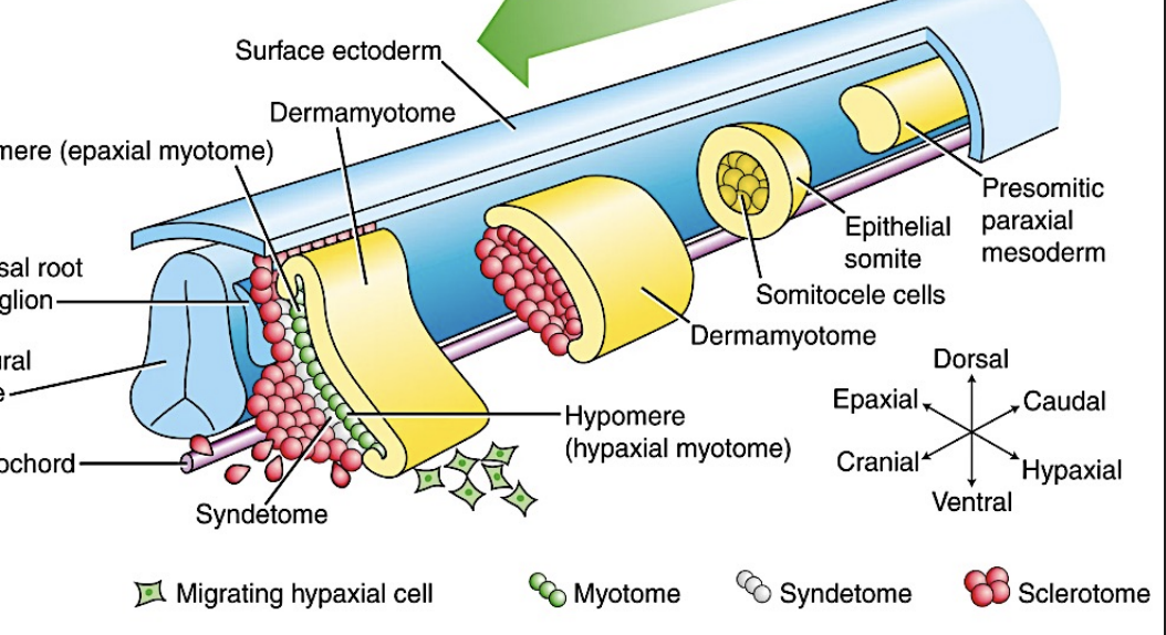
Wave 1 (early): Somite differentiation makes 2 fates
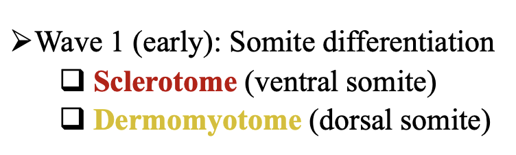
Wave 2 (late)
which part of the somite becomes sclerotome?
ventral part

which part of the somite becomes dermamyotome?
dorsal
process of Differentiation of Somites begins when?
in week 4
what kind of signals specify the dermomyotome fate
Wnt
where do these wnt signals come from?
dorsal neural tube and ectoderm
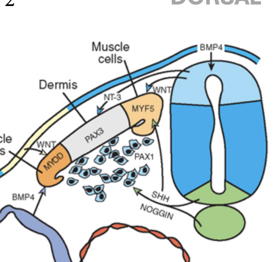
what signal helps maintain the dermomyotome fate and inhibit premature differentiation
BMP signlas
where are those BMp signals coming from?
the lateral plate mesoderm
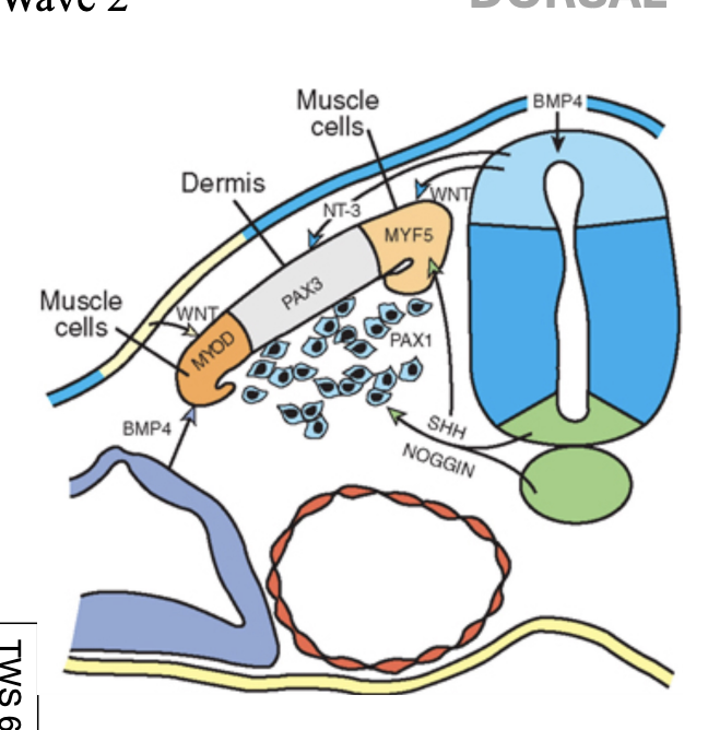
what signal makes sclerotome?
sonic hedgehog
where does teh SHH come from?
from the notochord and the floor plate of the neural tube
how is the “dorsal fate” of the sclerotome inhibited?
Noggin from the notochord and floor plate inhibits BMP signalling
what weird thing do scleorotome cells do?
undergo an epithelial-to mesenchymal transition, and migrate medially

sclerotome cells express sclerotome-specific markers: called — and —
Pax 1 and Pax9.
when is the somatocoele lost?
when they do the EMT
As the sclerotome cells move medially, they form what shape around the notochord and neural tube?
triangular mass
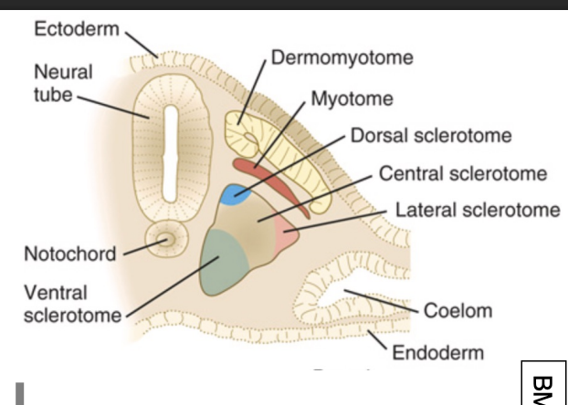
Dorsal Sclerotome will form the
neural arch of the vertebrae and spinal meninges
which scleorotome surrounds the notochord?
ventral and central???
forms the vertebral bodies of the vertebrae
Ventral and Central Sclerotome
Lateral Sclerotome forms what 3 things
costal processes
transverse process
ribs
Vertebral Body derived from
ventral sclerotome


whats the blue thing called?
vertebral foramen
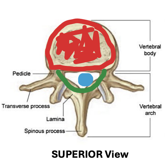
whats the green thing called?
Vertebral or Neural Arch
Vertebral or Neural Arch derived from what?
dorsal sclerotome
Anulus Fibrosis derived from
ventral sclerotome
Each sclerotome segregates into cranial and caudal halves, in a process referred to as—
resegmentation.
Cranial segment = — packed cells
loosely
Caudal segment = — packed cells
densely
The caudal half of one sclerotome fuses with the cranial half of the adjacent sclerotome to form a —
vertebra
The caudal half of one sclerotome fuses with the cranial half of the adjacent sclerotome to form a vertebra during the — week.
5th (they are still mesenchymal at this time)
Chondrification centres form during the —week
6th
how many chondrification centres form within the centrum?
2
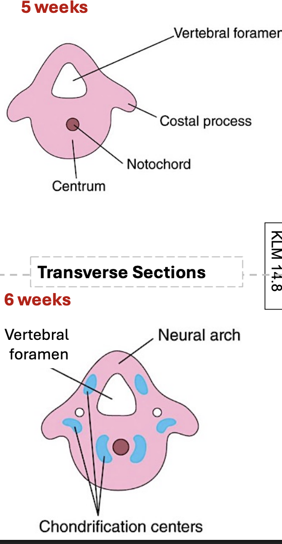
Ossification centres start to form during the— week.
7th
Ossification begins in the —
centrum.
Ossification of the neural arches begins in the—week
8th
Ossification is complete by ~—years of age
25
A failure of one of the chondrification centers to form in the centrum
Hemivertebra (scoliosis!!!) leads to a curve in the spine
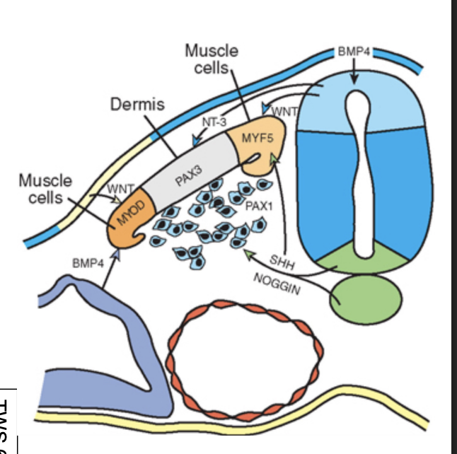
The dermomyotome gets subdivided into 3:
which sections migrate and form the myotome?
dorsal medial lip (DML) and ventral lateral lip (VLL)
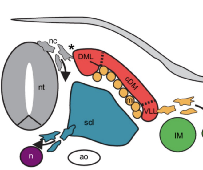
what forms the dermatome?
central dermatome (cDM)
The dorsal neural tube secretes -
neurotrophin 3 (NT-3
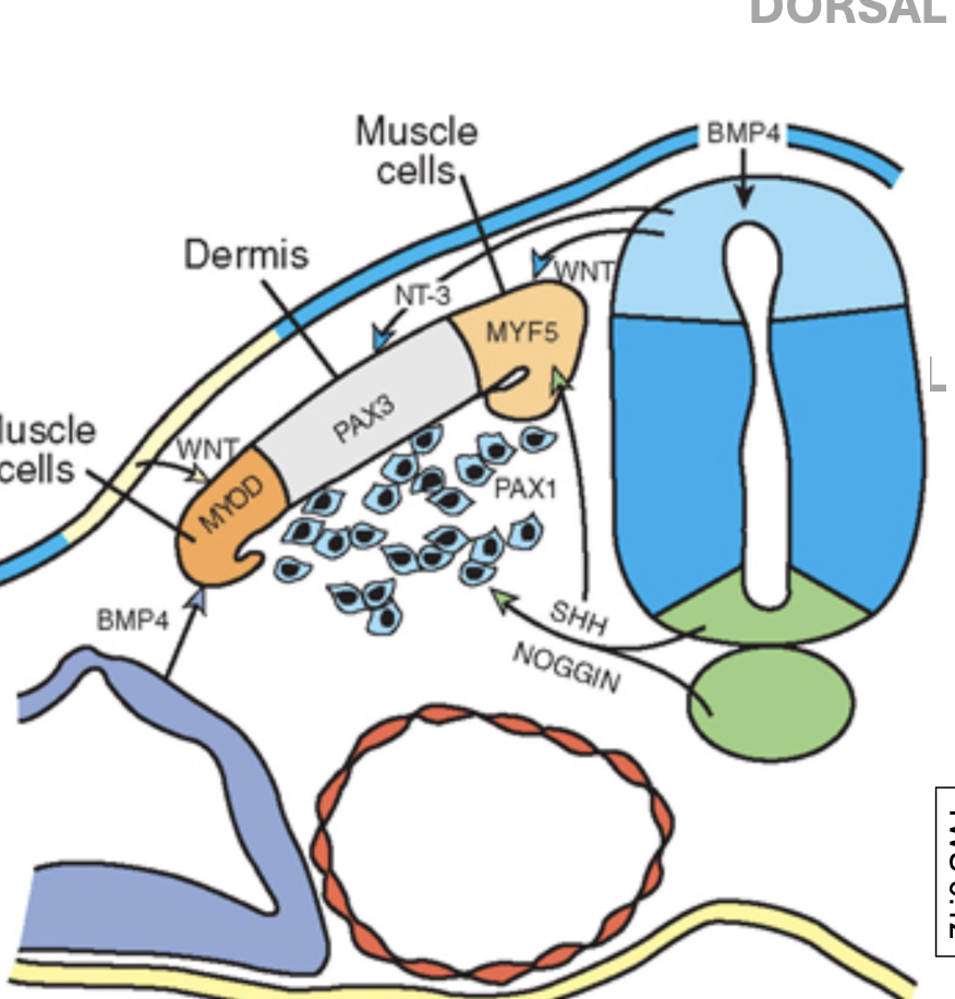
NT3 induces the expression of — in the central dermatome (cDM) specifying the dermatome fate
Pax 3
The dermatome cells undergo an —transition and spread beneath the ectoderm to form the — of the back .
epithelial-to - mesenchymal
dermis
Prior to formation of the myotome, around how many cells migrate to the limb bud?
30-100
which cells migrate to the limb bud?
the ones in the ventral lateral lip (most laterla ones, it makes sense)
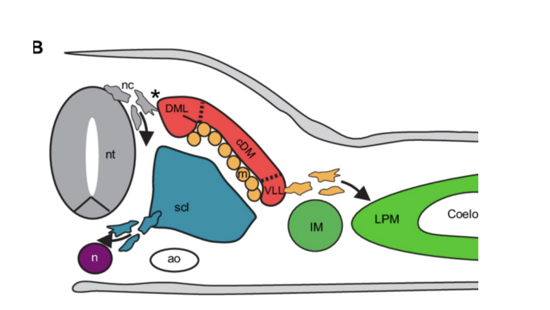
ventral lateral lip (VLL) migrate to the limb bud where they will contribute to — of the limb
skeletal muscles
Dorsal medial lip make what?
epaxial msucles of the back cayuse they are closest to the vertebra so it makes sense
the dorsal medial lip expresses an epaxial specific muscle marker called-
MYF5
what induces the cells to express MYF5?
SHH (notochord and floor plate) and WNTs (dorsal neural tube and overlying ectoderm)
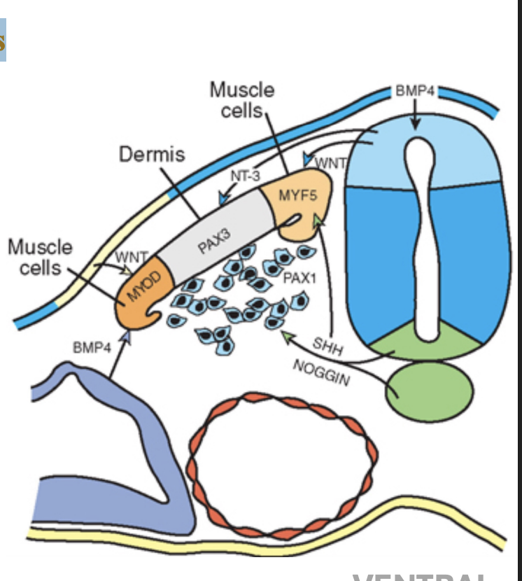
VLL expresses a hypaxial specific muscle marker, called —
MYO-D
what causes them to express MYO-D?
WNTs (dorsal neural tube and overlying ectoderm) and BMPs (lateral plate mesoderm),
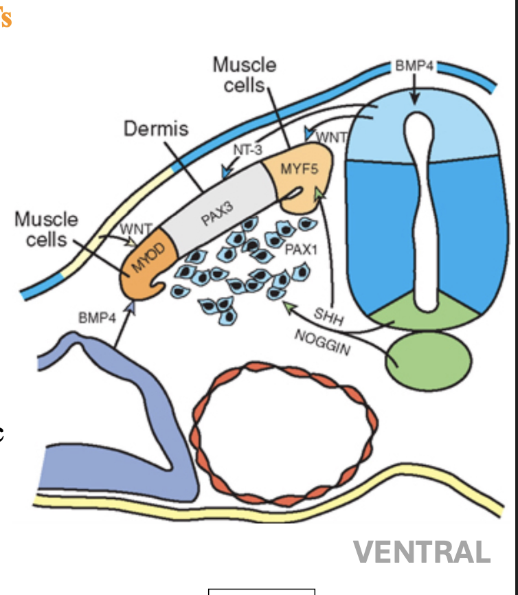
Ventral lateral Lip will make
hypaxial msucles of the trunk