Chapter 13 MCQ + Short essay
1/14
There's no tags or description
Looks like no tags are added yet.
Name | Mastery | Learn | Test | Matching | Spaced | Call with Kai |
|---|
No analytics yet
Send a link to your students to track their progress
15 Terms
(1) c
(2) f
(3) e
(4) g
(5) b
(6) e
(7) i
(8) a
(9) g
(10) h
(11) d
(b) feel no sensation on left side of her body.
(d) motor control
(1) G
(2) W
(3) W
(4) G
(5) W
(6) G
(7) W
(a) visual association area.
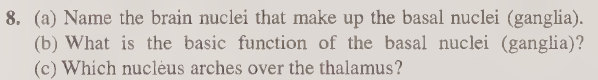
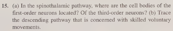
(b) The hemisphere most involved in most people's ability to draw is the right hemisphere. Drawing requires spatial reasoning, holistic perception, and recognizing shapes and proportions — functions dominated by the right cerebral hemisphere.
(c) The labeled regions and their major functions:
Primary motor cortex — Controls voluntary movement of specific body parts (body mapped topographically).
Premotor cortex — Plans and coordinates movements before the primary motor cortex executes them.
Somatosensory association area — Integrates and interprets sensory information from the body (touch, pain, temperature, position).
Primary visual area — Located in the occipital lobe; receives and processes raw visual information from the eyes.
Anterior association area (prefrontal cortex) — Higher-order functions including reasoning, planning, judgment, and personality.
Broca's area — Located in the left frontal lobe; controls the production of speech and language (damage causes expressive aphasia).
![<p><strong>(b)</strong> The hemisphere most involved in most people's ability to draw is the <strong>right hemisphere</strong>. Drawing requires spatial reasoning, holistic perception, and recognizing shapes and proportions — functions dominated by the right cerebral hemisphere.</p><p class="font-claude-response-body break-words whitespace-normal leading-[1.7]"><strong>(c)</strong> The labeled regions and their major functions:</p><p class="font-claude-response-body break-words whitespace-normal leading-[1.7]"><strong>Primary motor cortex</strong> — Controls voluntary movement of specific body parts (body mapped topographically).</p><p class="font-claude-response-body break-words whitespace-normal leading-[1.7]"><strong>Premotor cortex</strong> — Plans and coordinates movements before the primary motor cortex executes them.</p><p class="font-claude-response-body break-words whitespace-normal leading-[1.7]"><strong>Somatosensory association area</strong> — Integrates and interprets sensory information from the body (touch, pain, temperature, position).</p><p class="font-claude-response-body break-words whitespace-normal leading-[1.7]"><strong>Primary visual area</strong> — Located in the occipital lobe; receives and processes raw visual information from the eyes.</p><p class="font-claude-response-body break-words whitespace-normal leading-[1.7]"><strong>Anterior association area (prefrontal cortex)</strong> — Higher-order functions including reasoning, planning, judgment, and personality.</p><p class="font-claude-response-body break-words whitespace-normal leading-[1.7]"><strong>Broca's area</strong> — Located in the left frontal lobe; controls the <em>production</em> of speech and language (damage causes expressive aphasia).</p>](https://assets.knowt.com/user-attachments/e7b1e593-632f-47f1-bf7c-14e052d0c1e5.png)
(a) Brain nuclei that make up the basal nuclei (ganglia):
The basal nuclei consist of the caudate nucleus, putamen, and globus pallidus. The caudate and putamen together are sometimes called the striatum. The subthalamic nucleus and substantia nigra are also closely associated and often included.
(b) Basic function of the basal nuclei:
The basal nuclei are involved in the control and coordination of voluntary motor activity — specifically, they help regulate the initiation, smoothness, and stopping of movement. They suppress unwanted movements and fine-tune motor commands coming from the cortex. They are also involved in procedural learning and habit formation.
(c) Which nucleus arches over the thalamus?
The caudate nucleus arches (curves) over the thalamus, following the C-shaped curve of the lateral ventricles.
(a) Where is the limbic system located?
The limbic system is located on the medial (inner) border of the cerebral hemispheres, forming a ring around the top of the brainstem. It sits between the cerebral cortex and the hypothalamus/brainstem — essentially the "inner edge" of the cerebrum.
(b) Structures that make up the limbic system:
The main structures include the hippocampus, amygdala, cingulate gyrus, hypothalamus, fornix, and mammillary bodies. The olfactory bulbs and parts of the thalamus are also closely associated with it.
(c) How is the limbic system important in behavior?
The limbic system is often called the "emotional brain" and plays a critical role in:
Emotions — the amygdala is central to fear, aggression, and emotional responses
Memory — the hippocampus is essential for converting short-term memories into long-term ones
Motivation and drives — influences hunger, sexual drive, and pleasure
Survival behaviors — fight-or-flight responses, feeding, and reproduction
Smell and emotion connection — olfactory input feeds directly into the limbic system, which is why smells can trigger strong emotional memories
Essentially, the limbic system links conscious thought with emotional and instinctive responses, heavily influencing how we feel and behave.
Sonnet 4.6

(a) Location of the reticular formation:
The reticular formation is a diffuse network of neurons located in the central core of the brainstem, running through the medulla oblongata, pons, and midbrain. It forms a loosely organized "netlike" core that extends from the spinal cord up to the thalamus and hypothalamus.
(b) What does RAS mean, and what is its function?
RAS stands for the Reticular Activating System.
It is the part of the reticular formation that projects upward to the cerebral cortex. Its key functions include:
Consciousness and arousal — it keeps the brain awake and alert; it is essentially the brain's "on switch" for consciousness
Sleep-wake cycles — it regulates transitions between sleep and wakefulness
Attention and focus — it filters incoming sensory information, deciding what reaches conscious awareness and what gets ignored
Alertness to stimulation — sudden or novel stimuli activate the RAS, causing the cortex to "snap to attention"
Damage to the RAS can result in coma or permanent unconsciousness, highlighting how critical it is for maintaining awareness. General anesthetics work partly by suppressing RAS activity.
(a) Four ways the CNS is protected:
Bone — the brain is enclosed in the skull (cranium) and the spinal cord is protected by the vertebral column
Meninges — three layers of connective tissue membranes (dura mater, arachnoid mater, and pia mater) that wrap the brain and spinal cord
Cerebrospinal fluid (CSF) — acts as a liquid cushion, absorbing shocks and preventing the brain from hitting the skull
Blood-brain barrier (BBB) — a selective barrier formed by tight junctions in brain capillaries that prevents harmful substances, pathogens, and many toxins from entering brain tissue
(b) CSF formation, drainage, and pathway:
Formation: CSF is produced by the choroid plexuses — specialized capillary networks located in the walls of the ventricles (mainly the lateral ventricles and the third ventricle).
Pathway:
CSF flows from the lateral ventricles → through the interventricular foramina (foramina of Monro) → into the third ventricle → through the cerebral aqueduct (aqueduct of Sylvius) → into the fourth ventricle → exits through openings (foramina of Luschka and Magendie) → enters the subarachnoid space surrounding the brain and spinal cord
Drainage: CSF is reabsorbed into the venous blood through arachnoid granulations (villi) that project into the dural venous sinuses, particularly the superior sagittal sinus.
If drainage is blocked, CSF accumulates and causes hydrocephalus (increased intracranial pressure).
(a) Superior and inferior boundaries of the spinal cord:
Superior boundary: The spinal cord begins at the foramen magnum (the opening at the base of the skull), where it is continuous with the medulla oblongata of the brainstem.
Inferior boundary: The spinal cord ends at approximately the L1–L2 vertebral level (first or second lumbar vertebra) in adults, forming a tapered tip called the conus medullaris.
(b) Why a lumbar tap can hit the cauda equina or filum terminale but NOT the conus medullaris:
A lumbar spinal tap (lumbar puncture) is performed by inserting a needle into the subarachnoid space between vertebrae L3–L4 or L4–L5.
The key point is that the spinal cord ends at L1–L2, well above where the needle is inserted. Below L1–L2, the vertebral canal does not contain the spinal cord itself — instead it contains:
The cauda equina — a bundle of loose spinal nerve roots that hang down from the conus medullaris like a "horse's tail," floating freely in CSF
The filum terminale — a thin fibrous thread that anchors the conus medullaris to the coccyx
Because these nerve roots float freely in CSF and can be easily pushed aside by the needle rather than being pierced, the risk of serious damage is minimal. The solid spinal cord (conus medullaris) is safely out of reach above the insertion site, so it cannot be punctured.
(a) Spinothalamic pathway — neuron locations:
First-order neurons: Their cell bodies are located in the dorsal root ganglia (just outside the spinal cord). They receive sensory input (pain, temperature, crude touch) from receptors in the body and synapse in the dorsal horn of the spinal cord.
Third-order neurons: Their cell bodies are located in the thalamus (specifically the ventral posterior nucleus). They receive signals from second-order neurons and project up to the primary somatosensory cortex of the cerebrum.
(b) Descending pathway for skilled voluntary movements — the Corticospinal (Pyramidal) Tract:
Upper motor neuron (1st order):
Originates in the primary motor cortex (precentral gyrus)
Fibers descend through the internal capsule → into the cerebral peduncles of the midbrain → through the pons → to the medulla oblongata
At the medulla, about 90% of fibers decussate (cross) at the pyramidal decussation
They then descend as the lateral corticospinal tract in the spinal cord
Lower motor neuron (2nd order):
Cell bodies are located in the anterior horn of the spinal cord
Their axons exit via the ventral roots, travel through spinal nerves, and synapse directly on skeletal muscle fibers
This is why damage to the motor cortex on one side causes paralysis on the opposite side of the body — because of the decussation in the medulla.
(a) Sensory Homunculus:
Located in: the primary somatosensory cortex (postcentral gyrus), in the parietal lobe, just posterior to the central sulcus
Illustrates: the amount of cortical area devoted to receiving and processing sensory information (touch, pressure, pain, temperature) from each body part
Body representation: The body is mapped upside down — feet and legs at the top/medial surface, then trunk, arms, hands, face, and lips toward the lateral surface. Body parts with the most sensory receptors (like the hands, lips, tongue, and face) have disproportionately large representation, while areas with fewer receptors (like the back and trunk) are small
(b) Motor Homunculus:
Located in: the primary motor cortex (precentral gyrus), in the frontal lobe, just anterior to the central sulcus
Illustrates: the amount of cortical area devoted to controlling voluntary muscle movement in each body part
Body representation: Also mapped upside down, similar to the sensory homunculus. Body parts requiring fine, precise motor control (like the hands, fingers, thumb, lips, and tongue) have a large cortical area, while body parts with coarser movement (like the trunk and legs) are represented by a smaller area
Key distinction:
Both homunculi show that cortical representation is based on functional importance, not actual body size. The sensory homunculus reflects sensitivity, while the motor homunculus reflects precision of movement. They are mirror images of each other across the central sulcus.
Layers cut from skin to brain (in order):
Skin (scalp)
Subcutaneous connective tissue (dense, fatty layer)
Epicranial aponeurosis (galea aponeurotica — the fibrous sheet connecting the scalp muscles)
Loose connective tissue (areolar layer)
Periosteum (pericranium — connective tissue covering the outer skull bone)
Skull bone (cranium — typically consisting of outer compact bone, diploe/spongy bone, and inner compact bone)
Endosteum (periosteum lining the inner surface of the skull)
Dura mater (tough, outermost meningeal layer; two layers — periosteal and meningeal)
Subdural space (potential space)
Arachnoid mater (middle, avascular meningeal layer)
Subarachnoid space (contains CSF and blood vessels)
Pia mater (thin, innermost meningeal layer that clings directly to the brain surface)
Brain tissue (cerebral cortex)
The three meninges cut were the dura mater, arachnoid mater, and pia mater — as the question specifically highlights. The scalp layers and skull are also important to name for full credit.
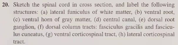
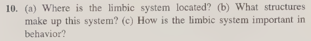
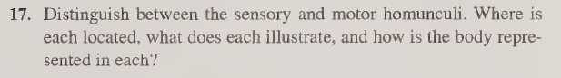
Here's a summary of each labeled structure:
(a) Lateral funiculus of white matter — the large white matter region on each side of the cord, between the dorsal and ventral horns. Contains ascending and descending tracts.
(b) Ventral root — carries motor (efferent) nerve fibers exiting the spinal cord to muscles.
(c) Ventral horn of gray matter — the anterior "wing" of the H-shaped gray matter; contains cell bodies of motor neurons.
(d) Central canal — the tiny central channel running the length of the cord, filled with CSF.
(e) Dorsal root ganglion — a swelling on the dorsal root just outside the cord; contains cell bodies of sensory (afferent) neurons.
(f) Dorsal column tracts — fasciculus gracilis (medial, carries signals from lower body) and fasciculus cuneatus (lateral, carries signals from upper body); both carry fine touch and proprioception upward.
(g) Ventral corticospinal tract — descending motor tract near the midline in the anterior white matter; carries voluntary motor commands (uncrossed fibers).
(h) Lateral corticospinal tract — the main descending motor tract in the lateral white matter; carries crossed voluntary motor commands for skilled movement.
![<p>Here's a summary of each labeled structure:</p><p class="font-claude-response-body break-words whitespace-normal leading-[1.7]"><strong>(a) Lateral funiculus of white matter</strong> — the large white matter region on each side of the cord, between the dorsal and ventral horns. Contains ascending and descending tracts.</p><p class="font-claude-response-body break-words whitespace-normal leading-[1.7]"><strong>(b) Ventral root</strong> — carries motor (efferent) nerve fibers exiting the spinal cord to muscles.</p><p class="font-claude-response-body break-words whitespace-normal leading-[1.7]"><strong>(c) Ventral horn of gray matter</strong> — the anterior "wing" of the H-shaped gray matter; contains cell bodies of motor neurons.</p><p class="font-claude-response-body break-words whitespace-normal leading-[1.7]"><strong>(d) Central canal</strong> — the tiny central channel running the length of the cord, filled with CSF.</p><p class="font-claude-response-body break-words whitespace-normal leading-[1.7]"><strong>(e) Dorsal root ganglion</strong> — a swelling on the dorsal root just outside the cord; contains cell bodies of sensory (afferent) neurons.</p><p class="font-claude-response-body break-words whitespace-normal leading-[1.7]"><strong>(f) Dorsal column tracts</strong> — fasciculus gracilis (medial, carries signals from lower body) and fasciculus cuneatus (lateral, carries signals from upper body); both carry fine touch and proprioception upward.</p><p class="font-claude-response-body break-words whitespace-normal leading-[1.7]"><strong>(g) Ventral corticospinal tract</strong> — descending motor tract near the midline in the anterior white matter; carries voluntary motor commands (uncrossed fibers).</p><p class="font-claude-response-body break-words whitespace-normal leading-[1.7]"><strong>(h) Lateral corticospinal tract</strong> — the main descending motor tract in the lateral white matter; carries crossed voluntary motor commands for skilled movement.</p>](https://assets.knowt.com/user-attachments/d8f034e3-1a2a-44e7-a910-995b87bc7897.png)