BIOL 2052 - Sensory systems: vestibular, auditory and touch
1/34
There's no tags or description
Looks like no tags are added yet.
Name | Mastery | Learn | Test | Matching | Spaced |
|---|
No study sessions yet.
35 Terms
sensory systems
have one for each of the 5 senses
can hep us to detect changes in internal/external environments which can help us to survive
sensory organs send info to the CNS and the sensory info sent to the cortical structures via the subcortical strictures (thalamus)
transduction —> encoding —> processing
TRANSDUCTION
sensory cues are transduced into electrical signals
a sensory receptor is a protein in the csm
activation of this protein leads to a current across the membrane
current must surpass threshold —> allows us to filter out weak stimuli
stimulus strength is encoded by
amplitude of generator potential
frequancy of action potential
driven by mechanogated ion channels
vibrational cues can lead to change in shape of the membrane in the flattened cells
ENCODING
sensory neuron carries signal to the brain
PROCESSING
signal sent to the cortical areas which will give us overall perception of sensory cue
when we do this we have to gain info about:
modality
intensity
duration
location
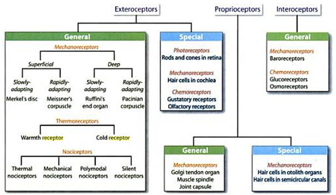
mechanoreceptors
4 different types
split into superficial and deep which further divides into rapidly adapting and slowly adapting
also specialised exteroceptors (mechanoreceptors in the cochlear)
mechanoreceptors that fall into the category of proprioceptors: in the golgi tendon organ, the otolith organs, hair cells and semi lunar canals
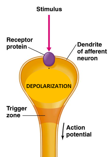
methods of signal transduction
SPECIALISED SENSORY NEURON
receptor protein embedded in membrane and responds to stimuli
E.g: pacinian cell
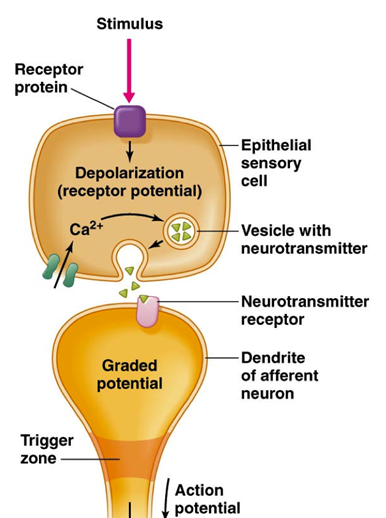
SPECIALISED EPITHELIAL RECEPTOR
has receptor protein that responds to the stimulus
depolarisation, influx of calcium, vesicle release
neurotransmitter release binds to receptor on sensory neuron membrane
E.g: a hair cell
tonic receptors vs phasic receptors (fast vs slow adapting)
TONIC RECEPTORS
slow adapting
stimulus is continuous during the duration of the stimulus
very consistent frequency
e.g: nociceptor
PHASIC RECEPTOR
when stimulus comes on initial burst of activity even though stimulus still there
when stimulus turns off theres another burst of activity
good at signalling continuous stimuli
E.g: some touch receptors
somatosensory system - fine touch
somatosensory afferents convey fine touch into central circuits
cell bodies in dorsal root ganglia
mechanosensory afferents are pseudo unipolar neurons
the cell bodies for the trigeminal neurons located in the trigeminal ganglia
somatic neurons organised into segments according to the region of the periphery they innervate
each segment recieves afferents from different regions called dermatomes (a region of skin innervated by the spinal nerve of a single dorsal root ganglion)
sensory afferents
PROPERTIES
A beta fibres
midrange sensory afferents
myelinated
involved in touch
4 types of receptor: merner, meissner, pacinian and ruffini cells
receptive field
an area of skin over which stimulation will cause a change in the rate of action potentials
size can vary depending on where they are in the body
greater the density of receptors = small receptive field
2 point discrimination
the abaility to distinguish between 2 points of touch
if 2 points within the same receptove field then they are percieved as one
when one point of touch is percieved then it is known as having a low spatial accuity
will vary across the body
CORTICAL MAPS
somatosensory cortex in post central gyrus
certain regions of the body have mcuh larger region in the brain dedicated to recieving their signals
transmission of signals - touch
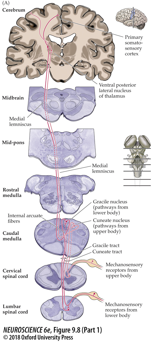
transmission of signal for touch goes through dorsal column of medial leminiscal system
separated into 1st, 2nd and 3rd order neurons
gracile nucleus recieves input from the pathways from the lower body
cuneate nucleus recieves input from pathways in the upper body
TRIGEMINOTHALAMIC SYSTEM
signals from the face go through the trigeminothalamic pathway
its different but does go through the thalamus
synapse between 1 and 2 is the principal nucleus of the trigeminal complex
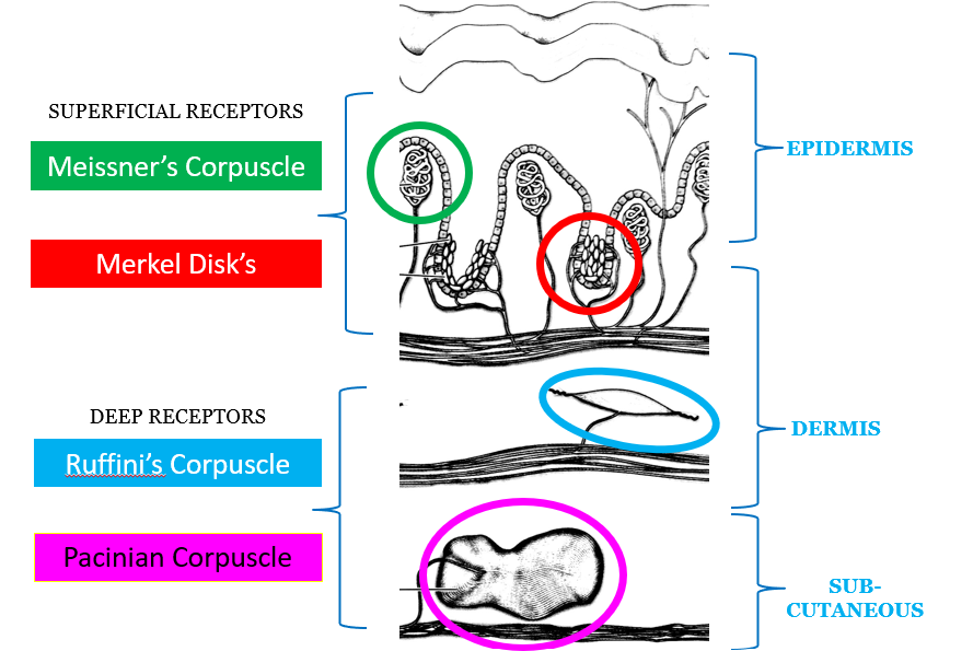
touch receptors
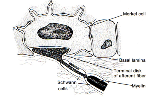
MERKEL DISKS
tonic
sensory function is shape and texture
stimulated by edges and points and curves
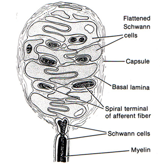
MEISSNERS CORPUCULE
phasic
motion detection, grip control
stimulated by skin motion
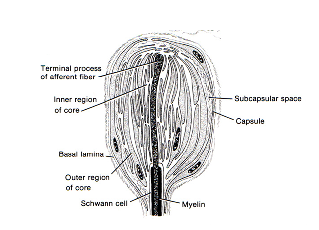
PACINIAN
terminal end of the afferent encapsulated
encapsulated alongside flattened cells
terminal processes of the afferent wind around collagen molecules
large receptive fields
phasic
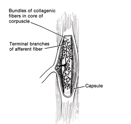
RUFFINI
large receptive field
tonic
good at responding to stretch as theres a collagen bundle that the receptor is stretched around
sensory function: hand shape, finger position
stimulated by skin stretch
auditory system
detection of movement of air molecules
sound waves caused by repeating patterns of compressed air and less compressed air
human ear
INNER EAR
houses hair cells that respond to mechanical cues
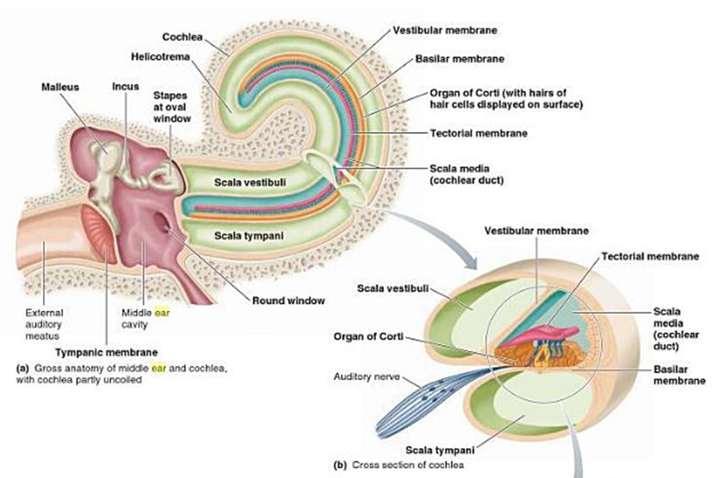
contains the cochlear, made up of three structures
scala vestibuli
scala tympani
scala media
MIDDLE EAR
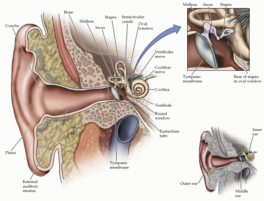
mallius, incus, stapes
transfers mechanical movement across space
important fr amplifying signal
goes from air environment to aqueous which has a high resistance to movement of molecules
large amount of signal is lost as its transferred to the oval window of cochlear
we can still generate movement in inner ear due to amplification
OUTER EAR
made up of concha, pinna and meatus
hervest sound waves and channel them to the eardrum (tympanic memb)
eardrum sets out mechanical movement
helps amplify 2-5kHz frequencies and localise the source of the soundwaves
how does the inner ear amplify the signal
focuses force of large tympanic membrane down onto the much smaller oval window
lever action of the malleus incus
inner ear anatomy
organ of corti within the cochlear
made up of inner and outer hair cells
also contains supporting cells and tectorial membrane which sits over the hair cells
supporting cells and the hair cells sit on the basilar membrane
BASILAR MEMBRANE
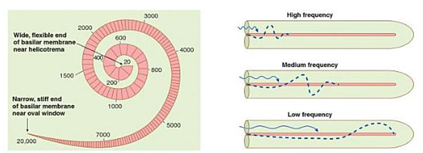
tapered structure - narrow at one end and gets progressively wider
different regions of basilar membrane will respond to different frequencies
waveforms will move along the basilar membrane until it causes undulation in the corresponding region
trandsuction of signal - auditory system
two pivot points for tectorial and basilar membrane sets up shearing force on hair cells when membrane starts to move
movement of basilar membrane converts to movement of stereocilia in 2 directions
when stereocilia move shortest to longest theres an opening of mechanogated ion channels which causes depolarisation
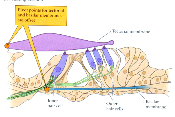
hair cells - auditory system
length of stereocilia increase in a stepwise fashion
stereocilia contain actin filaments and so are rigid
also anchored at pivot points
kinocilium is the longest cilia
have tight junctions which maintains the ionic gradient
they have synaptic ribbons - cytoskeletal adaptations which prime the vesicles for fast release
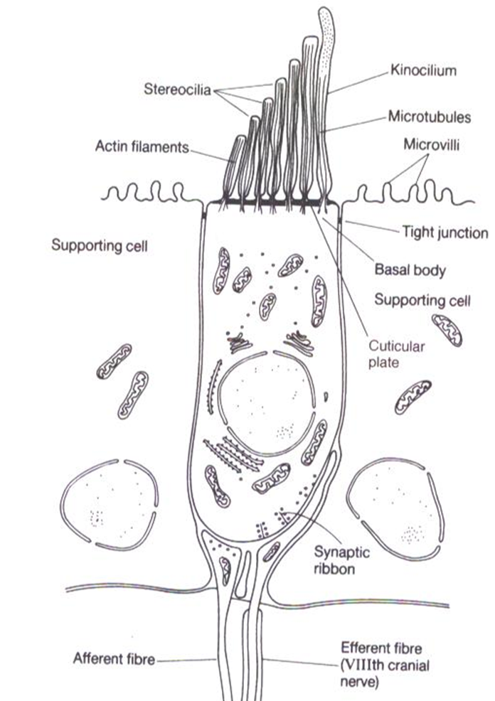
is a specialised type of epithelial cell which speaks to afferent neurons that speak to the CNS
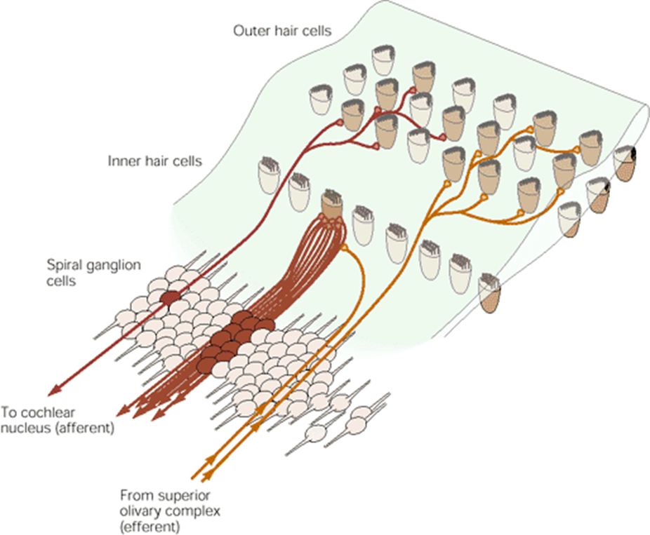
the outer hair cells recieve signals from the CNS whilst the inner hair cells send the signals to the CNS
hair cells are attached to bipolar neurons - one projection is sent to a hair cell whilst the other is sent to the CNS
the cell bodies lie in the spiral ganglia
tip links - auditory system
link together tips of stereocilia
links together mechanogated ion channels in the membrane of the stereocilia
this means that when one stereocilia moves so do the others
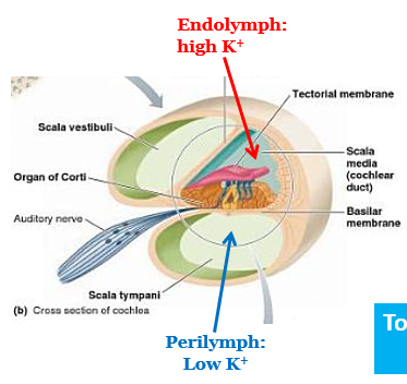
the ionic gradient of the auditory system (endonuclear potential)
depolarisation in the hair cell is caused by K+
K+ gradient caused by high concenytration in the endolymph and a low concentration in the perilymph
when the hair cell is at rest there’s a tonic release at basal level
sound waves will cause the release of neurotransmitter
hair cells moving to the right causes the release of neurotransmitter whilst hair cells moving to the left will cause hyperpolarisation
labelled line coding - auditory system
each auditory afferent is tuned to a specific frequency
a single neuron will respond maximally to a very specific stimulus
outer hair cells of the auditory system
primary role is to amplify cochlear signal
can change shape in response to depolarisation/hyperpolarisation
depolarised = contraction
hyperpolarised = relaxed
amplifies motion of bascillar membrane to amplify inner hair cells —> CNS
major pathways - auditory system
bipolar nerve cells have cell bodies in spiral ganglion
integration between two sides of the head occurs in the superior olive
projections from superior olive —> interior colliculus —> thalamus —> auditory cortex where theres a map of where inputs go based off frequency
how is localisation encoded in the auditory system
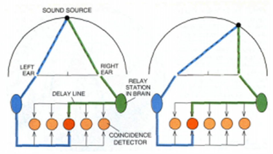
to get from one side of the hed to the other theres a time delay
instead we have delay lines
the signal must travel further to get to the coincidence detector so signals from left and right occurs at the same time
if the sound is coming from in front of us then the coincidence detector that has the maximal response is in the middle
each neuron has a specific unique location
the action potentials will converge on an MSO that responds most strongly if their arrival is coincident
intraural time difference is the difference in time between reaching left and right ear
vestibular system
located in the ear - same region as the inner ear
coordinated reflexive movements like balance and posture
integrates info from the proprioceptive system
controls head position, self motion and spatial orientation
structure and function of the vestibular system
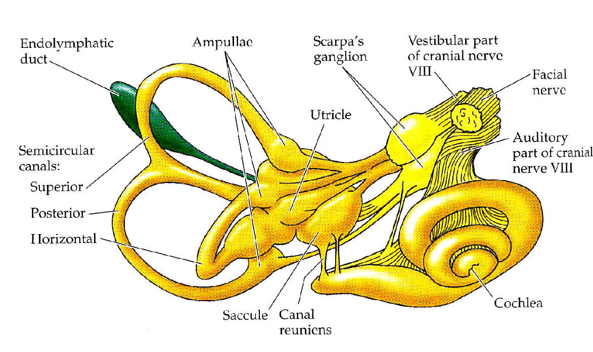
series of interconnected chambers
semi-circular canals give you info about rotational movement
vestibular part of cranial nerve 8 sends info back to the brain
scarpas ganglion holds the cell bodies of the neurons that send signal back to the brain
they are fluid filled chambers - the same fluids as in the auditory system
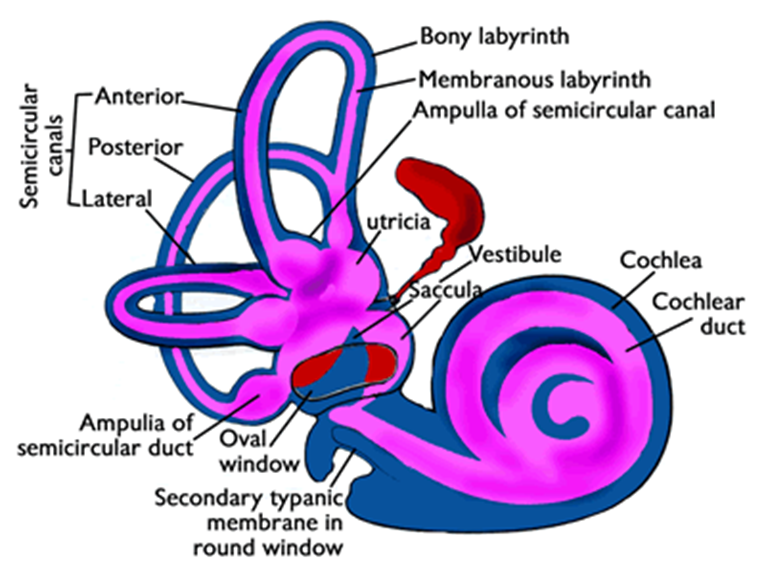
endolymph - inside the membranous labyrinth
perilymph - between the membranous labyrinth and the bony labyrinth
vestibular navigation
there are 3 axes, 2 types of movement
translational movement —> up, down, left, right
rotational movement —> eg roly poly
translational movement detected by otolith organs, rotational movements detected by semi-circular canals
vestibular hair cells
hair cells in ottolith organs and semi-circular canals
the movement of fluid in the semi circular canals results in teh movement of hair cells
there are no inner and outer hair cells, there is only one type
cytoskeletal ribbon synapses result in the fast release of glutamate at the basal membrane
hair cells communicate with afferent neurons with no efferent inputs
there are also tight junctions to maintain the ion gradients
tonic release of neurotransmitter which results in basal level of activity
tip links attached to mechano-gated ion channels —> activation causes influx of K+ which causes depolarisation
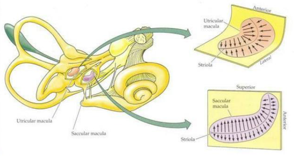
otolith organs
the utricle and the saccule
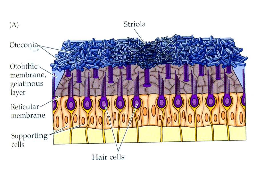
sensory epithelium called the maculla
the otolith membrane on top of the stereocilia
crystals of calcium carbonate sit in the gel otolith membrane and act to increase the mass so that it responds to gravitational force
when walking the hairs tilt backwards as the gel on top of the hair cell has a large mass and moves slower than the fluid
vestibular system - striola
is an axis of symmetry -- all the hair cells are orientated towards the striola
stereocilia are mirror imaged around the striola which creates a differential of electrical activity either side
when one side is depolarised the other is hyperpolarised
the utricle detects motion in the horizontal plane whilst the saccule detects movements in the vertical plane
orientation of the saccular and utricular maccula
saccular maccula orientated vertically and the utricular maccula orientated horizontally
stereocilia must move away from the saccule for depolarisation and nust move towards the utricle for depolarisation
the utricle responds to left, right sideways head tilts whilst the saccule responds to translational movements in the vertical plane
both of them will detect front back translational movements and front back head tilts
semi-circular canals
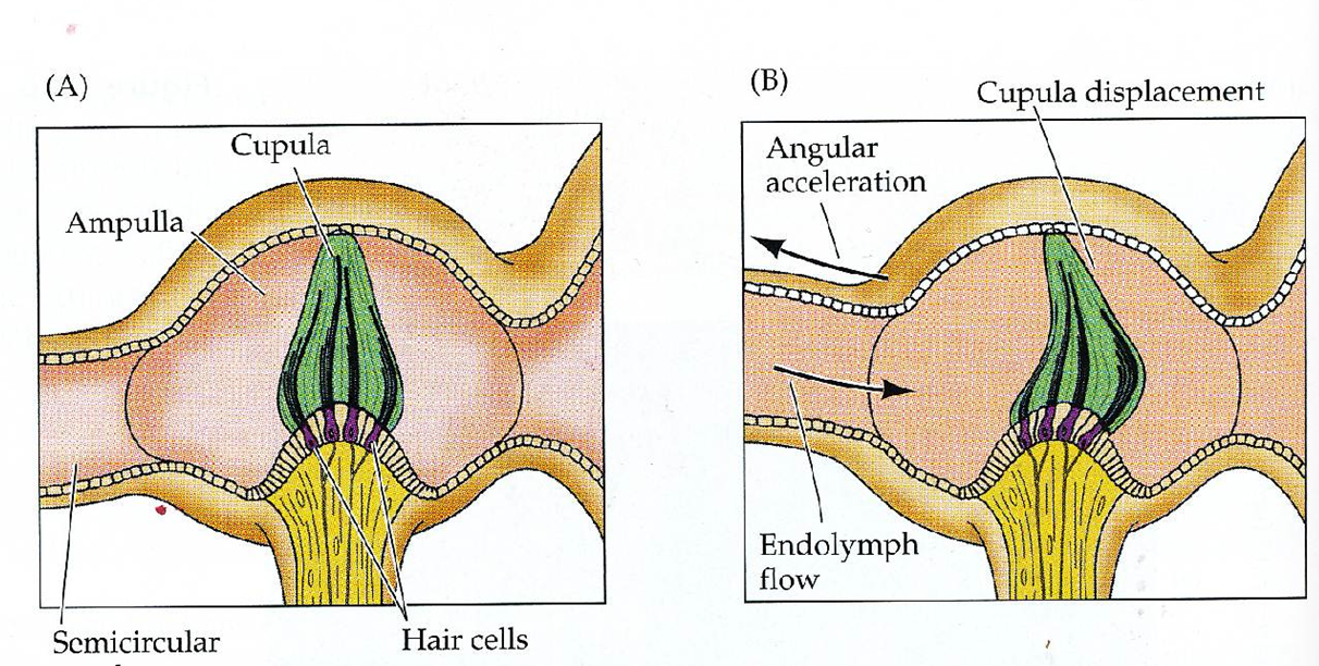
superior, posterior and horizontal
stereocilia embedded in the cupula
the cupula is a barrier that spans across the base of the semi circular canals at the ampluae
provides information about acceleration and deceleration
it is a gelatonous structure which will return back to the baseline position at rest
this results in electrical activity which will plateau rather than in the otolith organs where the signal is constant
when the endolymph moves it causes movement in the cupula
all stereocilia are arranged in the same plane —> theres no axis of symmetry
however there are pairs of stereocilia either side of the head
equilibrium pathways in vestibular system
vestibular nuclei recieves input from the visual, cutaneous, vestibular, proprioceptive systems
sends output to spinal
vestibulo occular reflex
reflex that controls the movement of eye sockets when the head moves
the reflexive eye movements move the eye in the opposite direction to the head
this stabilises the gaze when the eye is moving
is a reflex —> no conscious control
the eye muscles one side of the eye contract whilst the eye muscles on the other side relax which is controlled by movement of fluid in the semi-circular canal
vestibular spinal reflex
goes through lateral and medial pathway of vestibular system
signal is generated by the ottolith organs
few synapses
for maintenance of posture
ascending pathway projects ventral posterior nucleus of the thalamus
can also receive input from the muscle and touch
thalamic nuclei then projects to the vestibular cortex
allows you to integrate stimuli to respond to inputs and change body orientation