BIO Lab Practical 2
1/54
There's no tags or description
Looks like no tags are added yet.
Name | Mastery | Learn | Test | Matching | Spaced | Call with Kai |
|---|
No analytics yet
Send a link to your students to track their progress
55 Terms
What were the two main activities during the Heart Lab?
Exploring heart structure via models/diagrams and recording/interpreting an ECG/EKG.
What are the three primary cardiovascular parameters explored in this lab?
Blood pressure (BP), Pulse, and Mean Arterial Pressure (MAP).
What are the two main areas of focus regarding the lymphatic system in this lab?
Gross anatomy using models/diagrams and microanatomy of lymphatic organs.
What are the two divisions of the respiratory cycle?
Inspiration (inhalation) and Expiration (exhalation).
What is the fundamental rule for airflow in the body?
Air flows from areas of higher pressure to areas of lower pressure.
What is the relationship between the volume and pressure of a container?
There is an inverse relationship; increasing volume decreases pressure, and decreasing volume increases pressure.
What serves as the 'container' for the lungs?
The thoracic cavity.
How does the body generate airflow?
By changing the volume of the thoracic cavity to create pressure differences.
Which muscles contract during inspiration at rest?
The diaphragm and external intercostal muscles.
What happens to thoracic cavity volume and pressure during inspiration?
Volume increases and pressure decreases.
Do the lungs actively participate in changing their own volume during breathing?
No, the lungs themselves have nothing to do with creating the pressure changes.
Which muscles relax during expiration at rest?
The diaphragm and external intercostal muscles.
What happens to thoracic cavity volume and pressure during expiration?
Volume decreases and pressure increases.
What physical property of the lungs aids in expiration?
Elastic recoil.
Is expiration always a passive process?
It is usually passive, but muscles can be used to decrease chest volume more quickly.
generalized blood vessel flow
heart --> arteries --> arterioles --> capillaries --> venules --> veins --> heart
blood pressure is the highest...
arteries
blood pressure is the lowest...
veins
endothelium
innermost lining of blood vessels
which vein is it common to draw blood
median cubital vein
what is the function of the hepatic portal vein?
It carries blood from the gastrointestinal tract and spleen to the liver.
what is pulse?
alternating surges of pressure in an artery that occur within each contraction and relaxation of the left ventricle
What is blood pressure
force of blood against arterial walls
what blood vessel of the arm is used to take BP measurements?
brachial artery
how to calculate mean arterial pressure?
diastolic pressure + 1/3 pulse pressure (systolic bp - diaastolic bp)
how to calculate cardiac output?
heart rate x stroke volume
why is the lymphatic system a one way system?
fluid flows in one direction, has one way valves, return system only, do not have complete circulation
what is the function of lymphatic vessels?
carry lymph from peripheral tissues to the venous system
what is lymph?
a colorless fluid containing white blood cells, that bathes the tissues and drains through the lymphatic system into the bloodstream.
what factors allow the flow of lymph?
skeletal muscle contractions and tissue hydrostatic pressure
cisterna chyli
an enlarged pouch on the thoracic duct that serves as a storage area for lymph moving toward its point of entry into the venous system
right lymphatic duct
receives lymph from the right upper part of the body
where are lymph nodes clustered?
cervical, axillary, and inguinal regions
four processes that must occur in order for the respiratory system to fulfill this role
pulmonary ventilation, external respiration, transport of respiratory gases, internal respiration
largest laryngeal cartilage
thyroid cartilage
structual and functional units of the lungs...
alveoli
flow of air through respiratory system
nostril, nasal cavity, nasopharynx, oropharynx, laryngopharynx, larynx, trachea, main (primary) bronchi, lobar (secondary) bronchi, segmental (tertiary) bronchi, bronchioles, alveoli
type of cartilage found in the trachea
c-shaped cartilage
- lined by ciliated pseudostratified columnar epithelium
groups of alveoli
alveolar sacs
airway that leads from the respiratory bronchioles into the alveolar sacs...
alveolar duct
the right lung is....
shorter, liver is in the way
tidal volume
The amount of air inhaled and exhaled in one cycle of quiet breathing, normally about 500 mL.
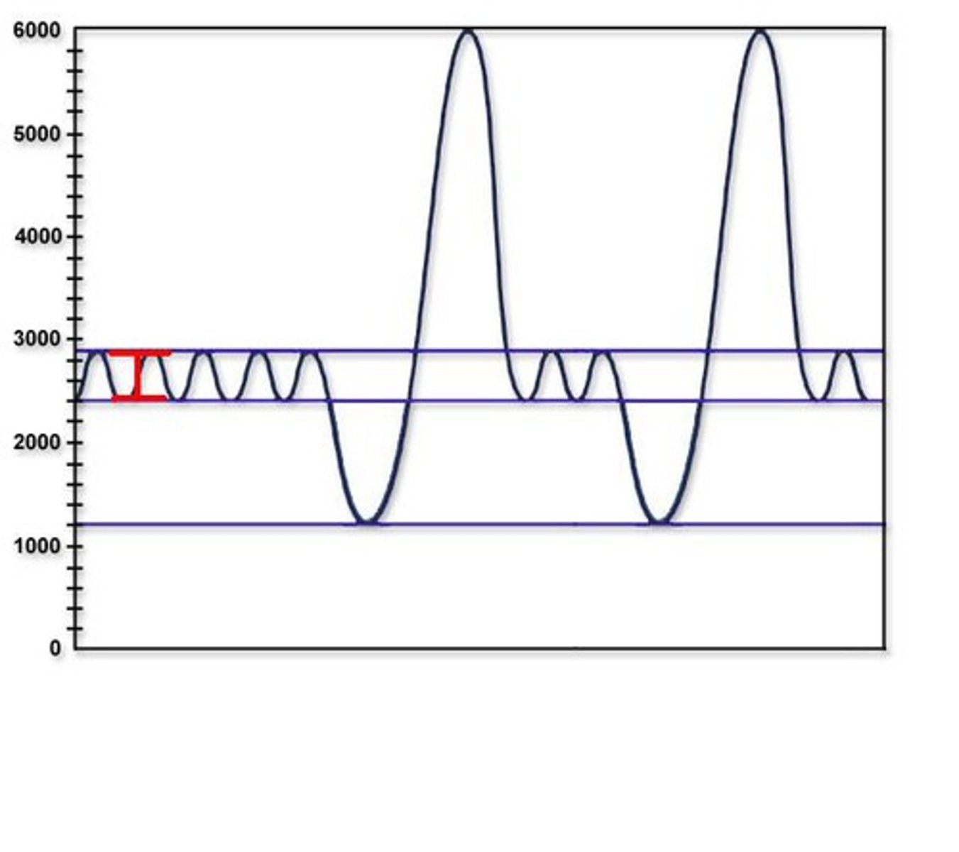
inspiratory reserve volume
The amount of air that can be inhaled beyond tidal volume with maximum effort, typically about 3,000 mL.
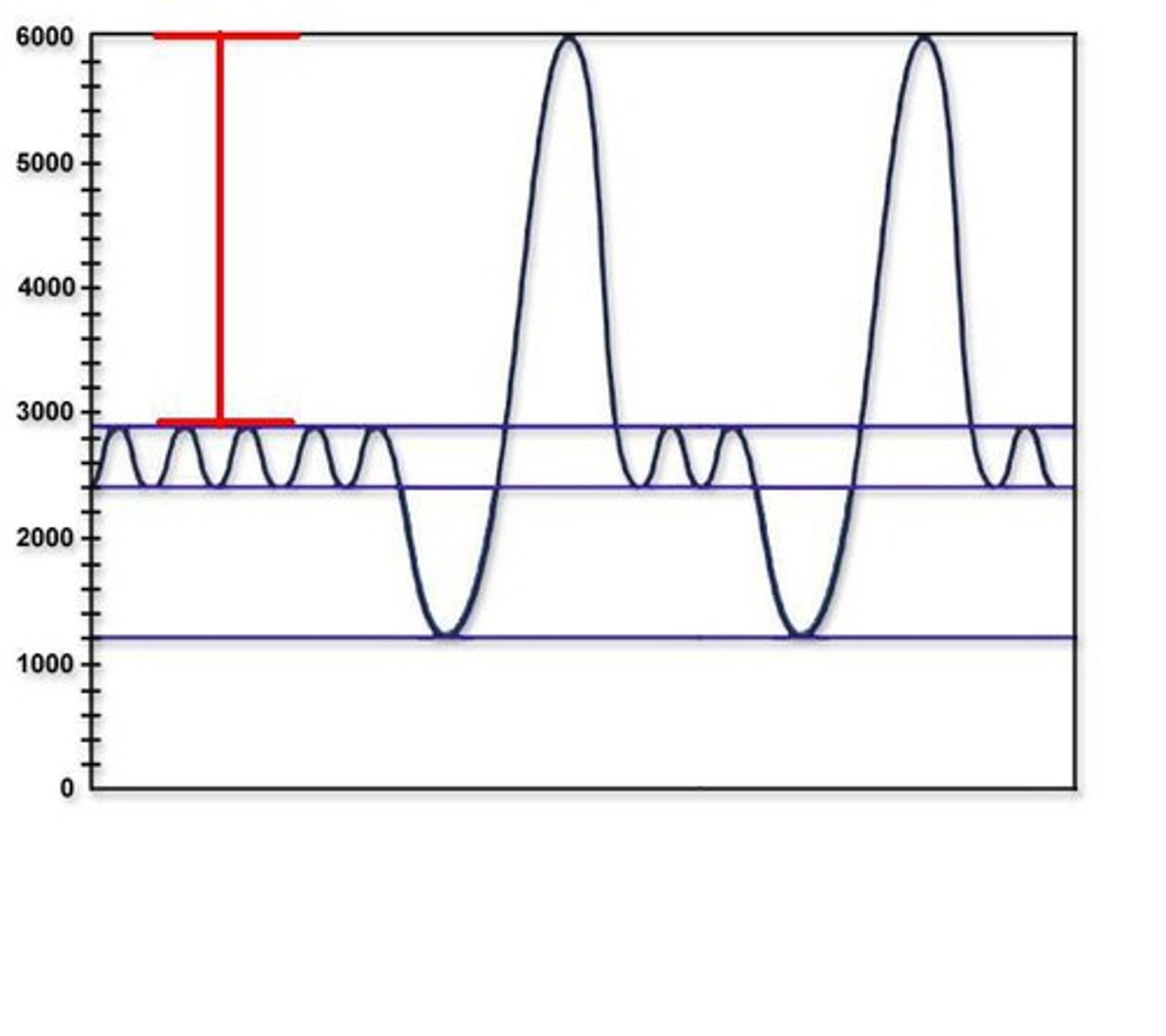
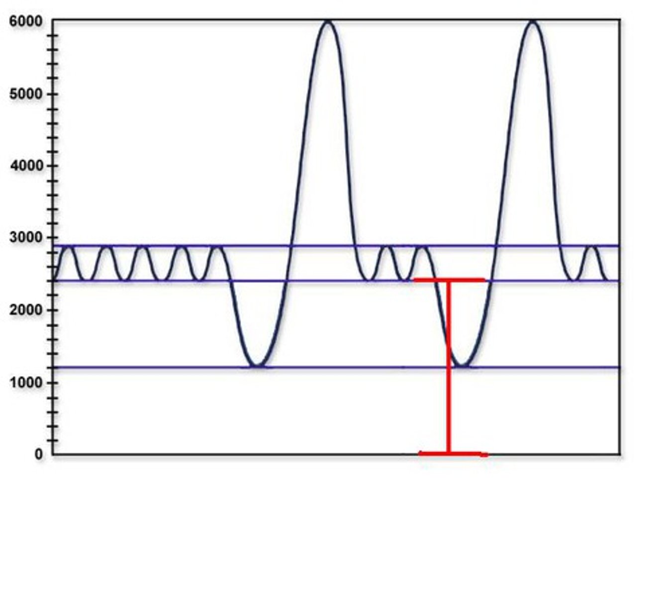
expiratory reserve volume
The amount of air that can be exhaled beyond tidal volume with maximum effort, about 1,200 mL.
residual volume
The amount of air remaining in the lungs after maximum voluntary expiration, typically 1,300 mL.
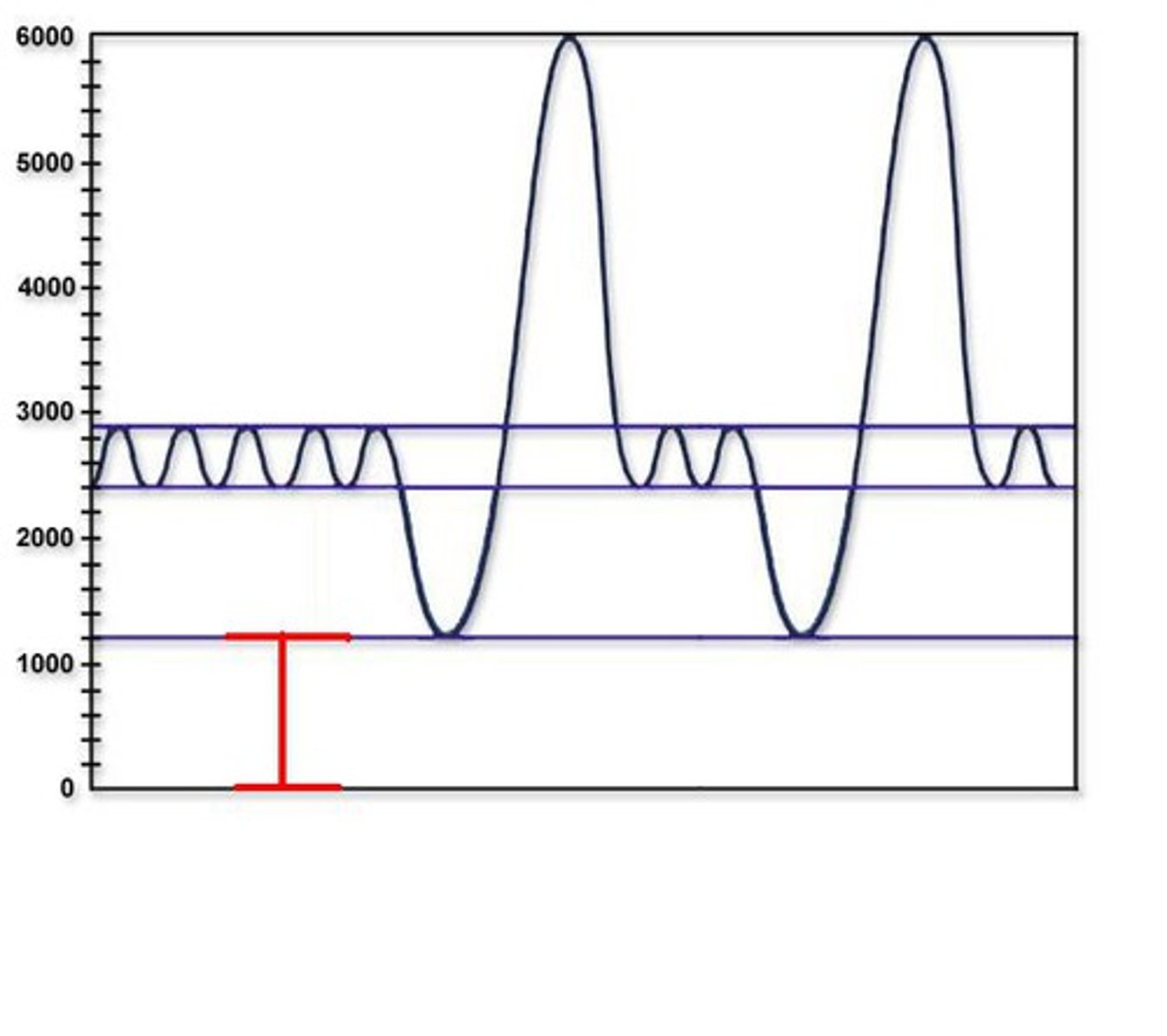
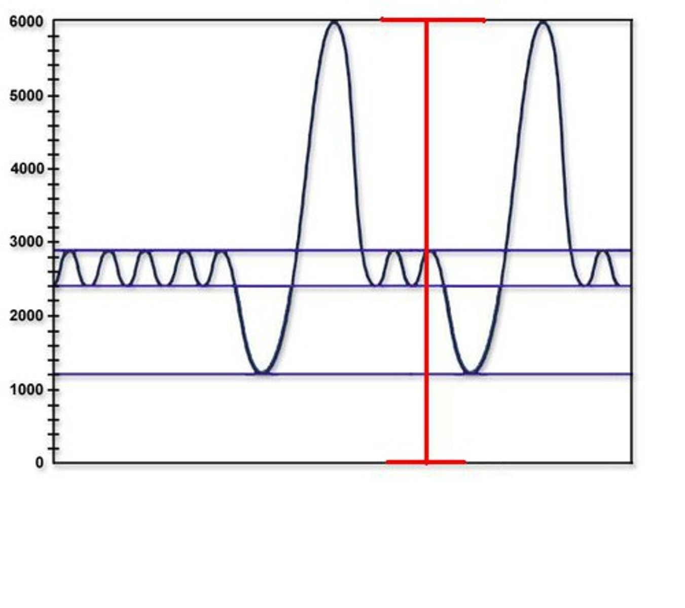
vital capacity
The maximum ability to ventilate the lungs in one breath, calculated as ERV + TV + IRV (4800 mL).
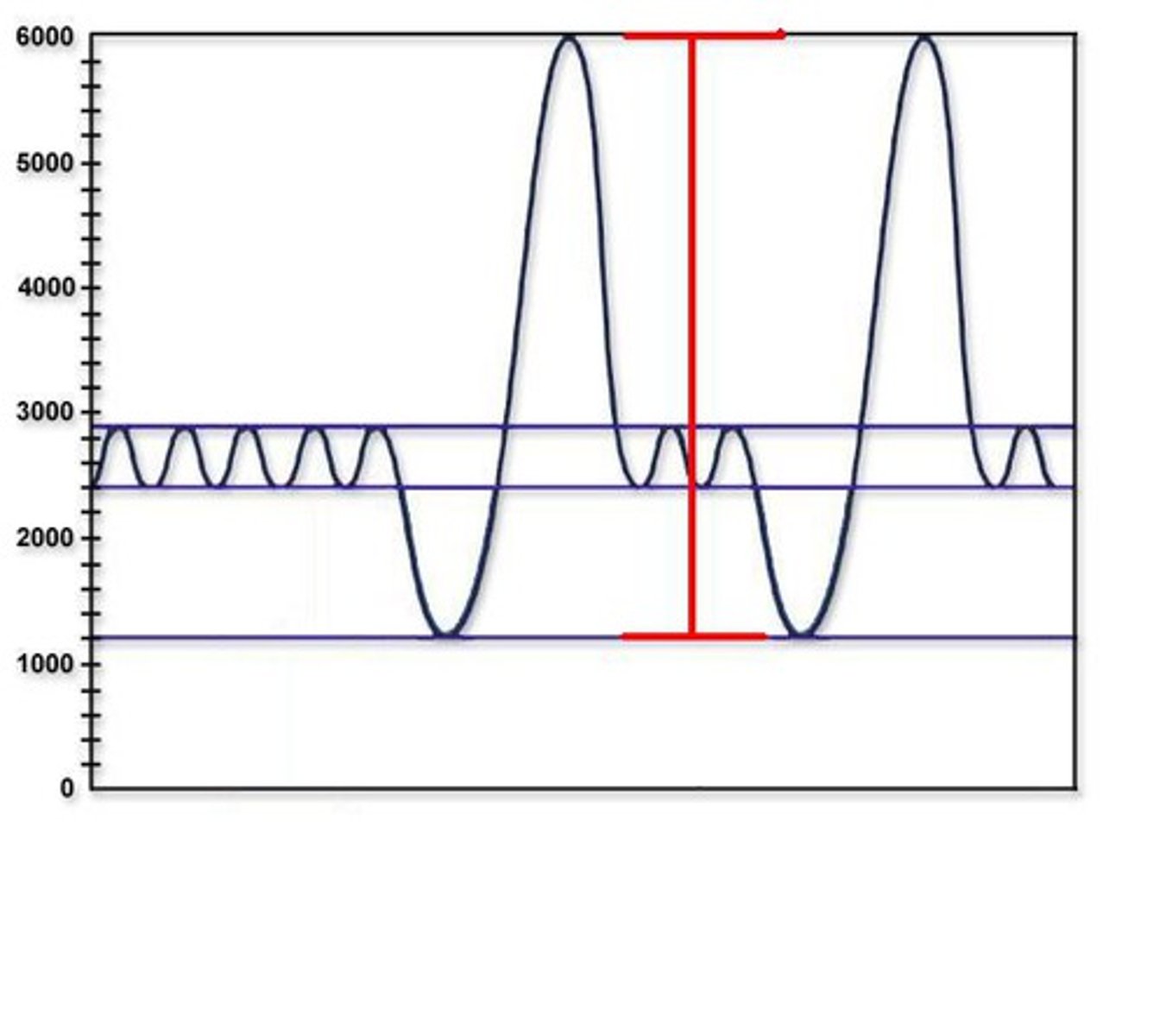
inspiratory capacity
The sum of tidal volume and inspiratory reserve volume (3600 mL).
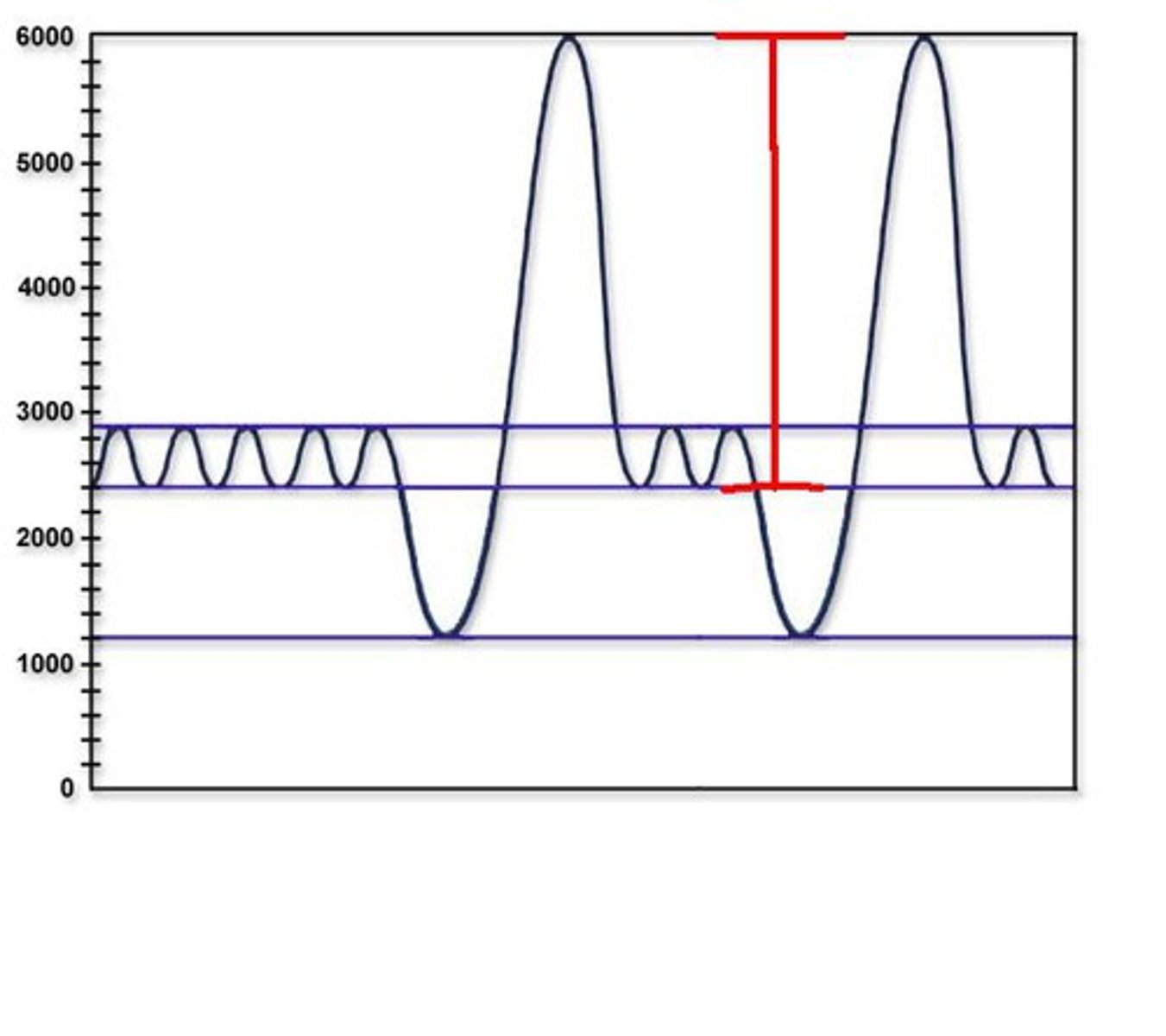
functional residual capacity
The sum of residual volume and expiratory reserve volume (2400 mL).
total lung capacity
The sum of residual volume and vital capacity (6000 mL).
minute ventilation
tidal volume x respiratory rate (L/min)
respiratory rate
number of breaths/delta T x 60 "min"
tonsil stone
mineralizations of debris within crevices of the tonsils, can cause bad breath and pain
plasma cells
Cells that develop from B cells and produce antibodies.
cytotoxic t cells
Cells created in the thymus that produce substances that attack infected cells in the body.
helper t cells
T cells that help the immune system by increasing the activity of killer cells and stimulating the suppressor T cells