Test 5: Anatomy and Physiology
1/126
There's no tags or description
Looks like no tags are added yet.
Name | Mastery | Learn | Test | Matching | Spaced | Call with Kai |
|---|
No analytics yet
Send a link to your students to track their progress
127 Terms
The Muscular System consists of what?
Only of skeletal muscles
Muscle organization and Function
Muscle organization affects power, range, and speed of muscle movement
Fascicles - Muscle cells (fibers) are organized in bundles (fascicles)
Classification of Skeletal Muscles
By the way fascicles are organized
By relationship of fascicles to tendon
Organization of skeletal muscle fibers
Four patterns of fascicle organization:
Parallel
Convergent
Pennate
Circular
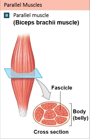
Parallel Muscles
Fibers parallel to the long axis of muscle
Example: Biceps Brachii
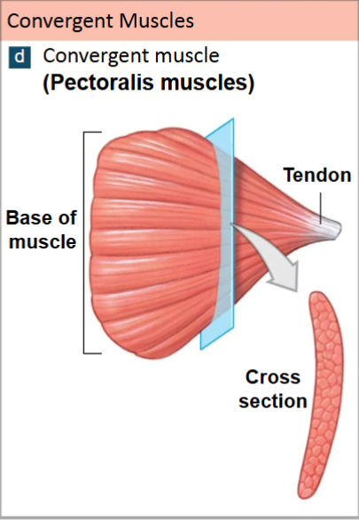
Convergent Muscles
A broad area converges on attachment site (tendon, aponeurosis, or raphe)
Example: Pectoralis Muscles
Pennate Muscles
Form an angle with the tendon
Example:
Unipennate (Fibers on one side of tendon) - Extensor digitorum
Bipennate (Fibers on both sides of tendon) - Rectus femoris
Multipennate (Tendon branches within muscle) - Deltoid
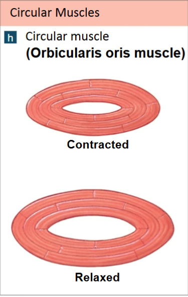
Circular Muscles (Sphincters)
Open and close to guard entrances of body
Example: Orbicularis oris muscle of the mouth
Skeletal motion
Skeletal muscles attach to skeleton, produce motion
Type of muscle attachment affects power, range, and speed of muscle movement
Levers
Mechanically, each bone is a lever (a rigid, moving structure)
-and each joint a fulcrum (a fixed point)
Muscles provide applied force (AF)
-Required to overcome load (L)
Functions of a lever
To change:
-direction of an AF
-distance and speed of movement produced by an AF
-effective strength of an AF
The Three Classes of Levers
Depend on the relationship between applied force, fulcrum and resistance
First-class lever
Second-class lever
Third-class lever
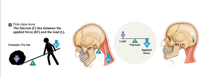
First-Class lever
Seesaw or teeter-totter is an example
Center fulcrum between applied force and load
force and load are balanced
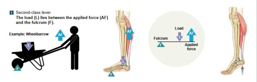
Second-Class lever
Wheelbarrow is an example
Center resistance between applied force and fulcrum
A small force moves a large weight
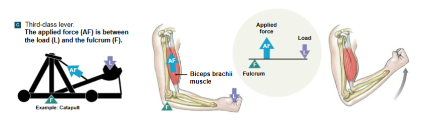
Third-Class lever
Most common levers in the body
Center applied force between load and fulcrum
greater force moves smaller load
maximizes speed and distance traveled
Origins and Insertions
Muscles have one fixed point of attachment (origin)
-and one moving point of attachment (insertion)
Most muscles originate or insert on the skeleton
origin is usually proximal to insertion
Actions
Movements produced by muscle contraction
Body movements
-Examples: flexion, extension, adduction, etc.
Described in terms of bone, joint, or region
Muscle Interactions
Muscles work in groups to maximize efficiency
Smaller muscles reach maximum tension first, followed by larger, primary muscles
Muscle terminology based on function
Agonist (or prime mover)
Antagonist
Synergist
Agonist (prime mover)
produces a particular movement
Antagonist
opposes movement of a particular agonist
Synergist
A smaller muscle that assists a larger agonist
Helps start motion or stabilize origin of agonist (fixator)
Muscle opposition
Agonists and antagonists work in pairs
-When one contracts, the other stretches
-Such as flexors-extensors, abductors-adductors, etc.
Effects of Aging
Skeletal muscle fibers become smaller in diameter
Skeletal muscles become less elastic
-Develop increasing amounts of fibrous tissue (fibrosis)
Decreased tolerance for exercise
Decreased ability to recover from muscular injuries
Cardiovascular system
Delivers oxygen and fuel
Removes carbon dioxide and wastes
Respiratory system
responds to oxygen demand of muscles
Integumentary system
Disperses heat from muscle activity
Nervous and endocrine systems
Direct responses of all systems
A muscles fiber is the same as
a muscle cell
Muscle tissue
A primary tissue type, divided into:
-Skeletal muscle tissue
-Cardiac muscle tissue
-Smooth muscle tissue
Skeletal muscles
Are attached to the skeletal system
Allow us to move
The muscular system
-Includes only skeletal muscles
Remember this about muscles
Shortening = tension = lifting the bone
Six functions of Skeletal Muscle Tissue
“Powerful Muscles Support Great Growth & Stability”
produce skeletal movement
maintain posture and body position
support soft tissues
guard entrances and exits
maintain body temperature
store nutrient reserves
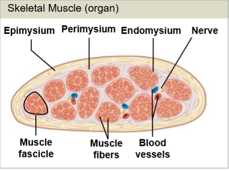
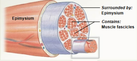
Skeletal Muscle
Muscle tissue (muscle cells or fibers)
Connective tissues
nerves
blood vessels
Organization of connective tissues
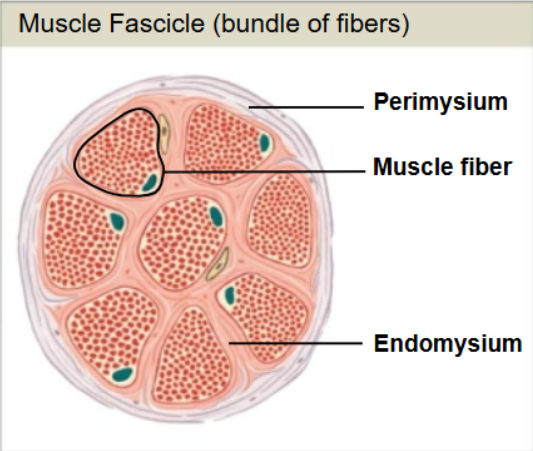
Epimysium
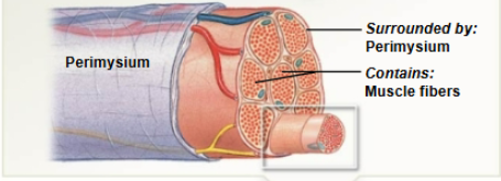
Perimysium
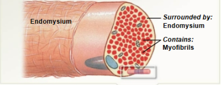
Endomysium
what does myo mean?
muscle
What does sarco mean?
flesh
plasma membrane of muscle cells
sarcolemma
Epimysium
Exterior collagen layer
connected to the deep fascia
Separates muscles from surrounding tissue
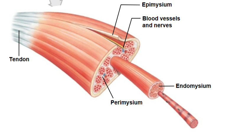
Perimysium
Surrounds muscle fiber bundles (fascicles)
Contains blood vessel and nerve supply to fascicles
Endomysium
Surrounds individual muscle cells (muscle fibers)
Contains capillaries and nerve fibers contacting muscle cells
Contains myosatellite cells (stem cells) that repair damage
Muscle attachments
Endomysium, Perimysium, and epimysium come together
-at ends of muscles
-Form connective tissue attachment to bone matrix
-i.e., tendon (bundle) or aponeurosis (sheet)
Put these in order:
Myofibrils
bundle of muscle cells/fibers
organ
Bundle of fascicles
organ
bundle of fascicles
bundle of muscle cells/fibers
myofibrils
The sarcolemma is associated with which one: Bundle of fascicles, bundle of muscle cells/fibers, or myofibrils?
Bundle of muscle cells/fibers
The Sarcoplasmic Reticulum (SR), the Terminal Cisternae (TC), and the T tubules are associated with which step one: Bundle of fascicles, bundle of muscle cells/fibers, or myofibrils?
Myofibrils
Blood vessels and nerves
Muscles have extensive vascular systems that
-Supply large amounts of oxygen
-Supply nutrients
-carry away wastes
Skeletal muscles are voluntary muscles, controlled by nerves of the central nervous system (brain and spinal cord)
Action potential
electrical current
Neurotransmitter
Chemical messenger
-Acetylcholine: Neurotransmitter that controls muscles
Skeletal Muscle cells
very long
develop through fusion of mesodermal cells (myoblasts)
become very large
contain hundreds of nuclei
The sarcolemma
the cell membrane of a muscle fiber (cell)
Surrounds the sarcoplasm (cytoplasm if muscle fibers)
A change in transmembrane potential begins contractions
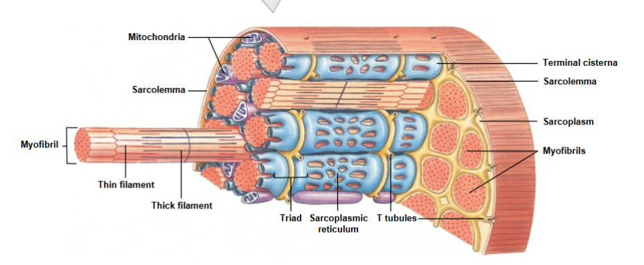
The Transverse tubules (T tubules)
transmit action potential through cells
allow entire muscle fiber to contract simultaneously
have same properties as sarcolemma

Myofilaments are associated with
Actin - Troponin/Tropomyosin
Myosin - Thick filament
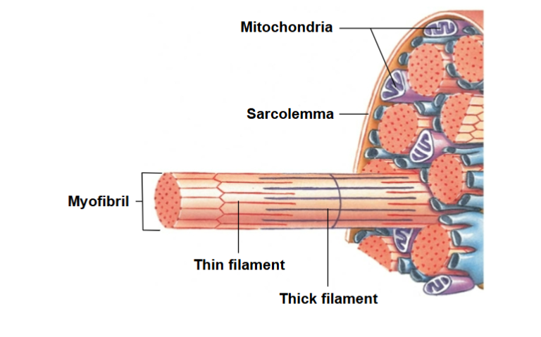
Myofibrils
lengthwise subdivisions within muscle fiber
made up of bundles of protein filaments (myofilaments)
Myofilaments are responsible for muscle contraction
Types of filaments
Thin Filaments - made of the protein actin
Thick filaments - made of the protein myosin
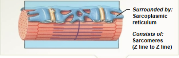
The Sarcoplasmic Reticulum (SR)
A membranous structure surrounding each myofibril
helps transmit action potential to myofibril
similar in structure to smooth endoplasmic reticulum
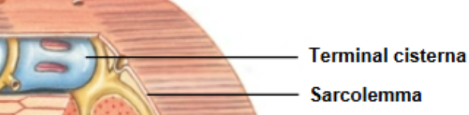
Forms chambers (terminal cisternae) attached to T tubules
The blue part of the image
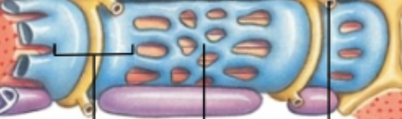
What does the terminal cisternae store?
Stores calcium
Triad
formed by one T-tubule and two terminal cisternae
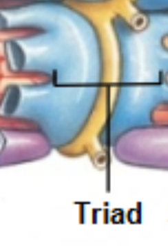
Terminal Cisternae
concentrate Ca2+ (via ion pumps)
Release Ca2+ into sarcomeres to begin muscle contraction
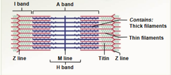
*Sarcomeres
* The contractile unit of muscle *
structural units of myofibrils
form visible patterns within myofibrils
A striped or striated pattern within myofibrils
-alternating dark, thick filaments (A bands) and light, thin filaments (I bands)
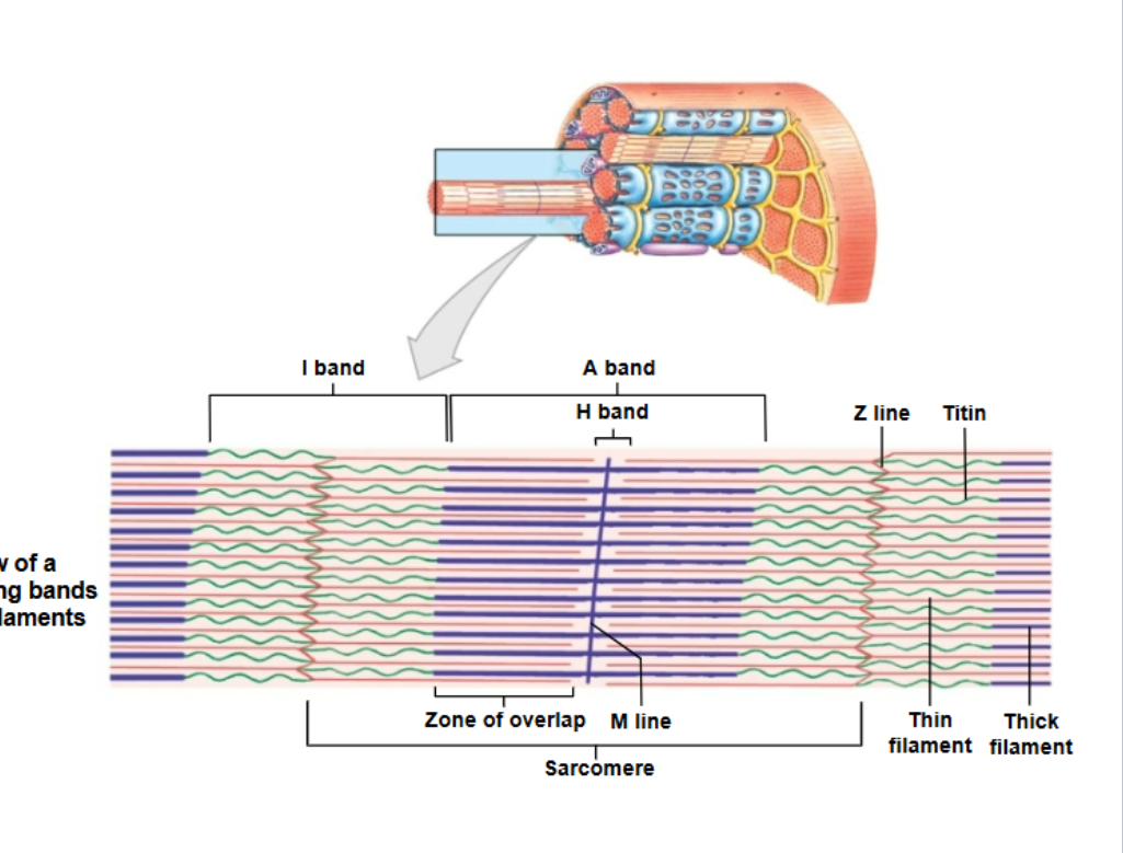
Sarcomeres: The A Band
Made up of the
M line:
-The center of the A band
-At midline of sarcomere
H Band:
-The area around the M line
-Has thick filaments but no thin filaments
Zone of overlap:
-The densest, darkest area on a light micrograph
-Where thick and thin filaments overlap
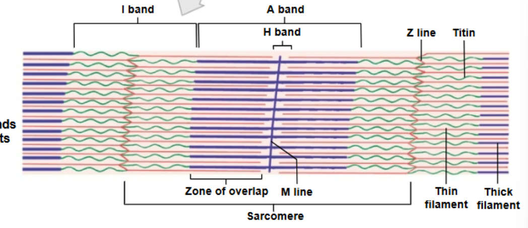
Sarcomeres: I Band
Made up of the
Z lines:
-The centers of the I bands
-At two ends of sarcomere
Titin:
-Are strands of protein
-Reach from tips of thick filaments to the Z line
-Stabilize the filaments

What does this image represent?
Skeletal muscle
What does this image represent?
Muscle Fascicle
What does this image represent?
Muscle Fiber
What does this image represent?
Myofibril
What does this image represent?
Sarcomere
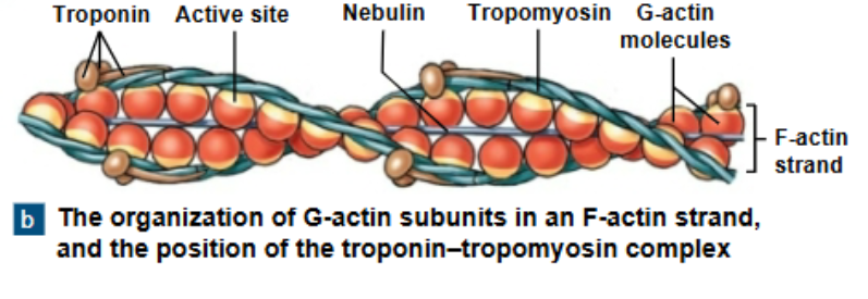
Thin filaments (F-Actin = filamentous actin)
-is two twisted rows of globular G-actin
-The active sites of G-actin strands bind to myosin

Thin filaments (Nebulin)
-Holds F-actin strands together

Thin Filaments (Tropomyosin)
Trop “rope” omyosin
-is a double strand
-prevents actin-myosin interaction

Thin filaments (Troponin)
-A globular protein
-binds tropomyosin to G-actin
-controlled by Ca2+
Initiating Contraction
Ca2+ binds to receptor or troponin molecule
Troponin - Tropomyosin complex changes
Exposes active site of F-Actin
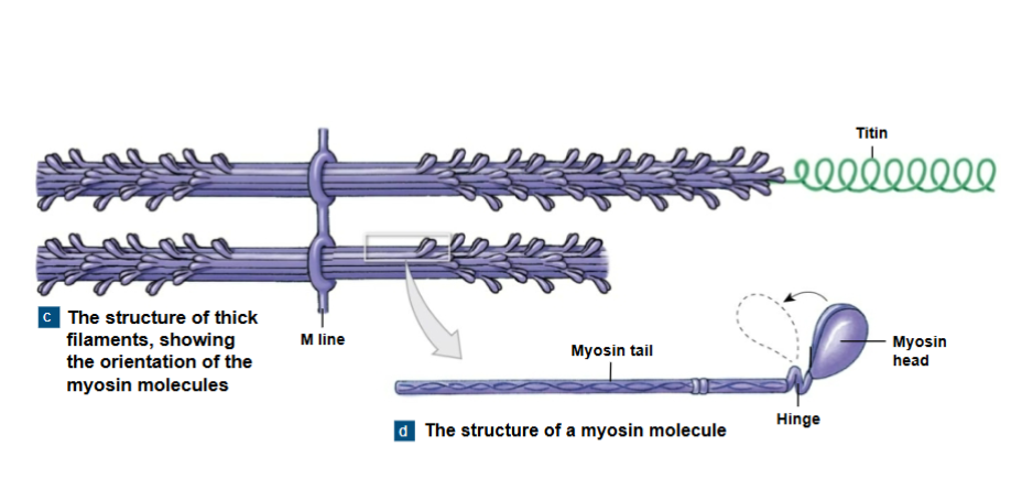
Thick filaments
The myosin molecule
The tail:
-binds to other myosin molecules
The head:
-made of two globular protein subunits
-reaches the nearest thin filament
Myosin Action
During contraction, myosin heads:
-interact with actin filaments, forming cross-bridges
-Pivot, producing motion
*Sliding filament theory
-thin filaments of sarcomere slide toward M line, alongside thick filaments
-the width of A zone stays the same
-Z lines move closer together
Skeletal muscle contraction
The process of contraction:
Neural stimulation of sarcolemma
-causes excitation-contraction coupling
Muscle fiber contraction
-interaction of thick and thin filaments
Tension production
The control of skeletal muscle activity
The Neuromuscular junction (NMJ)
-special intercellular connection between the nervous system and skeletal muscle fiber
-controls calcium ion release into the sarcoplasm
Excitation-Contraction coupling
Action potential reaches a triad
-releasing Ca2+
-Triggering contraction
Requires myosin heads to be in “cocked” position
-loaded by ATP enegry
The contraction cycle:
Contraction cycle begins
active-site exposure
cross-bridge formation
myosin head pivoting
cross-bridge detachment
myosin reactivation
Fiber shortening
As sarcomeres shorten, muscle pulls together, producing tension
Muscle shortening can occur at both ends of the muscle, or at only one end of the muscle
-This depends on the way the muscle is attached at the ends
Relaxation
Contraction duration
Depends on:
-Duration of neural stimulus
-number of free calcium ions in sarcoplasm
-availability of ATP
Ca2+ concentrations fall
Ca2+ detaches from troponin
Active sites are re-covered by tropomyosin
Rigor Mortis
A fixed muscular contraction after death
Caused when:
-Ion pumps cease to function; ran out of ATP
-Calcium builds up in the sarcoplasm
Tension production by muscle fibers
Tension Production by Muscle Fibers
As a whole, a muscle fiber is either contracted or relaxed
Depends on:
-The number of pivoting cross-bridges
-The fiber’s resting length at the time of stimulation
-The frequency of stimulation
A single muscle contraction
Twitch
Latent period (twitches)
The action potential moves through sareolemma
causes Ca2+ release
Contraction phase (twitches)
Calcium ions bind
tension builds to peak
Relaxation phase (twitches)
• Ca2+ levels fall
• active sites are covered and tension falls to resting levels

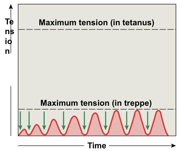
Treppe
• A stair-step increase in twitch tension
• repeated stimulations immediately after relaxation phase
-Stimulus frequency 50 /second
Causes a series of contractions with increasing tension
Increase in peak tension

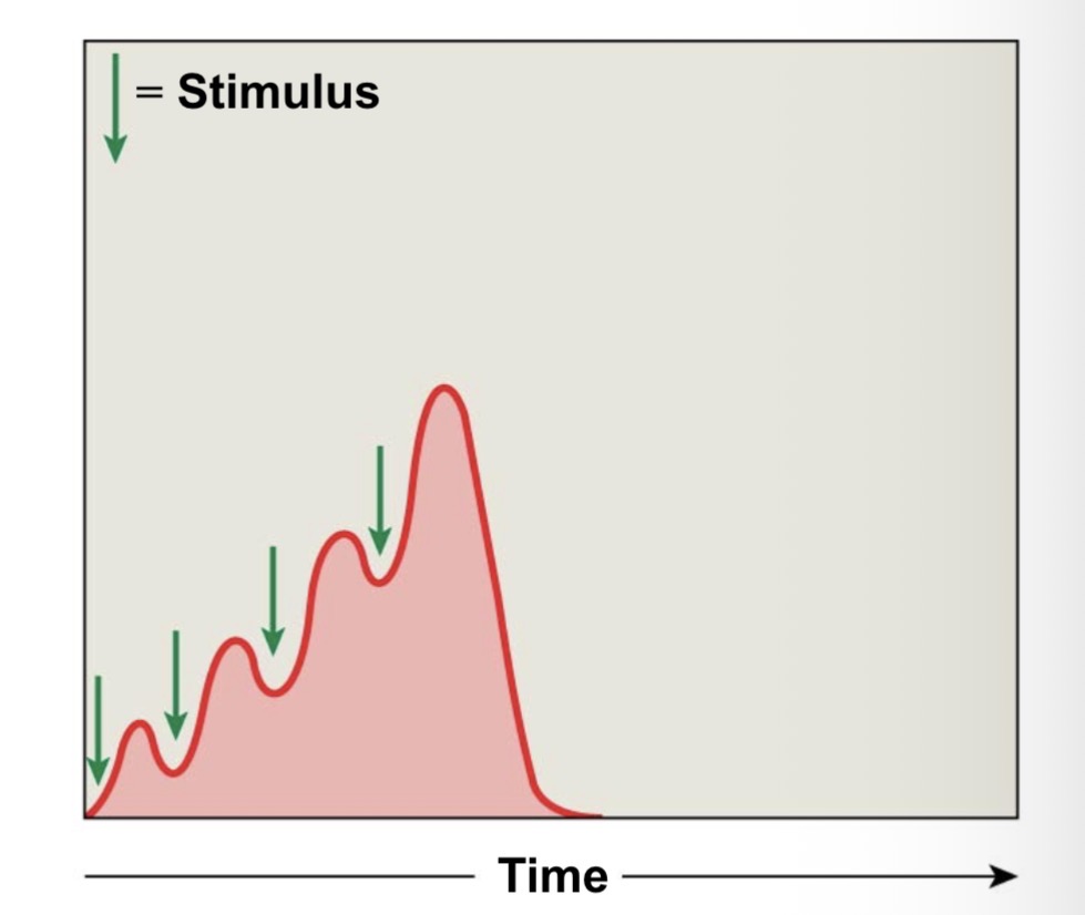
Wave summation
• increasing tension or summation of twitches
• repeated stimulations before the end of relaxation phase
-stimulus frequency 50/seconds
Causes increasing tension or summation of twitches
Occurs when successive stimuli arrive before the relaxation phase
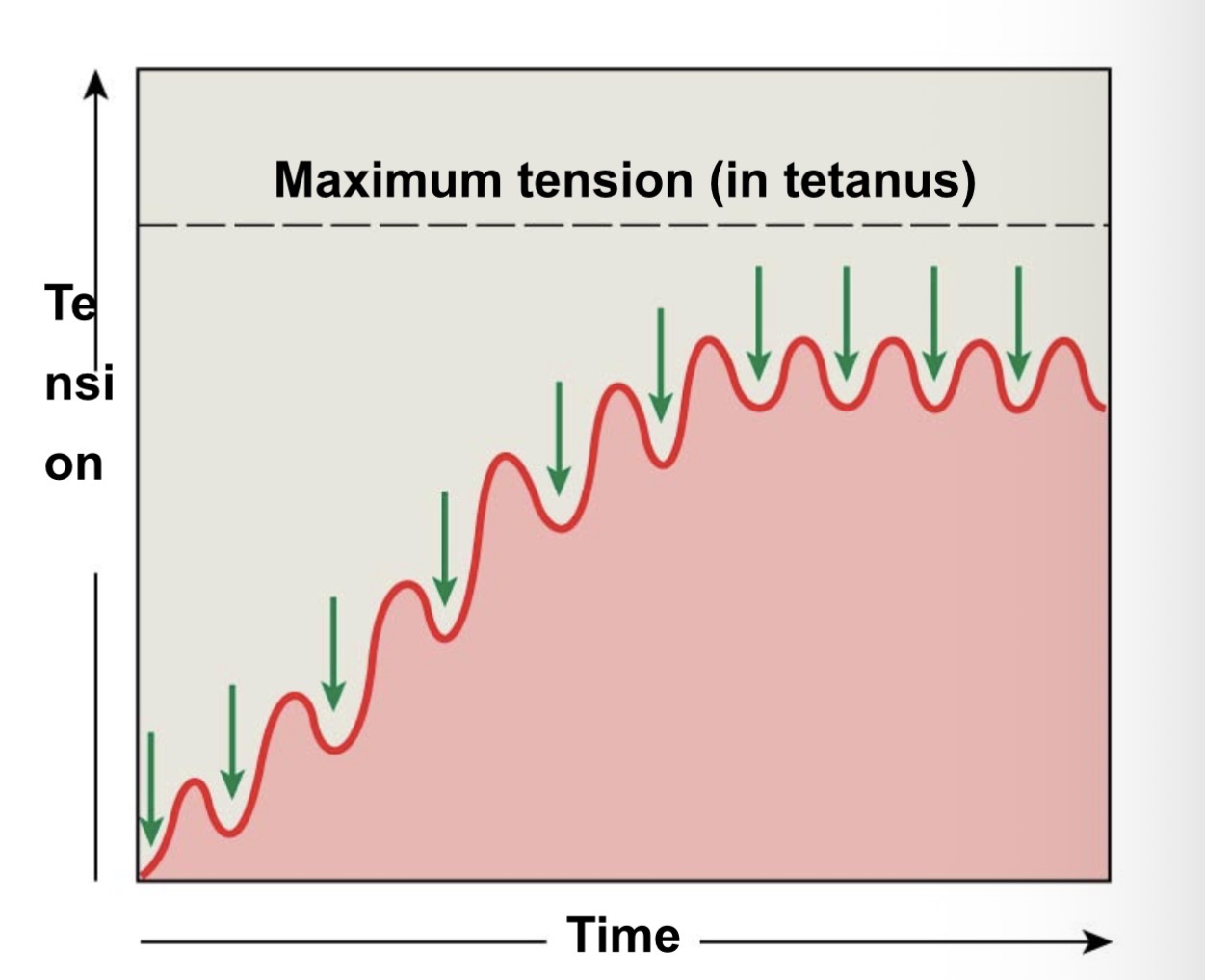
Incomplete tetanus
• Twitches reach maximum tension
• if rapid stimulation continues and muscle is not allowed to relax, twitches reach maximum level of tension.


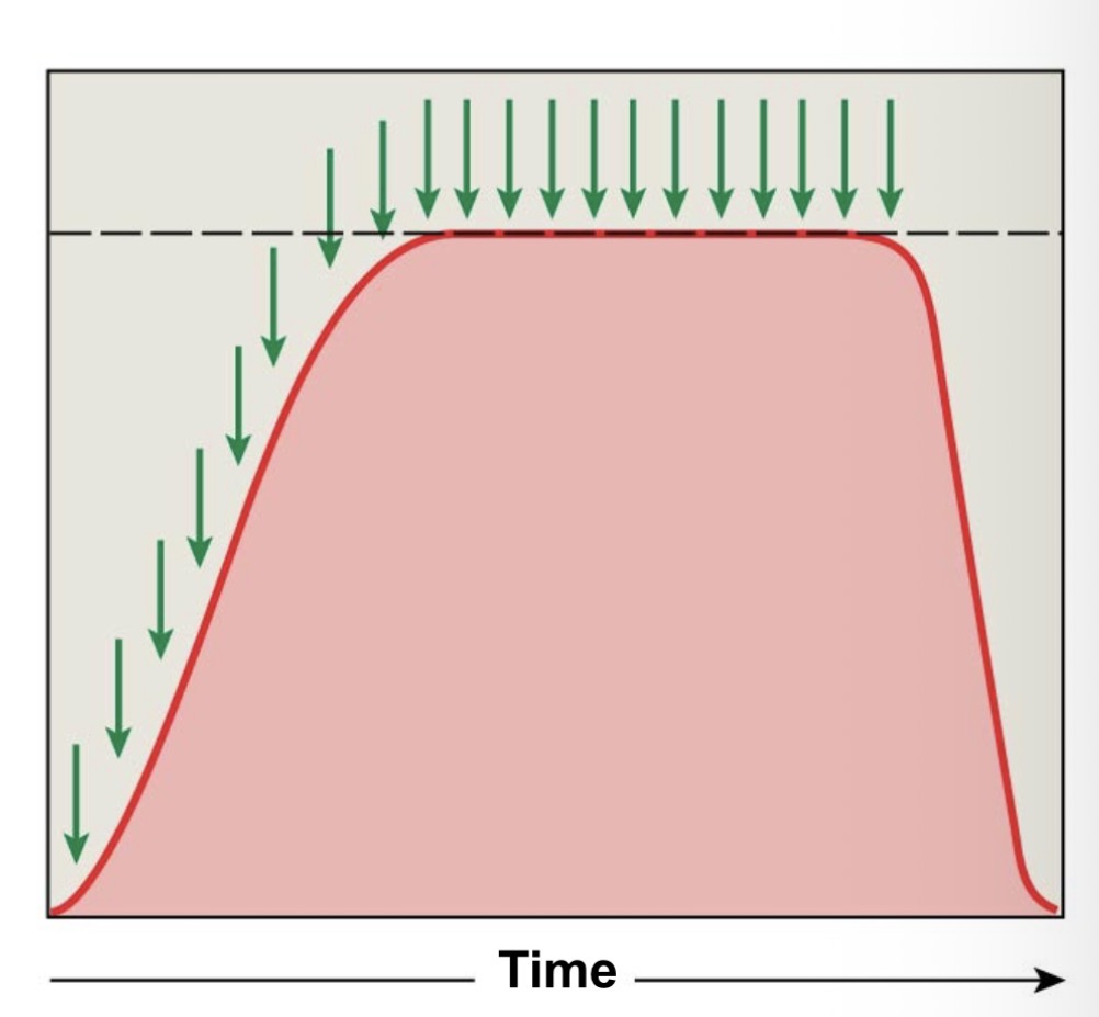
Complete tetanus
If stimulation frequency is high enough, muscle never begins to relax, and is in continuous contraction.
Tension production by skeletal Muscles (depends on)
• internal tension produced by muscle fibers
•External tension exerted by muscle fibers on elastic extracellular fibers
Total number of muscle fibers stimulated
Motor units in a skeletal muscle
Contains hundreds of muscle fibers
that contract at the same time
• controlled by a single motor neuron
Recruitment (multiple motor unit summation)
In a whole muscle or group of muscles, smooth motion and increasing tension are produced by slowly increasing the size or number of motor units stimulated
Maximum tension
• achieved when all motor units reach tetanus
Can be sustained only a very short time
Sustained tension
•Less than maximum tension
•Allows motor units to rest in rotation
Muscle tone
The normal tension and firmness of a muscle at rest
Muscle units actively maintain body position, without motion
Increasing muscle tone increases metabolic energy used, even at rest

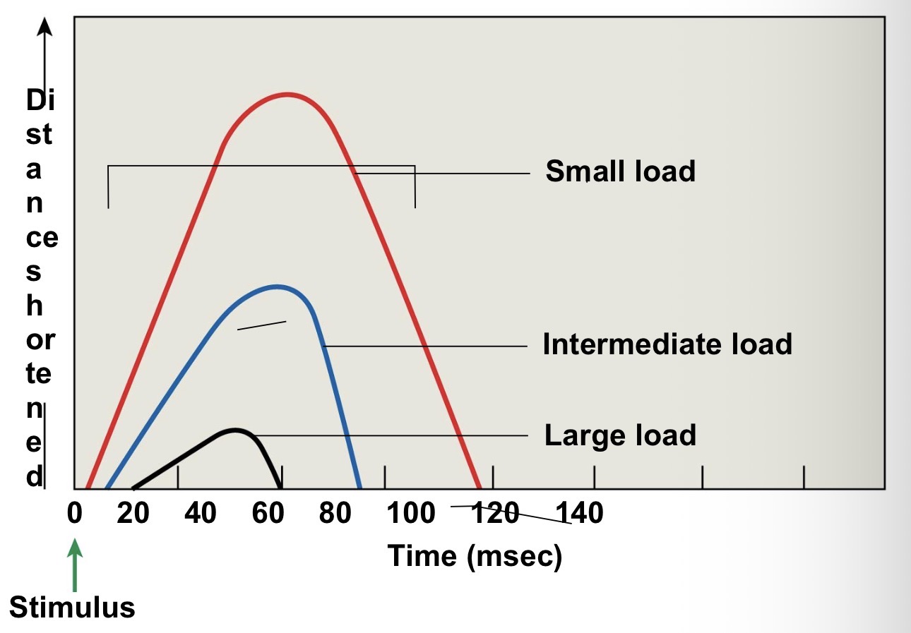
Load and speed of contraction
• Are inversely related
• the heavier the load (resistance) on a muscle:
-the longer it takes for shortening to begin
-And the less the muscle will shorten
Muscle relaxation and the return to resting length (elastic forces)
• the pull of elastic elements (tendons and ligaments)
Expands the sarcomeres to resting length
Muscle relaxation and the return to resting length (opposing muscle contractions)
• Reverse the direction of the original motion
•Are the work of opposing skeletal muscle pairs
Muscle relaxation and the return to resting length (gravity)
Can take the place of opposing muscle contraction to return a muscle to its resting state