Anatomy Quiz Four
1/75
There's no tags or description
Looks like no tags are added yet.
Name | Mastery | Learn | Test | Matching | Spaced |
|---|
No study sessions yet.
76 Terms
Adult brains have four regions:
Cerebral hemispheres
Diencephalon
Brain stem, consisting of:
Midbrain
Pons
Medulla
Cerebellum
Brain stem, consisting of :
Midbrain
Pons
Medulla
Brain stem has additional gray matter ____ scattered within _____ ______.
Brain stem has additional gray matter nuclei scattered within white matter.
Cerebral hemispheres (cerebrum) and cerebellum contain outer layer of ____ _____ called the _____.
Cerebral hemispheres (cerebrum) and cerebellum contain outer layer of gray matter called the cortex.
_______ and ________ also have scattered areas of gray matter nuclei amid white matter.
Cerebrum and cerebellum also have scattered areas of gray matter nuclei amid white matter.
Ventricles
Fluid-filled chambers that are continuous to one another and to central canal of spinal cord
Filled with cerebrospinal fluid (CSF)
Lined by ependymal cells (neuroglial cells)
Cerebral hemispheres
Form superior part of brain
Account for 83% of brain mass
Cerebral hemispheres
Surface markings:
Gyri: ridges
Sulci: shallow grooves
Fissures: deep grooves
Longitudinal fissure
Separates two hemispheres
Transverse cerebral fissure
Separates cerebrum and cerebellum
Several sulci divide each hemisphere into five lobes
Frontal
Parietal
Temporal
Occipital
Insula
First four are named after overlying skull bones
Insular lobe is buried under portions of temporal, parietal, and frontal lobes
Cerebral cortex
is “executive suite” of brain
the outer layer of the brain, made up of gray matter (neurons and their connections) that covers the cerebrum
Site of conscious mind : awareness, sensory perception, voluntary motor initiation, communication, memory storage, understanding
Thin (2–4 mm) superficial layer of gray matter
Composed of neuron cell bodies, dendrites, glial cells, and blood vessels, but no axons
Functional imaging (PET and MRI) of brain show specific motor and sensory functions are located in discrete cortical areas called domains
Site of conscious mind :
Awareness, sensory perception, voluntary motor initiation, communication, memory storage, understanding
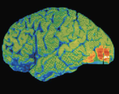
Seeing
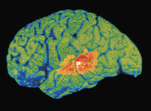
Hearing
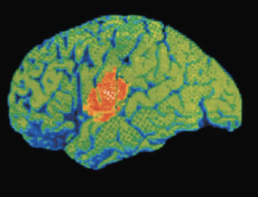
Speaking
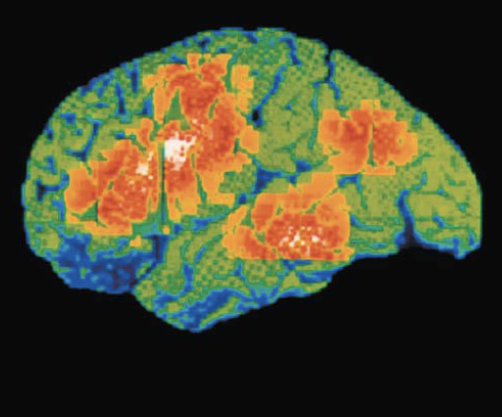
Thinking
Four general considerations of cerebral cortex:
Contains three types of functional areas:
Motor areas: control voluntary movement
Sensory areas: conscious awareness of sensation
Association areas: integrate diverse information
Each hemisphere is concerned with contralateral (opposite) side of body
Lateralization (specialization) of cortical function can occur in only one hemisphere
Conscious behavior involves entire cortex in one way or another
Cerebral Cortex
Motor areas
Located in frontal lobe, motor areas act to control voluntary movement
Cerebral Cortex
Primary (somatic) motor cortex
Located in precentral gyrus of frontal lobe
Pyramidal cells: large neurons that allow conscious control of precise, skilled, skeletal muscle movements
Pyramidal (corticospinal) tracts: formed from long axons that project down spinal cord
Somatotopy: all muscles of body can be mapped to area on primary motor cortex
Multimodal association areas
Receive inputs from multiple sensory areas
Send outputs to multiple areas
Allows us to give meaning to information received, store in memory, tie to previous experience, and decide on actions
Sensations, thoughts, emotions become conscious: makes us who we are
Broadly divided into three parts: anterior association area, posterior association area,and limbic association area
Cerebral Cortex
Lateralization of cortical functioning
Lateralization: division of labor between hemispheres
Cerebral dominance: refers to hemisphere that is dominant for language
90% of humans have left-sided dominance
Usually results in right-handedness
In other 10%, roles of hemispheres are reversed
Lateralization of cortical functioning (cont.)
Left and Right Hemisphere
Left hemisphere
Controls language, math, and logic
Right hemisphere
Visual-spatial skills, intuition, emotion, and artistic and musical skills
Hemispheres communicate almost instantaneously via fiber tracts and functional integration
Cerebral White Matter
Second of the three basic regions of cerebral hemispheres
Responsible for communication between cerebral areas, and between cortex and lower CNS
Consists of myelinated fibers bundled into large tracts
Classified according to direction they run: Association, commissural, and projection fibers
Basal Nuclei (Ganglia)
Third of the three basic regions of cerebrum
Each hemisphere’s basal nuclei include a:
Caudate nucleus
Putamen
Caudate nucleus + putamen = striatum
Globus pallidus
Functions of basal nuclei(Ganglia) are thought to:
Influence muscle movements
Play role in cognition and emotion
Regulate intensity of slow or stereotyped movements
Filter out incorrect/inappropriate responses
Inhibit antagonistic/unnecessary movements
Parkinson’s disease and Huntington’s disease are disorders of the basal nuclei
The Diencephalon
Consists of three paired gray-matter structures:
Thalamus
Hypothalamus
Epithalamus
Thalamus
Bilateral egg-shaped nuclei that form superolateral walls of third ventricle
Nuclei project and receive fibers from cerebral cortex
Thalamus
Main thalamic function
Is to act as relay station for information coming into cortex
Sorts, edits, and relays ascending input such as:
Impulses from hypothalamus for regulating emotion and visceral function
Impulses from cerebellum and basal nuclei to help direct motor cortices
Impulses for memory or sensory integration
Overall, it acts to mediate sensation, motor activities, cortical arousal, learning, and memory
Hypothalamus
Located below thalamus
Forms cap over brain stem
Contains many important nuclei
Hypothalamus
Functions
The hypothalamus is the main visceral control and regulating center that is vital to homeostasis
Chief homeostasis controls:
Controls autonomic nervous system
Examples: blood pressure, rate and force of heartbeat, digestive tract motility, pupil size
Initiates physical responses to emotions
The hypothalamus also:
Regulates body temperature: sweating or shivering
Regulates hunger and satiety in response to nutrient blood levels or hormones
Regulates water balance and thirst
Regulates sleep-wake cycles
Controls endocrine system functions such as:
Secretions of anterior pituitary gland
Production of posterior pituitary hormones
Epithalamus
Most dorsal portion of diencephalon
Contains pineal gland (body) - Secretes melatonin that helps regulate sleep-wake cycle
Brain Stem
Consists of three regions: midbrain, pons, medulla oblongata
Controls automatic behaviors necessary for survival
Contains fiber tracts connecting higher and lower neural centers
Medulla Oblongata
Respiratory centers
Generate respiratory rhythm
Control rate and depth of breathing
Various other centers regulate:
Vomiting
Hiccupping
Swallowing
Coughing
Sneezing
Cerebellum
Processes input from cortex, brain stem, and sensory receptors to provide precise, coordinated movements of skeletal muscles
Also plays a major role in balance
Cerebellar Processing
Cerebellum fine-tunes motor activity as follows:
1. Receives impulses from cerebral cortex of intent to initiate voluntary muscle contraction
2. Receives signals from proprioceptors throughout body, as well as visual and equilibrium pathways that:
Pathways continuously “inform” cerebellum of body’s position and momentum
Cerebellar cortex calculates the best way to smoothly coordinate muscle contraction
Sends “blueprint” of coordinated movement to cerebral motor cortex and brain stem nuclei
Brain Wave Patterns
Brain waves reflect electrical activity of higher mental functions
Normal brain functions are continuous and hard to measure
Electroencephalogram (EEG)
Electroencephalogram (EEG) records electrical activity that accompanies brain function
Electrodes placed on scalp measure electrical potential differences between various cortical areas EEG measures patterns of neuronal electrical activity generated by synaptic activity in cortex
Each person's brain waves are unique
Measures wave frequency in Hertz (Hz), numbers of peaks per second (1 Hz = 1 peak per second)
Can be grouped into four classes based on Hz:
Alpha, beta, theta,or delta waves
Alpha waves:
(8–13 Hz)—regular and rhythmic, low-amplitude, synchronous waves indicating an “idling” brain
Beta waves:
(14–30 Hz)—rhythmic, less regular waves occurring when mentally alert
Theta waves:
(4–7 Hz)—more irregular; common in children and uncommon in awake adults
Delta waves:
(4 Hz or less)—high-amplitude waves of deep sleep and when reticular activating system is suppressed, as during anesthesia; indicates brain damage in awake adult
Consciousness
Consciousness involves:
Perception of sensation
Voluntary initiation and control of movement
Capabilities associated with higher mental processing (memory, logic, judgment, etc.)
Classification by Stimulus Type
Mechanoreceptors
Respond to touch, pressure, vibration, and stretch
Classification by Stimulus Type
Thermoreceptors
Sensitive to changes in temperature
Classification by Stimulus Type
Photoreceptors
Respond to light energy (example: retina)
Classification by Stimulus Type
Chemoreceptors
Respond to chemicals (examples: smell, taste, changes in blood chemistry)
Classification by Stimulus Type
Nociceptors
Sensitive to pain-causing stimuli (examples: extreme heat or cold, excessive pressure, inflammatory chemicals)
Classification by Location
Exteroceptors
Respond to stimuli arising outside body
Receptors in skin for touch, pressure, pain, and temperature
Classification by Location
Interoceptors
Respond to stimuli arising in internal viscera and blood vessels
Sensitive to chemical changes, tissue stretch, and temperature changes
Sometimes cause discomfort but usually person is unaware of their workings
Classification by Location
Proprioceptors
Respond to stretch in skeletal muscles, tendons, joints, ligaments, and connective tissue coverings of bones and muscles
Inform brain of one's movements
Classification by Receptor Structure
Simple receptors of the general senses
General senses include tactile sensations (touch, pressure, stretch, vibration), temperature, pain, and muscle sense
No “one-receptor-one-function” relationship
Receptors can respond to multiple stimuli
Receptors have either:
Nonencapsulated (free) nerve endings or
Encapsulated nerve endings
Thermoreceptors
Cold receptors are activated by temps from 10 to 40°C
Located in superficial dermis
Heat receptors are activated from 32 to 48°C located in in deeper dermis
Outside those temperature ranges, nociceptors are activated and interpreted as pain
Nociceptors
Pain receptors triggered by extreme temperature changes, pinch, or release of chemicals from damaged tissue
Muscle spindles
Spindle-shaped proprioceptors that respond to muscle stretch
Tendon organ
Proprioceptors located in tendons that detect Stress
Joint kinesthetic receptors
Proprioceptors that monitor joint position and motion
Endoneurium
Loose connective tissue that encloses axons and their myelin sheaths (Schwann cells)
Perineurium
Coarse connective tissue that bundles fibers into fascicles
Epineurium
Tough fibrous sheath around all fascicles to form the nerve
Most nerves are mixtures of ____ and ____ fibers and ____ and ____ (visceral) fibers
Most nerves are mixtures of afferent and efferent fibers and somatic and autonomic (visceral) fibers
Nerves are classified according to the ______ they transmit impulses
Nerves are classified according to the direction they transmit impulses
Mixed nerves
Contain both sensory and motor fibers
Impulses travel both to and from CNS
Sensory (afferent) nerves:
Impulses only toward CNS
Motor (efferent) nerves
Impulses only away from CNS
Pure _____ (afferent) or ____ ____ (efferent) nerves are rare; most nerves are _____
Pure sensory (afferent) or pure motor (efferent) nerves are rare; most nerves are mixed
Types of fibers in mixed nerves
Somatic afferent (sensory from muscle to brain)
Somatic efferent (motor from brain to muscle)
Visceral afferent (sensory from organs to brain)
Visceral efferent (motor from brain to organs)
Ganglia
Contain neuron cell bodies associated with nerves in PNS
Vagus nerves
Only cranial nerves that extend beyond head and neck region
Most motor fibers are parasympathetic fibers that help regulate activities of heart, lungs, and abdominal viscera
Sensory fibers carry impulses from thoracic and abdominal viscera, baroreceptors, chemoreceptors, and taste buds of posterior tongue and pharynx
Spinal nerves
31 pairs of spinal nerves
All are mixed nerves named for point of issue from spinal cord
Supply all body parts except head and part of neck
8 pairs of cervical nerves (C1–C8)
12 pairs of thoracic nerves (T1–T12)
5 pairs of lumbar nerves (L1–L5)
5 pairs of sacral nerves (S1–S5)
1 pair of tiny coccygeal nerves (C0)
Sacral plexus
Arises from L4 to S4
Serves the buttock, lower limb, pelvic structures, and perineum
Sciatic nerve
Longest and thickest nerve of body
Innervates hamstring muscles, adductor magnus, and most muscles in leg and foot
Composed of two nerves: tibial and common fibular
Innervation of Visceral Muscle and Glands
Autonomic motor endings and visceral effectors are simpler than somatic junctions
Branches form synapses en passant (“synapses in passing”) with effector cells via varicosities
Acetylcholine and norepinephrine act indirectly via second messengers
Visceral motor responses are slower than somatic responses
Stretch reflex
Brain sets muscle’s length via stretch reflex
Example: knee-jerk reflex is a stretch reflex that keeps knees from buckling when you stand upright
Stretch reflexes maintain muscle tone in large postural muscles and adjust it reflexively
How stretch reflex works
Stretch activates muscle spindle
Sensory neurons synapse directly with α motor neurons in spinal cord
A motor neurons cause extrafusal muscles of
stretched muscle to contract
Functions of Nervous System