Identifying and describing lesions
1/21
There's no tags or description
Looks like no tags are added yet.
Name | Mastery | Learn | Test | Matching | Spaced |
|---|
No study sessions yet.
22 Terms
Steps for identifying and describing lesions
Patient interview
Examination
extraoral and intraoral
Documentation
record if soft/hard tissue is normal or not
Images
What are the steps of the patient interview
Reason for attendance
History of present condition
SOCRATES
Site, Onset, Character, Radiating, Associations, Time, Exacerbating, Severity
Medical history + Medication
Dental history
Oral Hygiene
Diet
Habits (smoking, drinking, drugs)
Social history
Steps of clinical examination
Extraoral
Intraoral
Special (diagnositic) tests
Steps of extraoral exam
general considerations
head and neck
lymph nodes
TMJ
muscles of mastication
mouth opening
Steps of intraoral exam
soft and hard tissues, lips, buccal mucosa, sulcus, hard/soft palate, tongue, floor of mouth, lingual mucosa
saliva
occlusion
periodontium
dentition
Frequent sites of oral cancer
commissure (corners of mouth)
retromolar pads
tonsillar pillars
lateral and ventral tongue
floor of mouth
5 main descriptions of soft tissue pathologies
Site
Morphology
Colour
Size
Consistency
Elevated lesion
Blisterforms and Non-blisterforms
Blisterforms
contain fluid, translucent in appearance, soft
<5mm in diameter = Vesicle
>5mm in diameter = Bulla
Pustules = any size, yellow in colour, contain pus
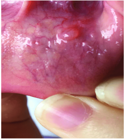
What lesion is this?
Vesicle
What lesion is this?
Bulla
What lesion is this?
Pustule
Non-blisterforms
solid with no fluid, opaque, firm
Papule = <5mm diameter
Nodule = >5mm but <20mm diameter
Tumor = >20mm diameter
Plaque = very slightly elevated, and usually >5mm in diameter
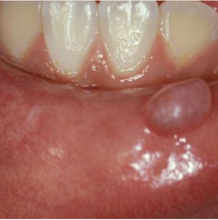
What lesion is this?
Mix of papules of nodules
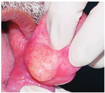
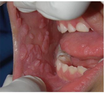
What lesion is this?
Tumor
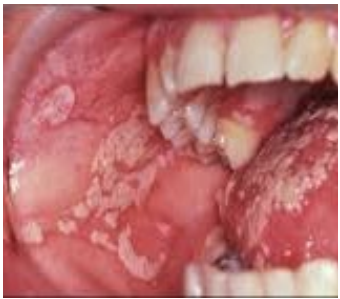
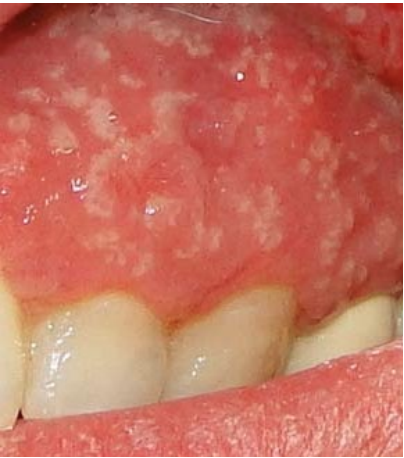
What lesion is this?
Plaque
Types of depressed lesions
ulcer
atrophy and scarring
pits
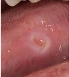
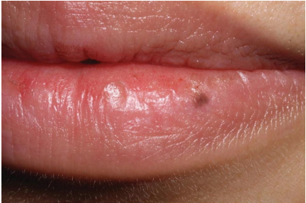
What lesion is this?
Ulcer
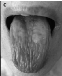
What lesion is this?
atrophy and scarring
Types of flat lesions
Macule (<10mm)
Patch (>10mm)
What lesion is this?
Macule
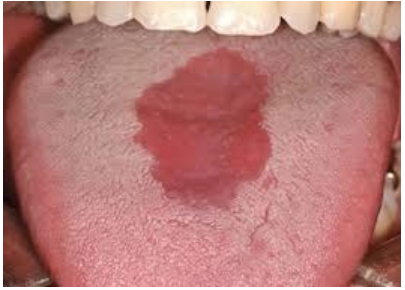
What lesion is this?
Patch