Lecture 2: Environmental Influences on The Endocrine System
1/37
There's no tags or description
Looks like no tags are added yet.
Name | Mastery | Learn | Test | Matching | Spaced | Call with Kai |
|---|
No analytics yet
Send a link to your students to track their progress
38 Terms
Factors Affecting the Endocrine System
The system responds rapidly to external changes in the environment e.g.
Light/ dark e.g. day length
Stress – e.g. predators – fight or flight
Temperature
Food supply
Factors have a direct influence
Pituitary Stalk
Connect the Pituitary Gland to The Hypothalamus
Anatomical link between the nervous system and endocrine system
Central regulatory component of the endocrine system
The endocrine gland (pituitary) is below the hypothalamus – key part of CNS
aka supraoptic-hypothalamic tract
Inputs to Hypothalamus
Hypothalamus receives information about changes in the environment e.g. cold, stress, puberty, dehydration, exercise, breastfeeding growth etc through neural inputs from other brain areas, including:
Limbic system; Brain stem; Reticular formation; Thalamus; Subthalamus; Basal ganglial; Retina; Neocortex (possibly)
These brain areas sense changes and signal the hypothalamus to integrate the information and produce a response via the pituitary gland.
Pituitary Gland
Master endocrine gland that takes (electrical) signals from neuronal/ chemical input to elicit a response
May influence other glands (indirect) or have a direct impact on physiological processes
Multiple functions - mediated by trophic effects
Consist of 2 regions
Anterior PG
Posterior PG
Anterior and Posterior Pituitary Gland
2 distinct regions of the Pituitary
Respond to, and produce, different hormonal products – ‘separate glands that share blood supply’
have no direct functional interaction with each other
Hypothalamus Pituitary Axis
Effect of Low Temperature (Hypothalamus-Pituitary Axis)
Stimuli detected by hypothalamus
Hypothalamus releases thyrotropin-releasing hormone (TRH)
TRH is sent to the anterior pituitary gland to stimulate the secretion of Thyroid-stimulating Hormone (TSH)
Trophic hormone influences the Thyroid Gland to produce Thyroid hormones
Results in
↑ metabolism
Heat production – body adapts to low temp
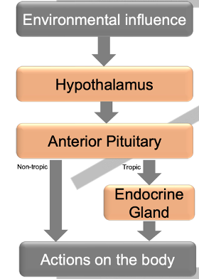
Trophic Effect
One gland i.e. anterior pituitary, has an effect on another endocrine gland which then acts on the body
Non-Trophic Effects
Anterior pituitary gland has direct action on the body
Posterior Pituitary Gland
Linked to the hypothalamus by long magnocellular neurons
Tissue present in mainly neuronal - single nerves that extend into the gland
‘like an extension of the brain’
Only stores and secretes oxytocin and ADH -
Involved in their secretion directly from supraoptic and paraventricular nuclei of the hypothalamus
Magnocellular Neurons
Cell bodies located in the 3rd ventricle and extend down the pituitary stalk and axons
Directly link axons to blood vessels in the PPG
Contained within the Supraoptic and paraventricular nuclei - Extend down into the gland
Releases oxytocin and ADH
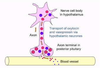
Synthesis and Transport of PPG Hormones
Hormones are produced by cell bodies within the hypothalamus;
Transported down long axons are stored in nerve endings in storage granules before release
Nuclei
A collection of neurons with a similar function close together
General Principle of Hypothalamic Control of PPG
Nerve cell bodies in the hypothalamus produce a hormone packaged into a granule that is transported to the nerve endings and is stored until released into the blood vessel
Form of neuroendocrine hormone control with oxytocin and vasopressin (ADH)
Secretion of Oxytocin and Vasopressin From PPG
Following stimulation hormone is released in blood vessels to be distributed around the body
Dysregulation of ADH or Oxytocin
Leads to diabetes insipidus or inapproprate secretion
Anterior Pitutiary Gland
Has many endocrine cell types and different releasing hormones from the hypothalamus will be released into the blood supply and will interact with cells to cause the release of other hormones
binds to receptors on specific cells
Once released, hormones are transported through the body through the main blood supply to target cells
has short parvocellular neurons connecting between the hypolathalamus and the gland
Hypothalamic Communication With APG
Gland communicates with the lateral wall of 3rd ventricle of the Hypothalamus
Small parvocellular neurons pass to median eminence
APG receives releasing hormone from the hypothalamus via the median eminence capillary network
No direct nervous connection can be demonstrated
Median Eminence
Contains the primary portal system of capillaries;
Parvocellular neurons linked to this capillary network are located above APG
Parvocellular Neurons
Receives and integrates signals from other brain regions
Site of releasing hormone synthesis
Release releasing hormones into the capillaries of the median eminence in the primary plexus to influence secretion from APG
Decides whether to secrete or inhibit the release of releasing hormones and then transmits to APG in response
Hypothalamic Portal Blood System
Portal capillary system of the human hypothalamus and anterior pituitary.
Neurohormones are released from the axon termini of the hypothalamic neurons into the primary plexus.
They are then transported through the vessels to the secondary plexus from which they move through fenestrations in the capillary walls to interact with specific receptors in target cells of APG.
Signal Amplification in Hormone Axis
Mediated by the production/ release of tropic and non-tropic hormones from APG e.g.
e.g. ng level of hormones from hypothalamus required to influence APG
peripheral hormone – short half-life- reaches target cells quickly
in APG larger levels (ug) are needed to travel and act on the endocrine gland
mg levels of hormones released by endocrine gland – travels distance in circulation to target; longer half-life
Feedback Loops
Essential feature of hormone-gland axis amplification regulation
Can be short or long loops
Peripheral hormones from the endocrine gland will have negative feedback on hormones produced by APG - stop production
Additional Role of Hypothalamic Releasing Hormones e.g. Thyrotropin Releasing Hormone
Regulates other systems
In hypothalamus - TSH release
Other brain regions – regulate appetite and mood
Orexins (Orexin A & B)
Aka hypocretins
Neuropeptide hormones
Released from cells in lateral and posterior hypothalamus
Promote wakefulness and eating
Secretion inhibited by glucose and leptin
Narcolepsy
Methods of Clinical and Experimental Demonstrations of Hypothalamic Pituitary Axis
In-vitro experiments
Historic in-vivo experiments
Case studies
Lesion Studies
Damaging specific areas of the hypothalamus or posterior pituitary can help identify their roles in hormone regulation
Electrophysiology
Recording electrical activity in the hypothalamus and posterior pituitary reveals neural signals involved in hormone release
Theory for In Vitro Demonstrations of HPA
Chemical messengers (i.e. “releasing hormones”), synthesised within the hypothalamus, stimulate the release of hormones from the pituitary gland into the general circulation
Tested by the measurement of the pituitary response to hypothalamic extracts in an in vitro pituitary system
Testing of In Vitro HPA
ACTH release from isolated anterior pituitary cells
Test tube with isolated APG cells releasing hormones e.g. ACTH
In test tube – receive no input/ stimulus from the environment; overtime amount of ACTH released decreases
Hypothalamic tissue added after day 5 and secretes factors that lead to increased ACTH release
Semi-permeable membrane can be added to show the communication is chemical, not physical
In Vitro HPA: Conclusions
Hypothalamic cells produce and release a soluble factor that can stimulate the release of ACTH from pituitary cells
Shows a chemical communication between hypothalamic and APG cells
Actions of Adrenocorticotropic Hormone (ACTH)
Released in response to CRH from the hypothalamus acting on APG corticotrope cells
Causes the release of glucocorticoids from the adrenal cortex
Negative feedback - cortisol feedbacks to the APG to inhibit corticotropes and ACTH
Blair Bell - Experimental Observations on The Pitutary
Brain surgery carried out in dogs to physically clamp infundibular stalk, located between the hypothalamus and pituitary,
Tied it off and physically prevented communication between the hypothalamus and APG, blocking secretions
Separation of the Infundibular Stalk Between Hypothalamus and Pituitary
Blood supply from Hyp to PG was therefore reduced or prevented – neural and chemical communication was prevented
Resulted in
Weight gain
Dystrophia adiposo-genitals
Genital and mammary atrophy
Anterior pituitary atrophy
Consequence of Infindubular Stalk Clamping on PRL, FSH and LH Production
Hypothalamus will secrete GnRH, stimulating the production of Prolactin, FSH and LH
Clamping blocks GnRH communication with APG
Interrupts FSH and LH production – no GnRH released
No inhibition of prolactin – excess secretion, producing morphological effects
Clinical Case Study of 16yr Old Craniopharyngioma Compressing PG and Hypothalamus
Tumour in bony tissue around PG - Prevent hypothalamus and PG communication
Resulted in:
Obesity
Failure to enter puberty
Small testes
Headaches
Visual abnormalities - tumor compresses optic nerve
Froelich’s disease (adiposogenital dystrophy) - secondary to tumour
visual disturbances, delayed puberty and breast development
Treatment via tumour removal
Mediation and Modulation of Actions Between Hypothalamus and Neuonons
Diverse chemical messengers within the brain mediate inputs between neurons and the hypothalamus, and may also modulate the actions of each other.
Brain adapts to the environment through neural connections with other neurons in the hypothalamus
Neurons in the hypothalamus then influence the PG to secrete or inhibit hormones which can go onto to have direct or indirect actions on the body through other endocrine glands
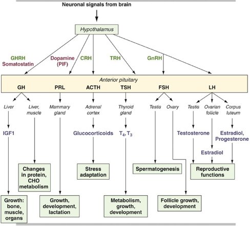
APG Secretions
Secretes a range of hormones in response to “releasing hormone” signals from the hypothalamus.
Hormones released:
ACTH
TSH
FSH
LH
GH
PRL