Lesson 25 - Forelimb radiography LA
1/133
There's no tags or description
Looks like no tags are added yet.
Name | Mastery | Learn | Test | Matching | Spaced |
|---|
No study sessions yet.
134 Terms
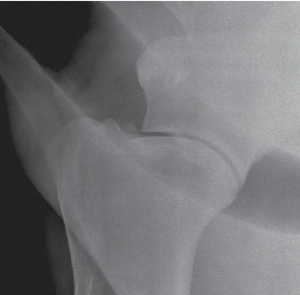
What view is this?
Medio-lateral view of shoulder
How to perform a lateral shoulder view radiograph
Distal limb is pulled cranially with detector at lateral aspect of joint in vertical position
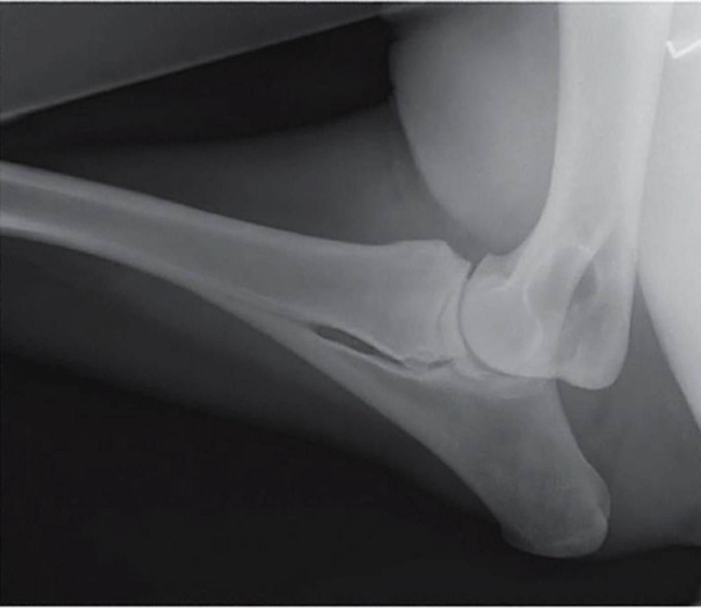
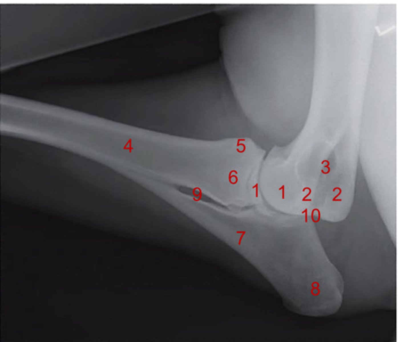
What view is this?
Medio-lateral view of elbow
What are the views for the elbow?
Lateral and cranio-caudal view
What are the views for the shoulder
Medio-lateral view
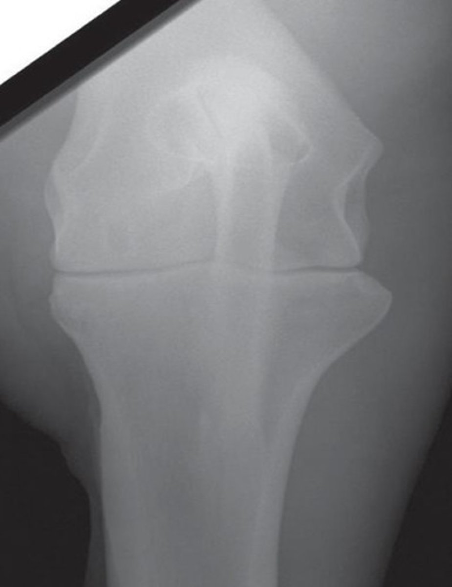
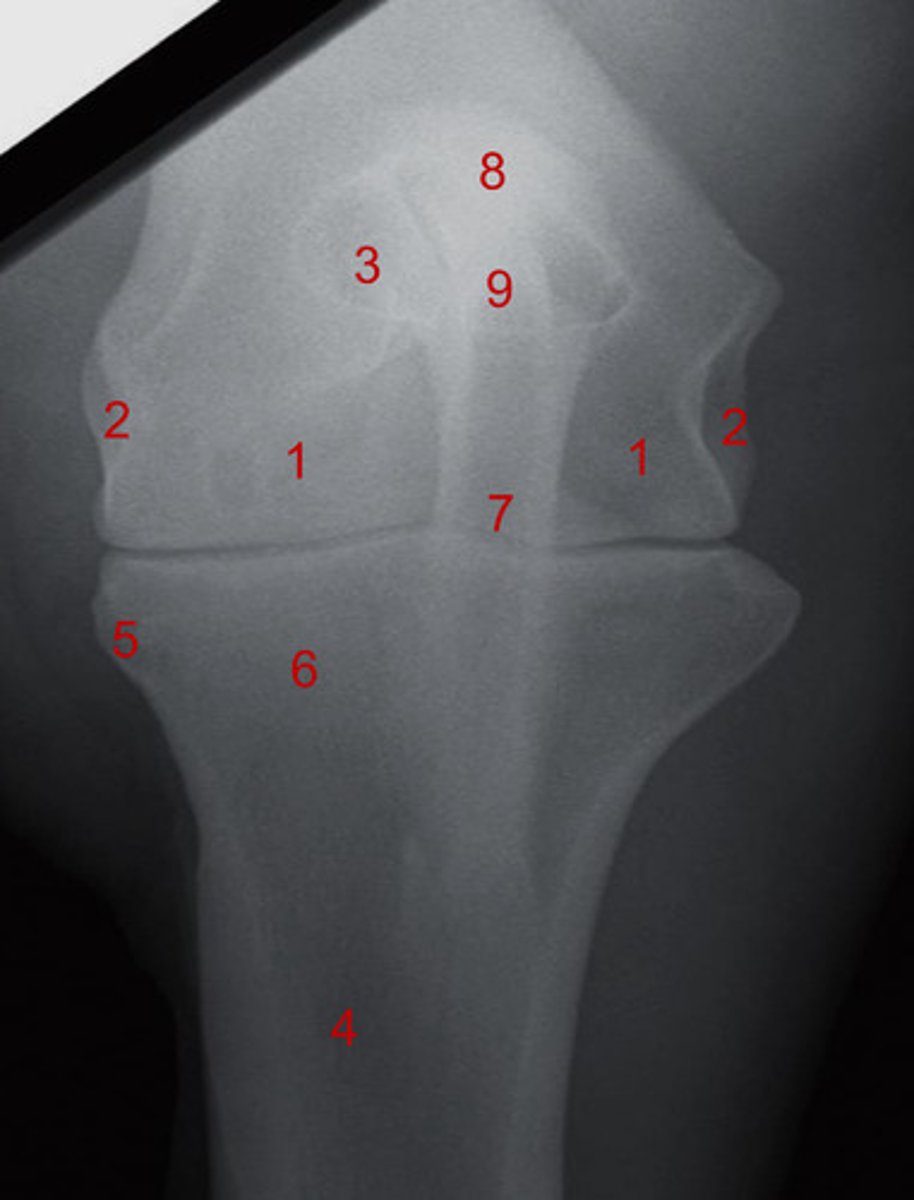
What view is this?
Cranio-caudal view of elbow
How many views are usually taken for the carpus?
Four routine views
What are the routine views for the carpus?
Latero-medial, dorso-palmer, dorsolateral-palmeromedial oblique, dorsomedial-palmarolateral oblique view
What are the additional views for the carpus?
Flexed lateral, tangential/skyline views
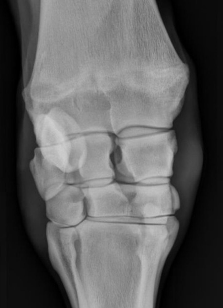
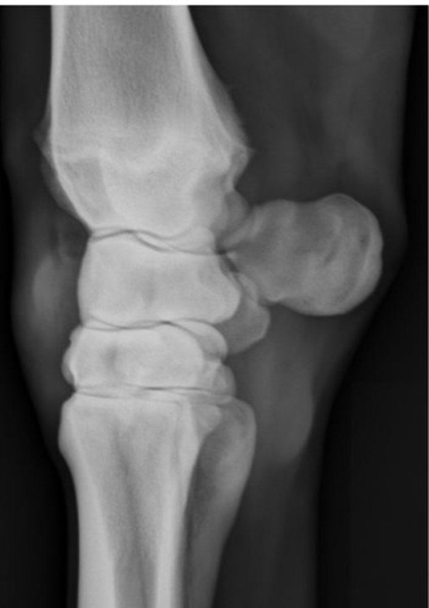
What view is this?
Dorso-palmer view of carpus
What view is this?
Latero-medial view of carpus
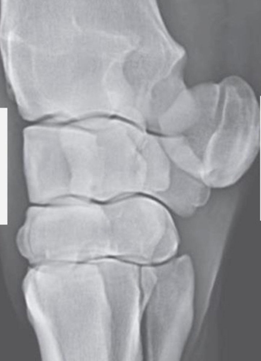
What view is this?
Dorsolateral-palmeromedial oblique view of carpus
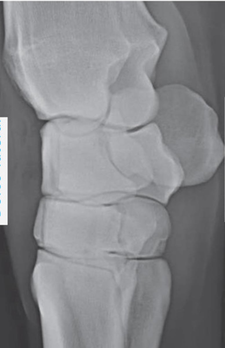
What view is this?
Dorsomedial-palmerolateral oblique view of carpus
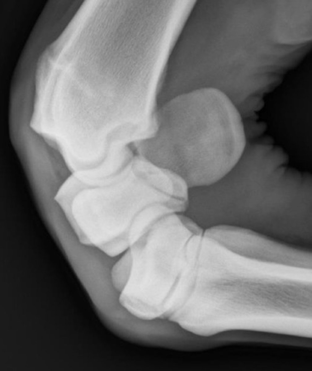
What view is this?
Flexed latero-medial view of carpus
What is another term for the skyline views of the carpus?
Dorsoproximal-dorsedistal oblique views
What is the position for taking a skyline view for the distal radius?
Flex leg so radius is perpendicular with beam angled 65-85 degrees downwards
What is the position for taking a skyline view for the proximal row of carpals?
Flex leg with distal limb pushed slightly cranial and metacarpus parallel to ground with beam angled 45-55 degrees downwards
What is the position for taking a skyline view for the distal row of carpals?
Flex leg with the metacarpus pushed slightly forward cranially with beam angled 30-35 degrees downwards
What are the views for the metacarpals?
Latero-medial, dorso-plantar, dorsolateral-palmaromedial oblique, dorsomedial-palmarolateral oblique views
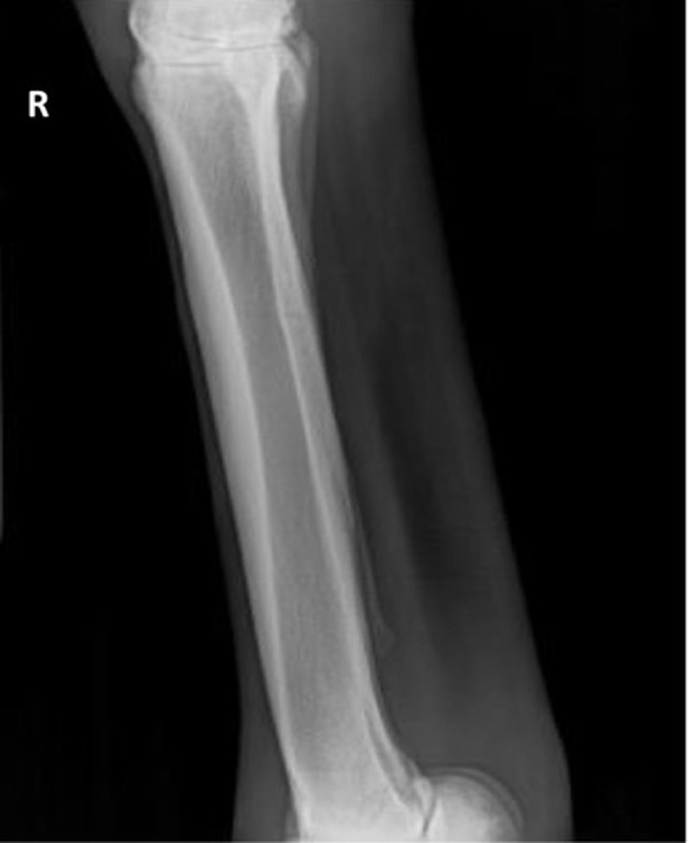
What is this view?
Latero-medial view of metacarpals
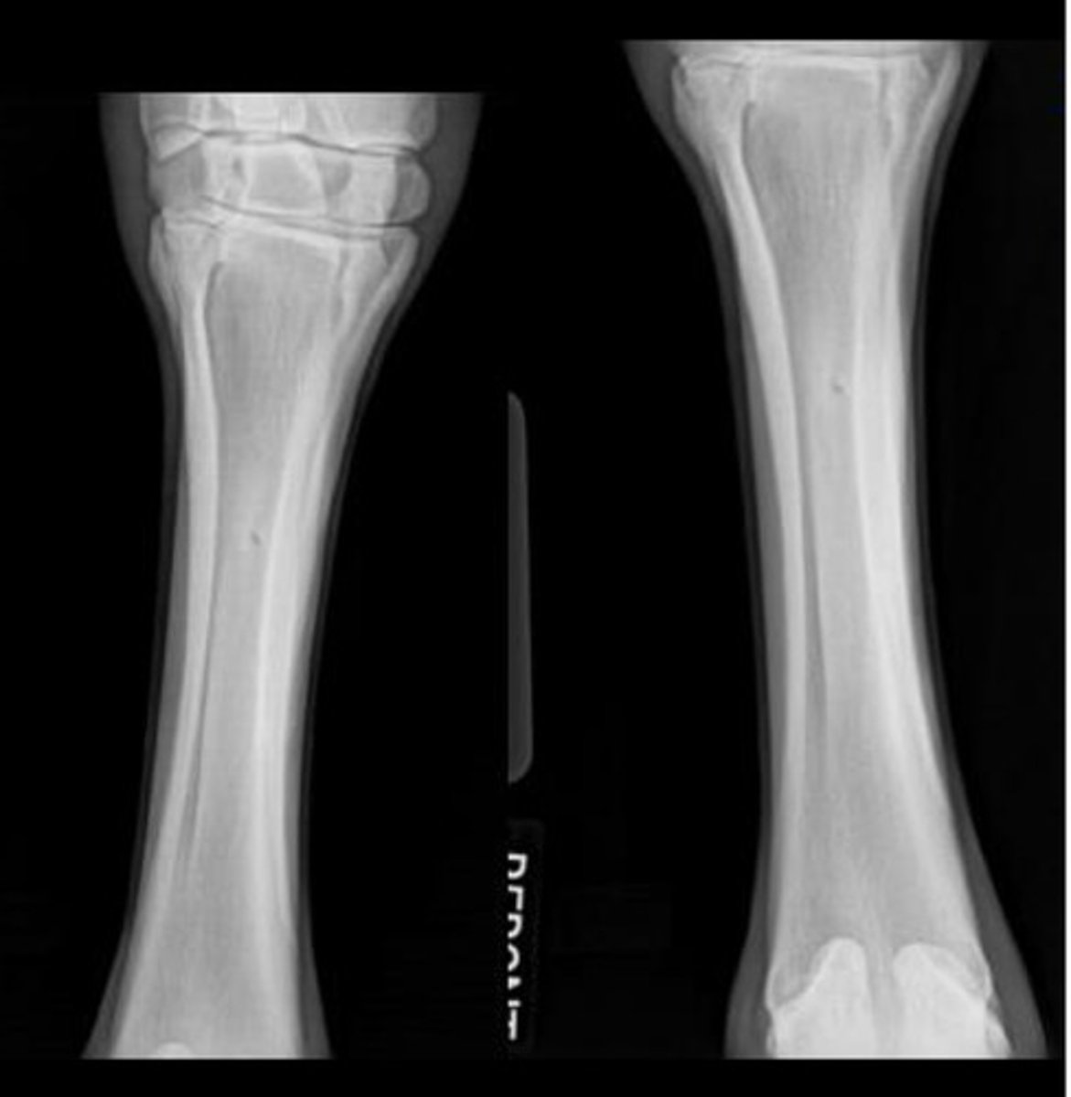
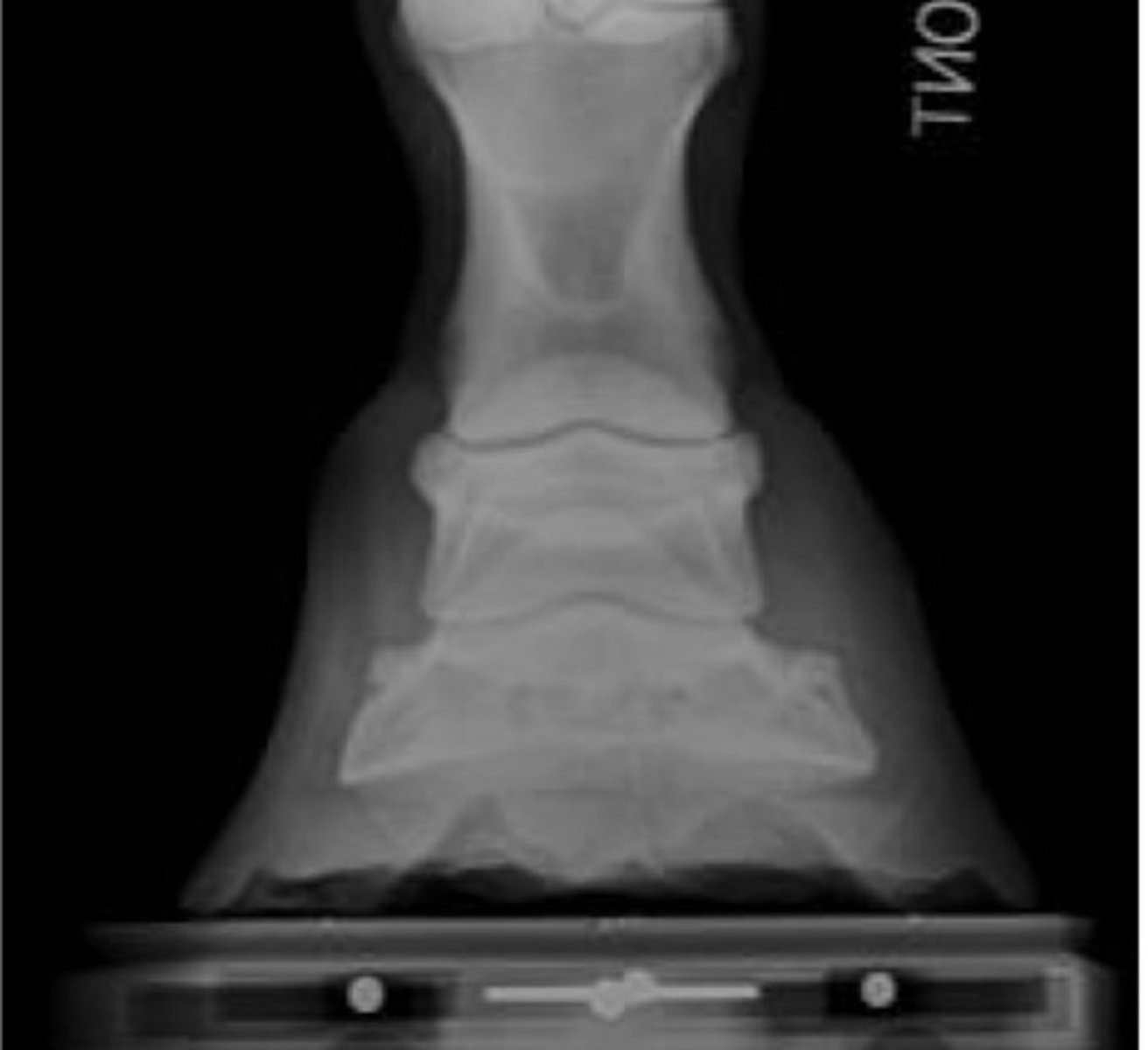
What is this view?
Dorso-palmar view of metacarpals
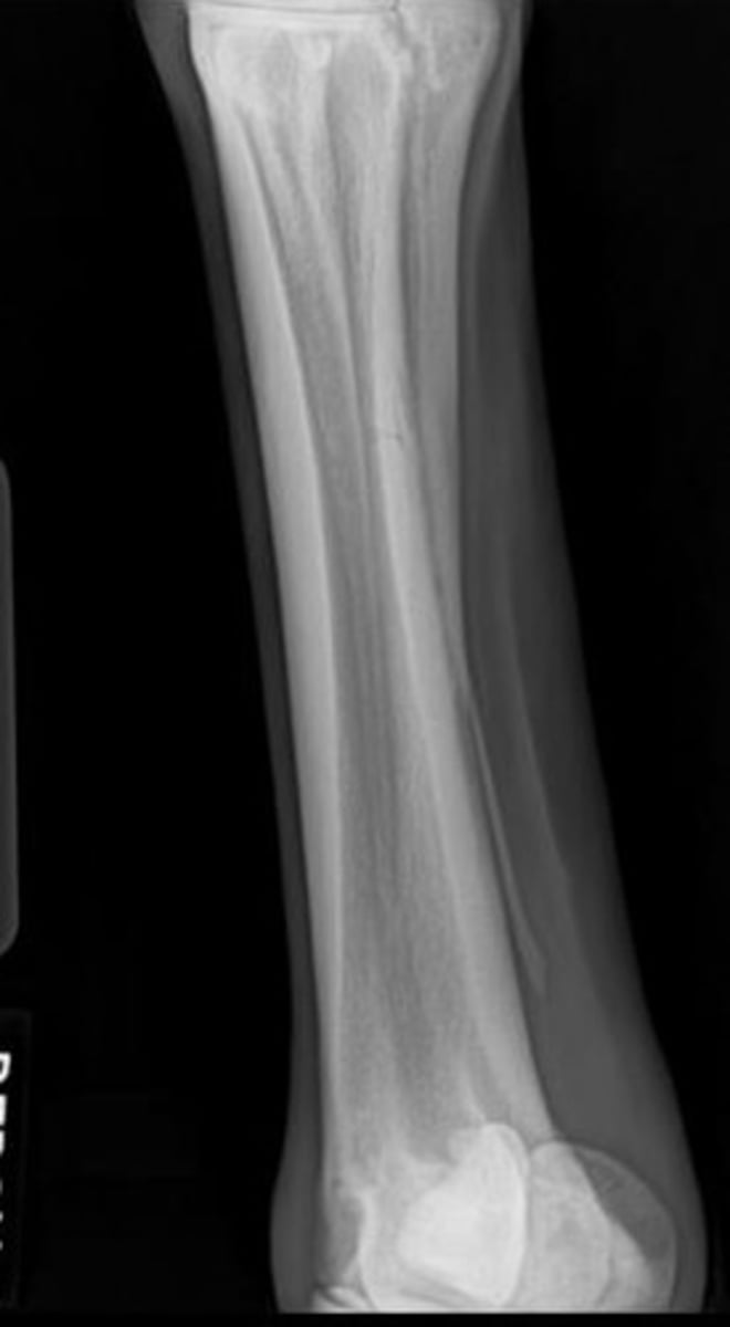
What view is this?
Dorsolateral-palmaromedial oblique view of metacarpals
What splint bone is visualized well in the dorsolateral-palmaromedial oblique view of metacarpals?
Metacarpal 4
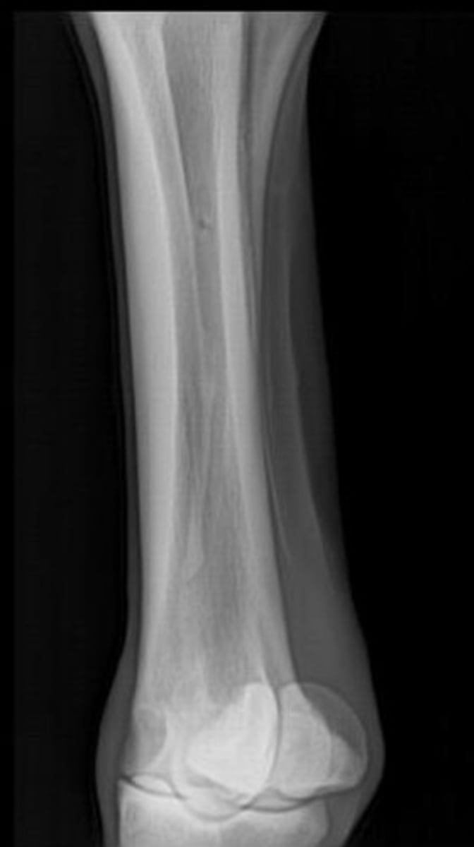
What view is this?
Dorsomedial-palmarolateral oblique view of metacarpals
What splint bone is visualized well in the dorsomedial-palmarolateral oblique view?
Metacarpal 2

What is 1?
Scapula

What is 2?
Supraglenoid tubercle

What is 3?
Glenoid cavity

What is 4?
Head

What is 5a?
Cranial lesser tubercle

What is 5b?
Caudal lesser tubercle

What is 6?
Intermediate tubercle

What is 7a?
Cranial greater tubercle
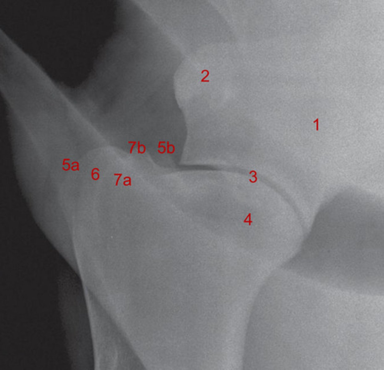
What is 7b?
Caudal greater tubercle

What is 1?
Condyles
What is 2?
Epicondyles

What is 3?
Olecranon fossa

What is 5?
Radial tuberosity

What is 7?
Ulna

What is 8?
Olecranon process

What is 9?
Anconeal process

What is 1?
Condyles

What is 2?
Epicondyles

What is 5?
Radial tuberosity

What is 8?
Olecranon process
What is 9?
Interosseous space

What is 10?
Anconeal process
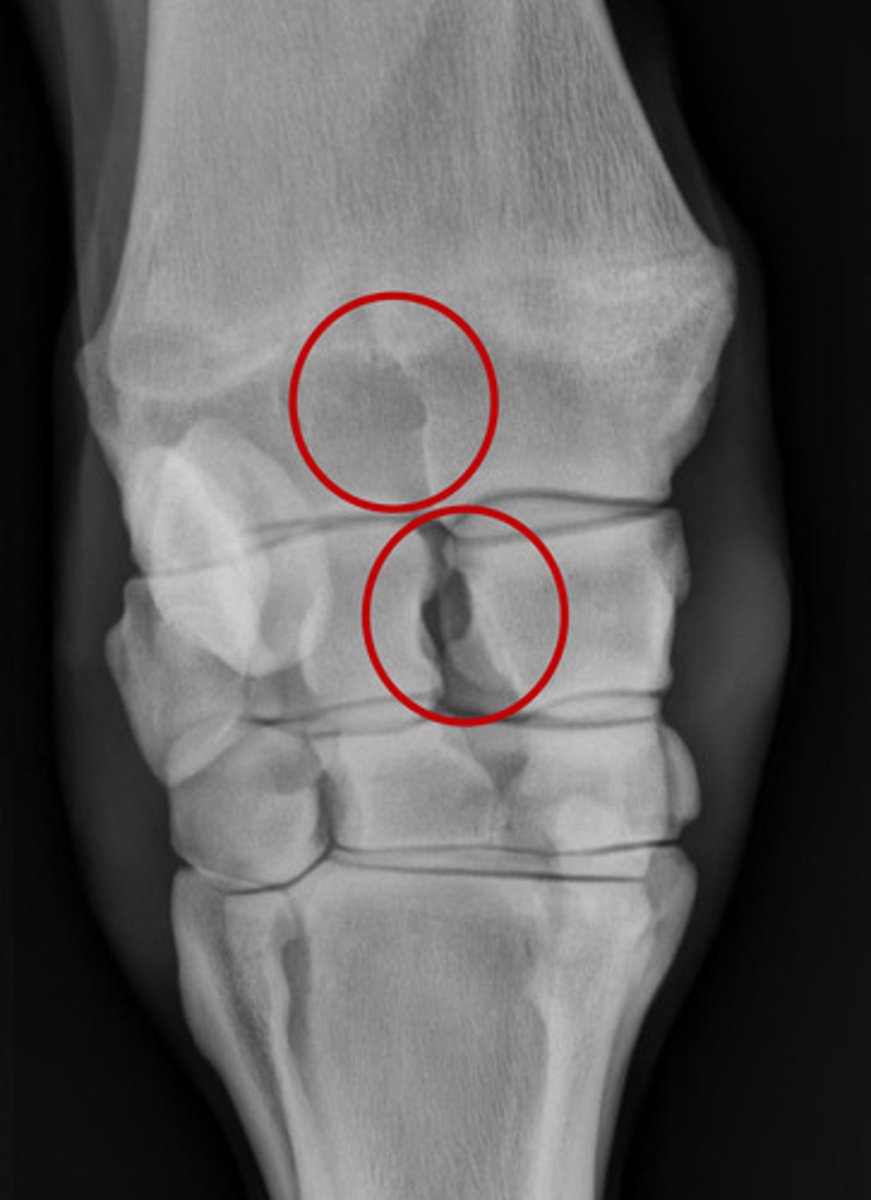
What causes the lucent zone in the upper red circle?
Depression in the caudal surface of the bone
Where is there a radiolucent canal in the carpus?
Between the radial and intermediate carpal bones
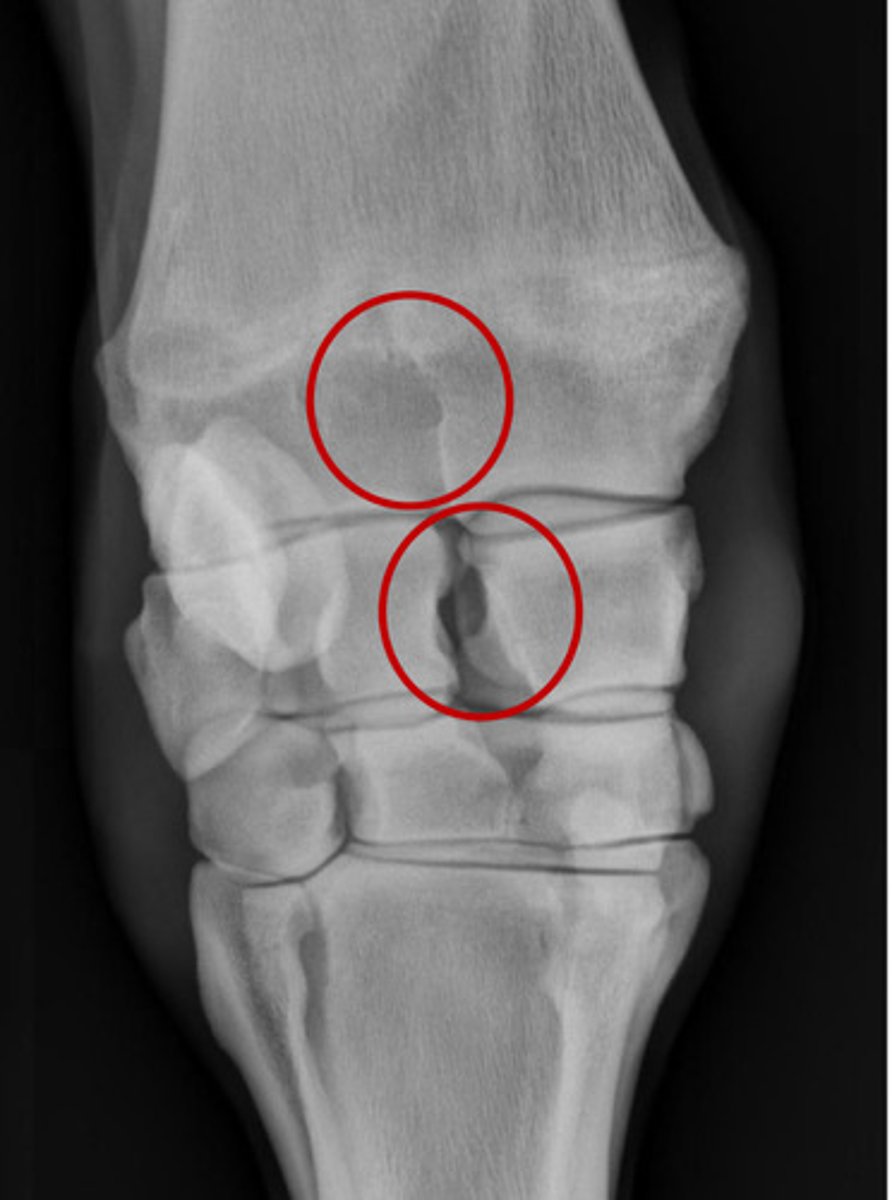
What is in the bottom circle?
Radiolucent canal

What is the blue line?
Antebrachiocarpal joint
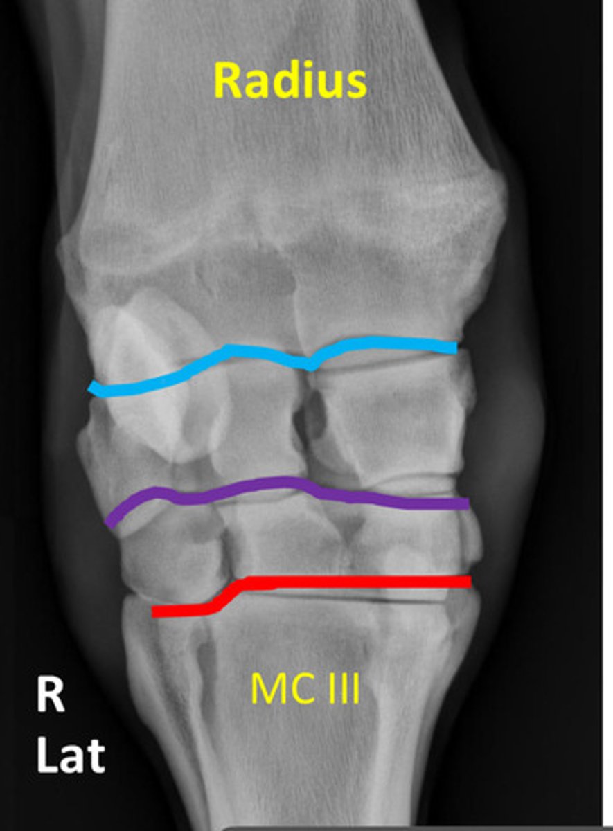
What is the purple line?
Middle carpal joint

What is the red line?
Carpometacarpal joint

What is highlighted in dark blue?
Radial carpal bone

What is highlighted in orange?
Intermediate carpal bone

What is highlighted in light blue?
Ulnar carpal bone
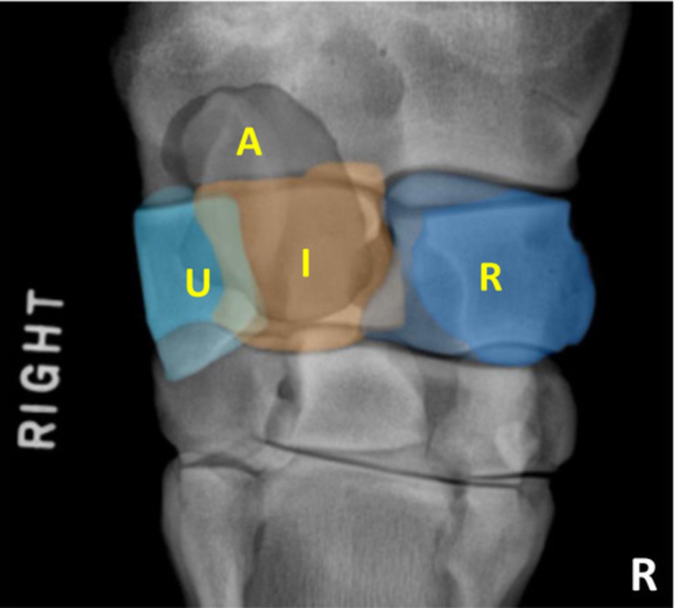
What is highlighted in gray?
Accessory carpal bone

What is highlighted in green?
Second carpal bone
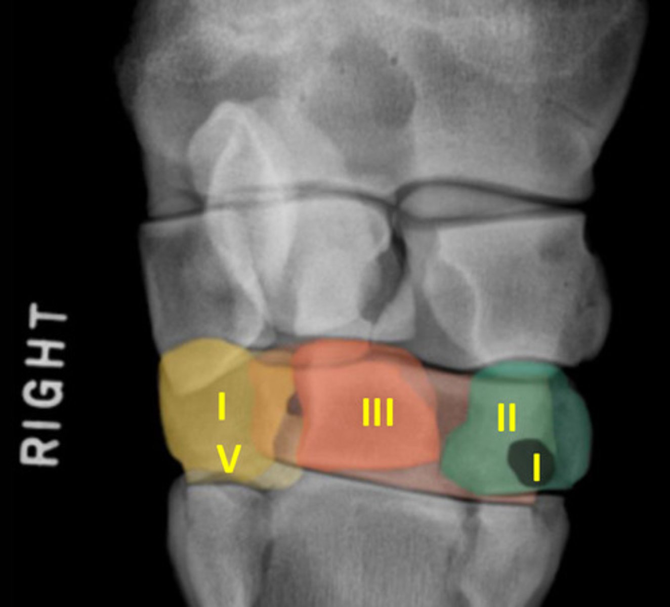
What is highlighted in orange/red?
Third carpal bone

What is highlighted in yellow?
Fourth carpal bone

What is the small black circle?
First carpal bone
Where is the cranial side in lateral and oblique images?
To the left
Which metacarpal is slightly more palmar?
Metacarpal four
How does the intermediate carpal bone move during flexion?
Moves proximally
How do you tell which splint bone is being viewed on the oblique views?
Metacarpal four does not articulate fully with C4 while metacapral two fully articulates with C2

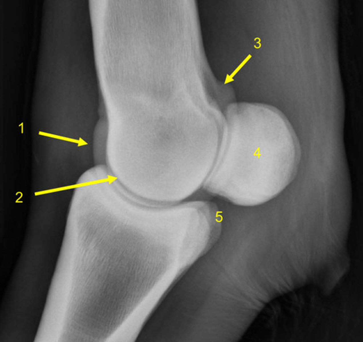
What is the middle arrow pointing to?
Medial facet of radial trochlea

What is the right arrow pointing to?
Intermediate facet of radial trochlea
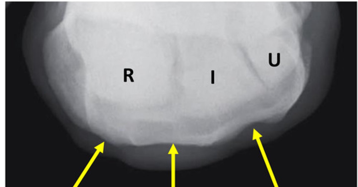
What is the left arrow pointing to?
Distal radius
Which proximal carpal bone is the largest?
Radial carpal bone
Which distal carpal bone is the largest?
Third carpal bone
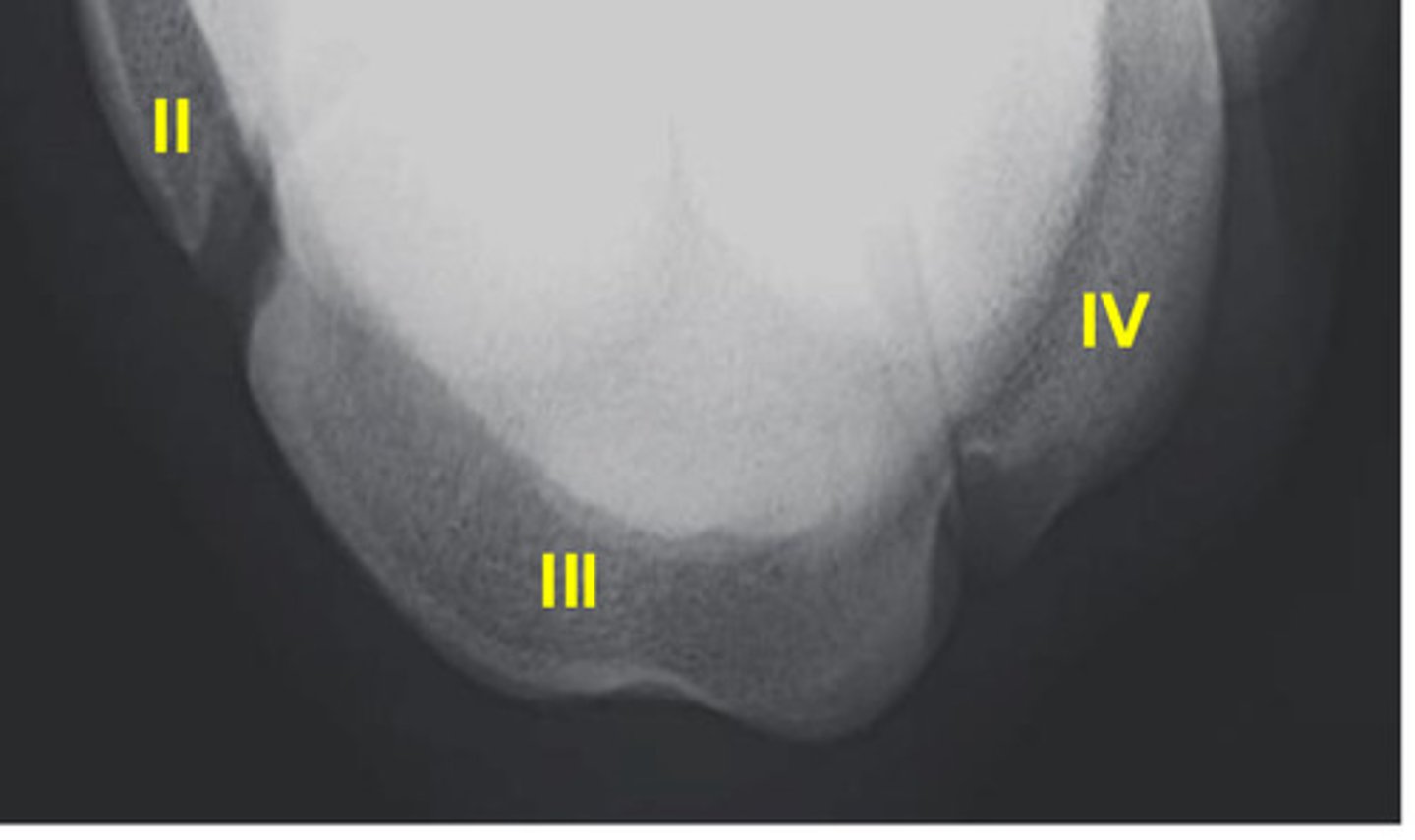
What is number II?
Second metacarpal bone

What is number III?
Third metacarpal bone

What is number IV?
Fourth metacarpal bone

What is 1?
Metacarpal three

What is 2?
Metacarpal two

What is 3?
Metacarpal four
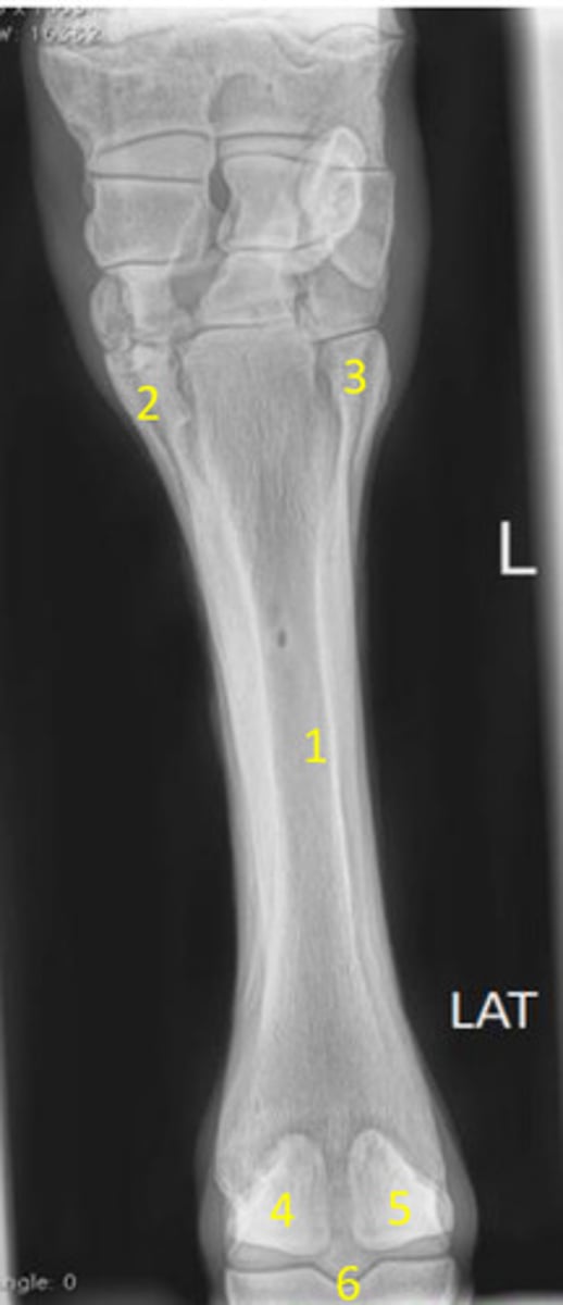
What is 4?
Medial Proximal sesamoid bone

What is 5?
Lateral proximal sesamoid bone

What is 6?
Fetlock joint

What is 1?
Metacarpal three

What is 2?
Metacarpal two

What is 3?
Metacarpal four
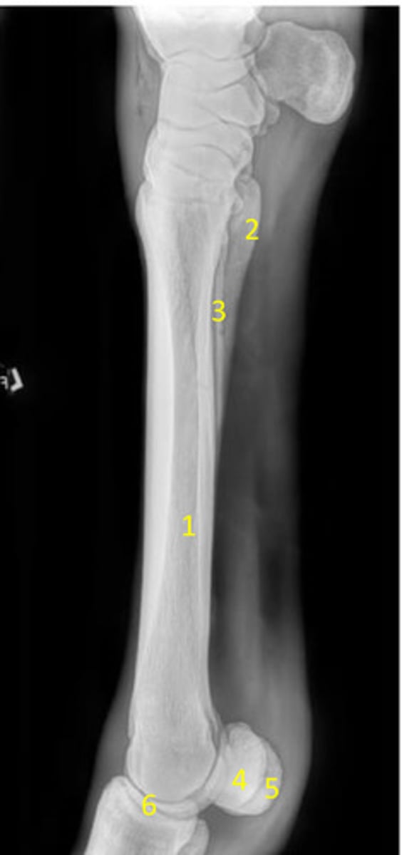
What is 4?
Medial proximal sesamoid bone

What is 5?
Lateral proximal sesamoid bone

What is 6?
Fetlock joint
What are the views for the fetlock joint?
DP, high DP, LM, flexed LM, DLPMO, DMPLO, DPrDDiO views
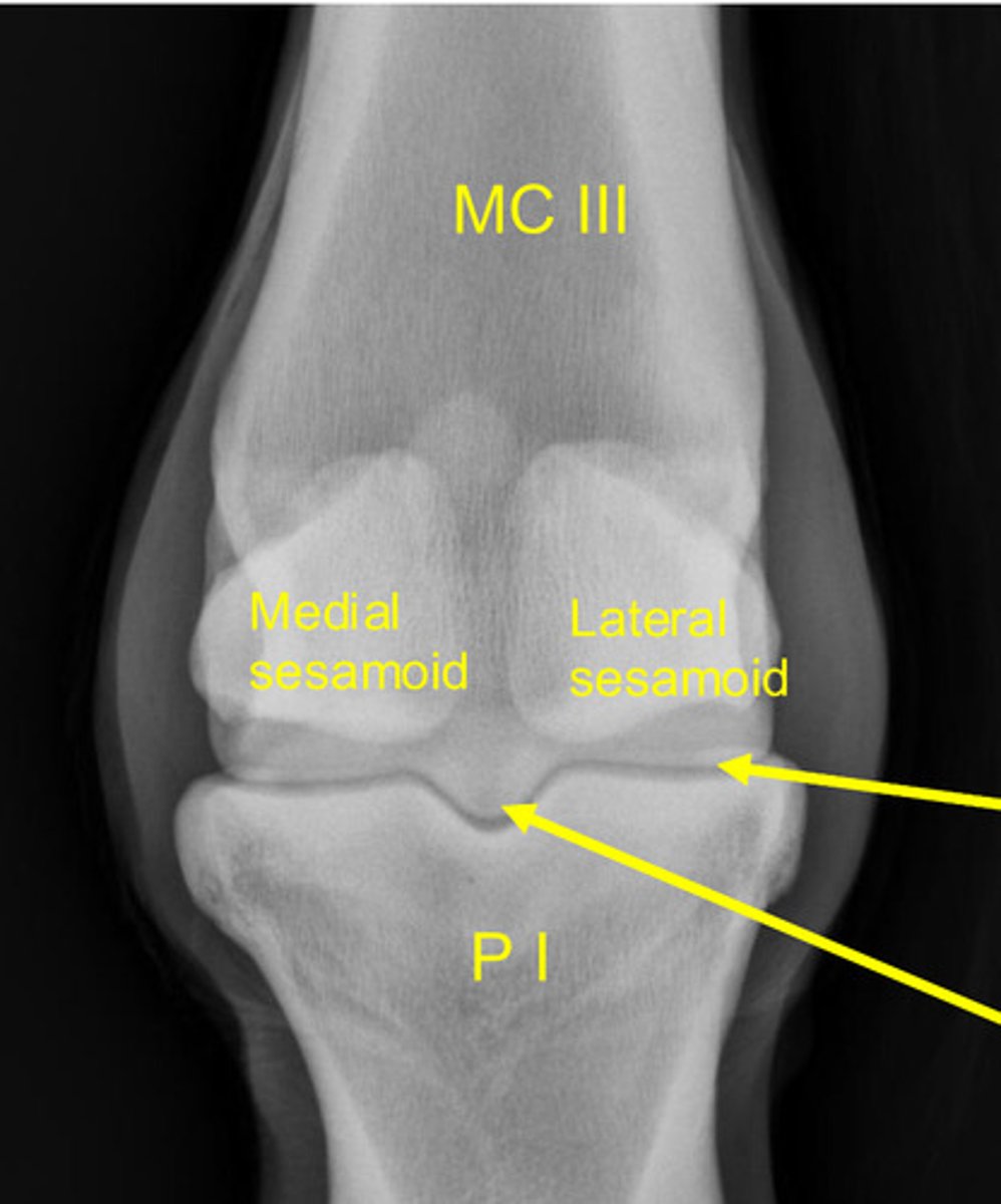
What is the bottom arrow pointing to?
Sagittal ridge
What is the position for a high DP of the fetlock joint?
Detector palmar and parallel to the pastern with beam angles distally 10-30 degrees
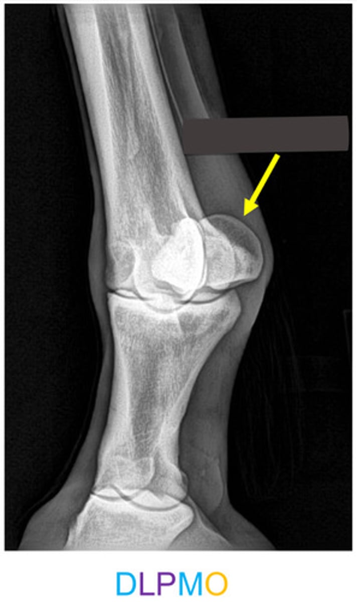
What is the arrow pointing to?
Lateral proximal sesamoid bone
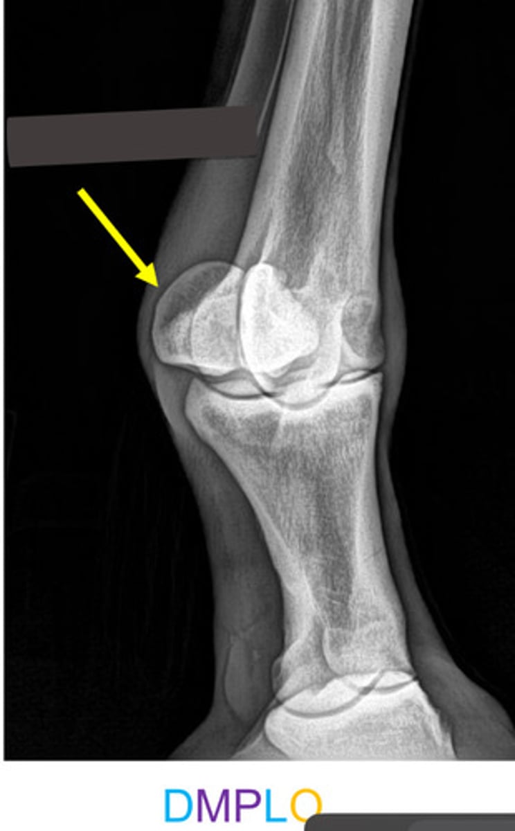
What is the arrow pointing to?
Medial proximal sesamoid bone
What is the purpose of a dorsoproximal-dorsodistal oblique view of the fetlock?
View the sagittal ridge
What is an important part of imaging the foot?
Stand on blocks
What view is this?
Latero-medial view
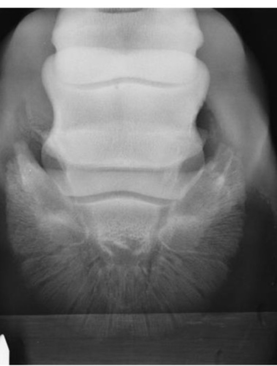
What view is this?
Dorso-palmar view
How is the dorsoproximal-palmarodistal oblique view obtained in the coffin joint?
Foot placed in upright foot holder
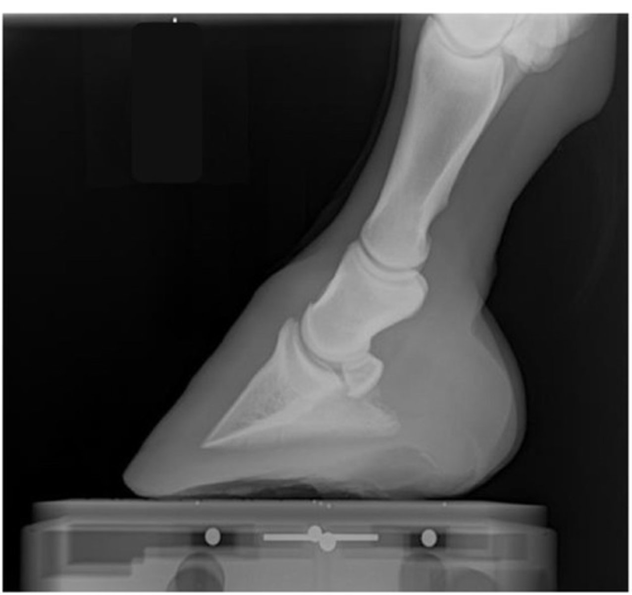
What view is this?
Dorsoproximal-palmarodistal oblique view of foot
What view of the foot results in distortion of P3?
High coronary / dorsoproximo 60 to palmarodistal oblique view
What is the function of the palmaroproximal-palmarodistal oblique view?
See navicular bone

What is 1?
Sagittal ridge
What is 2?
Condyles of metacarpal three