(Images) Merrill's Ch 1 Workbook
1/4
There's no tags or description
Looks like no tags are added yet.
Name | Mastery | Learn | Test | Matching | Spaced |
|---|
No study sessions yet.
5 Terms
Image A: Without compensating filter
Image B: With (Ferlic wedge) filter
30. Fig. 1.1 shows two images demonstrating differences in the range of brightness between anatomic structures of varying tissue densities. Compare these images and identify which image was exposed using a compensating filter and which image was exposed without a compensating filter. (The choices are "with filter" and "without filter.")
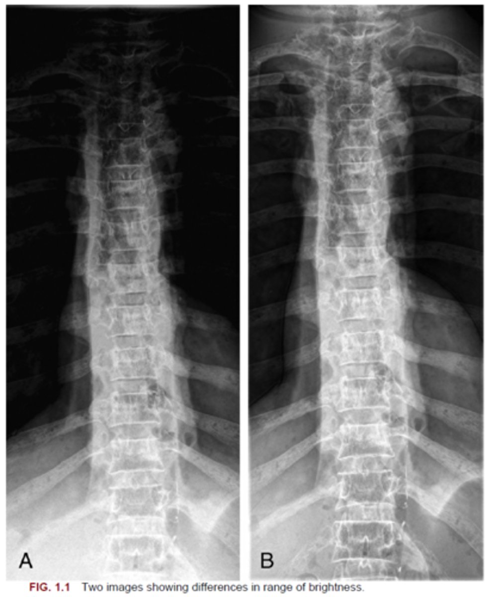
Image A
69. Fig. 1.2 shows two images of a PA projection of a chest. Which image (A or B) is correctly displayed?
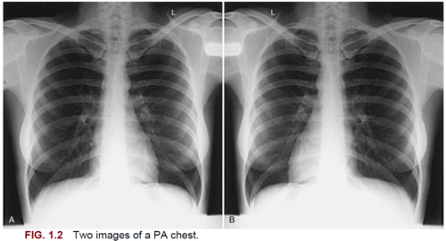
Image A
71. Fig. 1.3 shows two lateral projection chest radiographs. Which image (A or B) is correctly displayed as a left lateral chest radiograph?
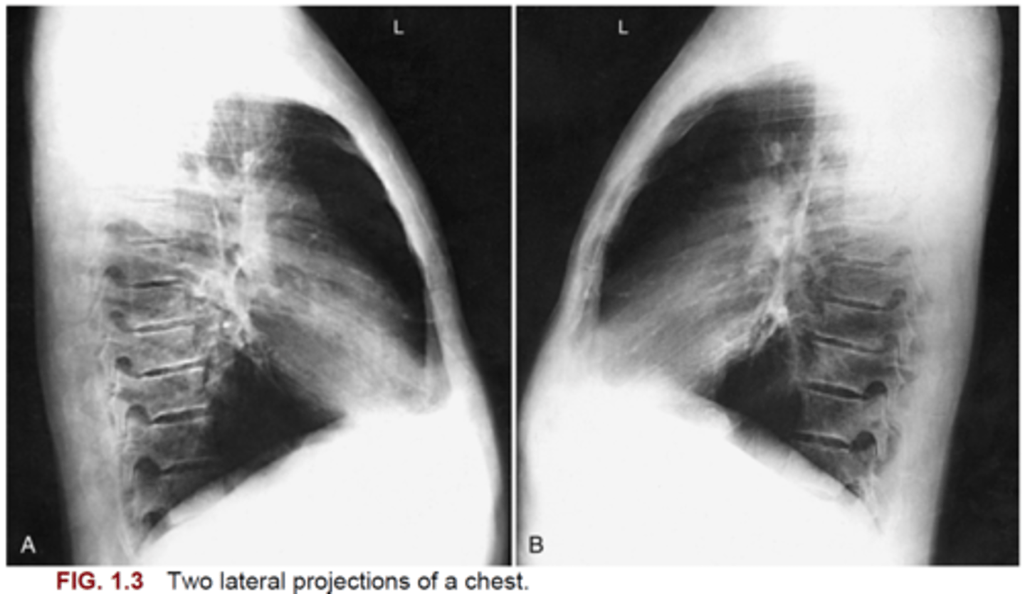
Image A
73. Fig. 1.4 shows two images of a PA left hand radiograph. Which image (A or B) is correctly displayed?

Image A
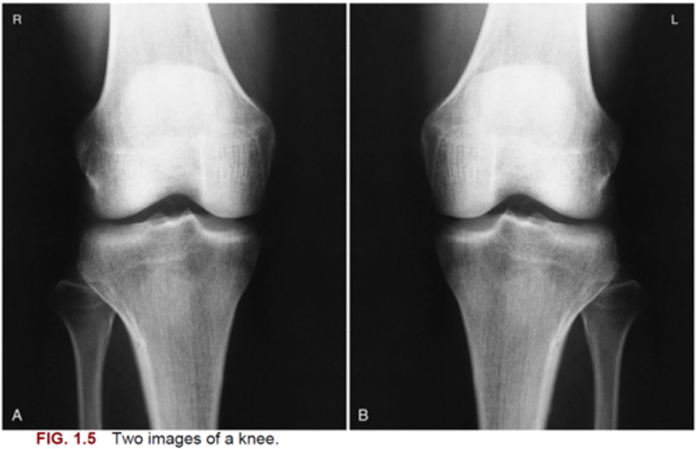
74. Fig. 1.5 shows two images of an anteroposterior (AP) right knee radiograph. Which image (A or B) is correctly displayed?