Bio 251 (Membrane Proteins) with complete expert curated questions and answers (with Diagrams)
1/41
There's no tags or description
Looks like no tags are added yet.
Name | Mastery | Learn | Test | Matching | Spaced |
|---|
No study sessions yet.
42 Terms
What do scramblases do?
- They are a type of transporter rpotein
- They randomly transfer phospholipids molecules from one monolayer to another
- random and symmetric distribution
What do flippases do?
- They are a type of transporter protein
- They remove specific phospholipids from one face of a membrane and move them to the other face
- Facilitate asymmetric distribution
How are membranes transported throughout a cell?
By vesicle fusing and budding
What happens to a membrane when they are transferred between cell compartments?
- they are transported by vesicle budding and vesicle fusion
- membranes retain their orientation during transfer (cytosolic side continues to face cytosol, lumen side faces the extracellular environment)
- embedded proteins also retain orientation
How are phospholipids and glycolipids distributed in the lipid bilayer of an animal cell's plasma membrane?
The phospholipids and glycolipids are distributed asymmetrically in the lipid bilayer of an animal cells plasma membrane.
Where are glycolipids found within a cell?
They are mainly located in the plasma membrane of a cell
On what face of the plasma membrane do glycolipids end up?
- They end up on the non-cytosolic face of the plasma membrane
- they face the outside of the cell
Where does the addition of sugar groups to a glycolipid happen?
The addition of sugar groups to a glycolipid happens in the Golgi apparatus.
What macromolecule makes up 50% of the mass of most plasma membranes?
Proteins makes up 50% of the mass of most plasma membranes.
Which macromolecule is more plentiful in plasma membranes?
- Lipid molecules are more plentiful in plasma membranes
- More lipids are present in the plasma membrane than proteins but since lipids don't weight a lot proteins make up 50% of the mass of the membrane.
What part of a membrane allows specific organelles to do their specific jobs?
- different membranes contain specific proteins (specific proteins are the part of a membrane that allow specific organelles to do their specific job)
- these specific proteins allow membranes to perform specific jobs
- different organelles will have different proteins associated with their membrane allowing them to do different jobs
Mitochondrial membranes contain which type of protein associated with which process?
- they are the only place/membrane that contains proteins associated with the electron transport chain
- Cellular respiration
ER membranes contain which enzymes and proteins? These enzymes and proteins are associated with which process?
- They contain Acyl transferase, Phosphatase, Choline phosphotransferase and Flippases
- These enzymes are associated with phospholipid production and phospholipid movement (being flipped from one face to another)
How can integral membrane proteins be removed from the lipid bilayer?
- They can be removed from the bilayer by disrupting the bilayer with detergents
- It is really hard to remove an integral membrane protein from the lipid bilayer
How can peripheral membrane proteins be released from the lipid bilayer?
- They can be released from the bilayer by disrupting protein-protein interaction
- Peripheral membrane proteins are easier to remove from the lipid bilayer than integral membrane proteins
What are the three types of Integral membrane proteins?
1) Transmembrane Proteins
2) Monolayer associated proteins
3) Lipid linked proteins
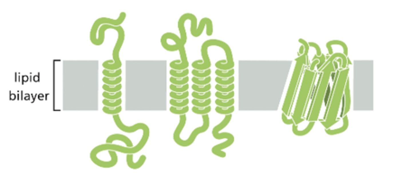
How are transmembrane proteins associated with the lipid bilayer?
- They are single pass or multi pass proteins (they pass through the bilayer once or multiple times)
- They are amphipathic proteins (the hydrophobic region is embedded in the interior of the bilayer and the hydrophilic regions are exposed to the aqueous environment)
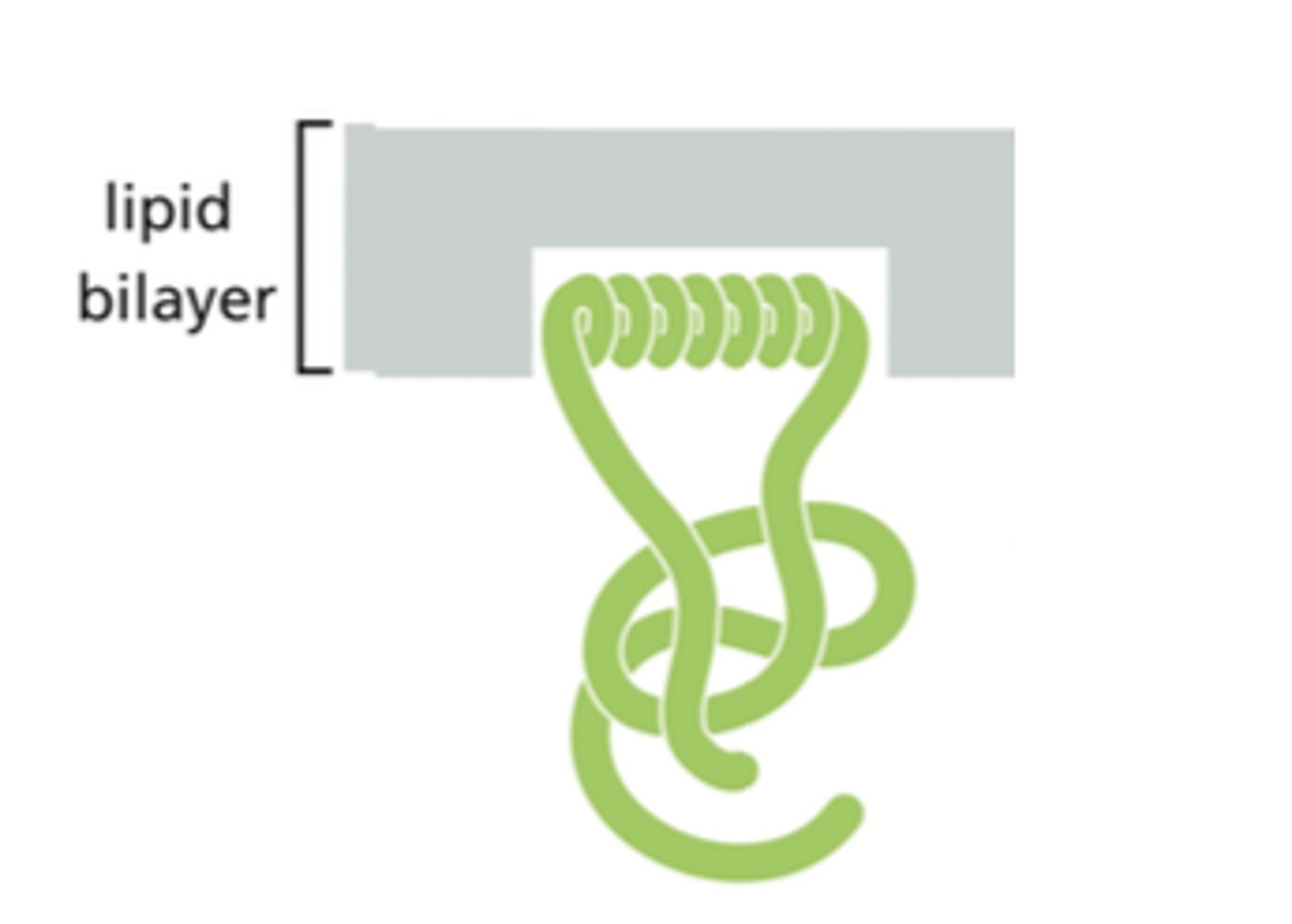
How are monolayer associated proteins associated with the lipid bilayer?
- They are associated with the cytosolic face (face of the bilayer inside of the cell) of the lipid bilayer
- Peripheral membrane proteins have an amphipathic A-helix!
How are lipid linked proteins associated with the lipid bilayer?
They are attached to the bilayer by covalent attachments to lipid molecules embedded in the bilayer

Where are peripheral membrane proteins located on the lipid bilayer
They are located on the surface of the bilayer
How do peripheral proteins interact with integral membrane proteins?
Peripheral proteins interact with the hydrophilic portions of integral membrane proteins
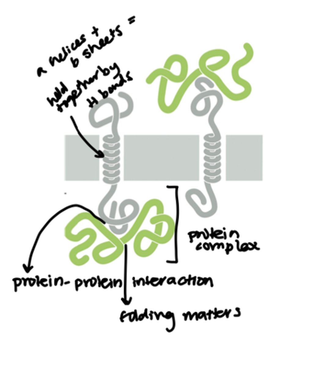
Does the protein folding of a peripheral protein matter? Why does it matter?
- Yes it does matter
- To be able to interact with the hydrophilic portion of the integral membrane proteins the peripheral proteins need to be folded in a specific way to be able to fit right with the integral membrane protein
Why is the orientation of a protein so important?
- The receptor protein needs to be inserted/orientated properly in the lipid bilayer
- The point of the receptor protein with the receptor site need to be extended in the extracellular fluid so that a ligand can bind to it
- If the part of the protein is pointing into the cytosol, a ligand from the extracellular environment will not be able to bind to it and get its message into the cell

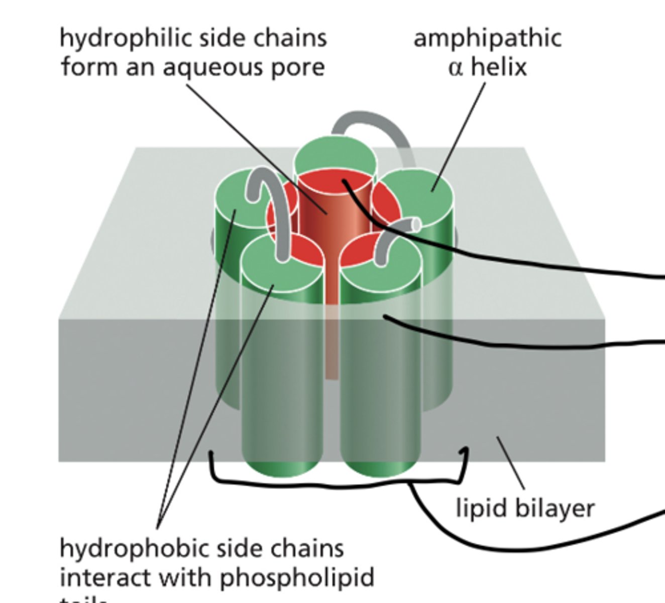
What is the structure of a transmembrane pore protein?
- It is a multi pass transmembrane protein (passes into the lipid bilayer multiple times)
- Is made up on multiple amphipathic A-helices that pack side by side to create a water-filled channel across the lipid bilayer
- The hydrophilic side chains of the A-helices point inward (make up the inside of the pore)
- The hydrophobic side chains of A-helices associate with the phospholipid tails of the bilayer
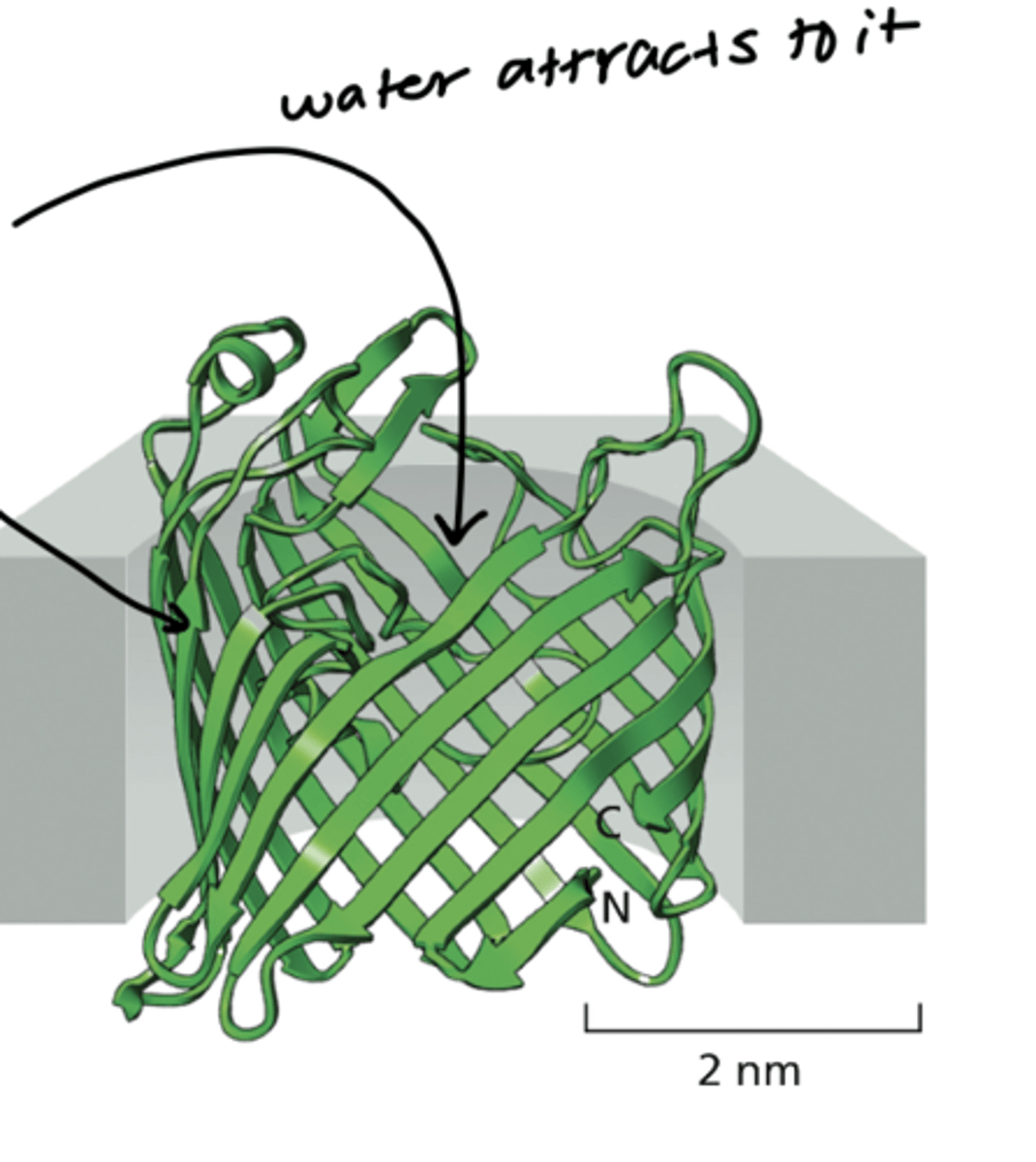
What is the makeup of a beta barrel?
- B pleated sheets form a cylinder
- The hydrophillic amino acids face the inside of the barrel
- he hydrophobic amino acids interact with the lipid bilayer core (the phospholipid tails)
- Since the hydrophilic amino acids make up the inside of the barrel it attracts water molecules (this is why the barrel is filled with water)
What are Porin proteins?
They are water-filled channels (water filled beta barrels) in the outer membrane of bacteria and mitochondria (mitochondria was originally an early bacteria that was engulfed)
How are membrane proteins solubilized in detergents?
1) SDS (a strong ionic detergent with a hydrophilic charged end and a hydrophobic end) displaces lipid molecules from around an integral membrane protein
2) Then a water soluble complex forms: Detergent surround the hydrophobic core of the integral membrane protein. The detergent's hydrophobic tails face the hydrophobic core of the protein
3) when the water soluble complex forms a water- soluble mixed lipid detergent micelle also forms from the displaced lipids and the detergent (SDS)
What is SDS (sodium dodecyl sulfate)?
- It is an ionic detergent
that removes integral membrane proteins from the lipid bilayer
- this is difficult to do
What is TX-100 (Triton X-100)
- It is a mild non-ionic detergent that removes integral membrane proteins from the lipid bilayer
- this is difficult to do
What is X-ray crystallography?
- It is a standard method used to determine the 3-D structures of many proteins
- It requires good protein crystals
- The proteins need to fold like how they would fold in nature/humans for an accurate picture to be taken by X-ray crystallography
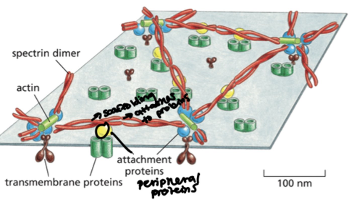
What structure reinforces the plasma membrane of animals cells?
- The under-lying cell cortex reinforces the plasma membrane of animal cells
- The cell cortex is a meshwork of filamentous proteins
- A meshwork of peripheral and integral membrane proteins associates with the inner surface of the plasma membrane
- Actin, myosin (which make up the cell cytoskeleton), integral proteins and peripheral proteins are associated with Spectrin dimers to complete the cell cortex
What is the three main functions of the cell cortex?
1) the meshwork supports the membrane
2) the meshwork helps maintain cell shape
3) the meshwork help the cell withstand stress
- this is especially important in red blood cells when they are being pushed and squeezed through time capillaries
- The RBC's need to keep their shape and withstand the stress/pressure being exerted on them. The cell cortex help the RBC accomplish all this.
How is protein movement within the membrane different from lipid movement within the membrane?
- Lipids can move freely within the membrane
- Some proteins can move freely
- Others are anchored to protein complexes and cannot move
What did the mouse cell and human cell experiment prove?
- It proved that some membrane proteins can move laterally in the lipid bilayer
1) a mouse cell with rhodamine (red) label membrane proteins was fused with a human cell with fluorescein (blue) labeled membrane proteins
2) 0 minutes after the cell fusion the red proteins stayed at the top of the cell and the blue proteins stayed at the bottom
3) after 40 minutes the red and blue proteins had dispersed and gotten mixed up --> this proves that some plasma membrane proteins can move laterally in the lipid bilayer
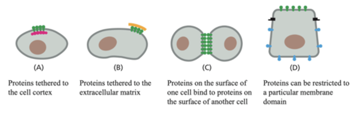
How can a cell restrict the movement of its membrane proteins?
1) Proteins can be tethered to the cell cortex (cytoskeleton?)
2) proteins can be tethered to the extracellular matrix (extracellular structures?)
3) proteins on the surface of one cell can bind to the proteins on the surface of another cell
4) proteins can be restricted to a particular membrane domain
What is an example of proteins that are restricted to particular domains of the plasma membrane?
- Epithelial cells in the gut
- Epithelial cells establish an apical-basal polarity (the differential distribution of phospholipids, protein complexes and cytoskeletal components between the various plasma membrane domains)
What is required for the proper functioning of a cell?
- The proper distribution of proteins is required for the proper functioning of a cell
Ex) A neuron needs the proper distribution of channels to depolarize and hyper-polarize. The receiving cell needs receptor proteins in the right places to receive the neuron signal.
What is a oligosaccharide?
It is polymer containing a small number, 10 - 3, monosaccharides
- monosaccharides are the building blocks of polysaccharides aka a polymer
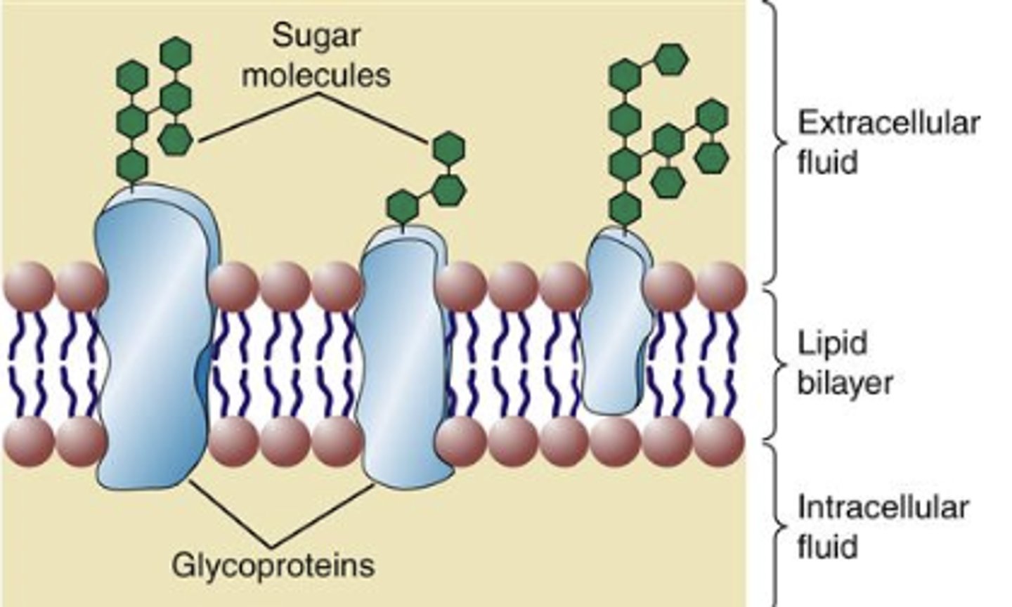
What is a glycoprotein?
- A glycoprotein is a protein with an oligosaccharide (carbohydrate group) attached and protruding on the outer surface of the cell membrane
- Glycoproteins are most prominent in plasma membranes
- They play a role in cell-cell recognitions
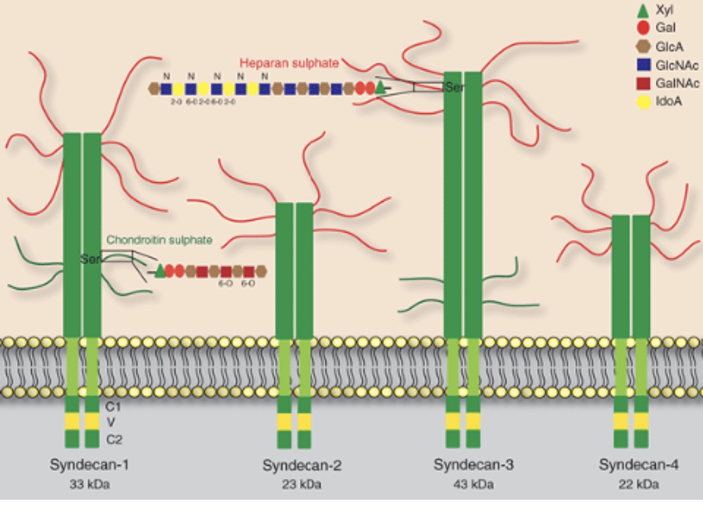
What is a proteoglycans?
- It is a core protein that has one of more polysaccharide side chains
- a polysaccharide is a long chain of sugars
What is the glycocalyx?
- It is the cell surface coat found in animal cells (it is a sugary coat )
- It is made up of carbohydrate groups of plasma membrane glycoproteins, glycolipids, and proteoglycans
- It helps protects the cell's surface from mechanical damage
- It attracts water molecules
- It creates a slimy surface
- It plays a role in cell-cell recognition
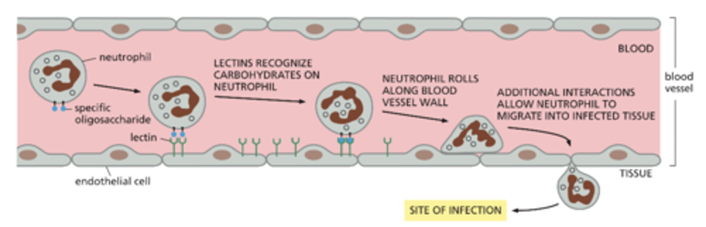
Describe the details behind the neutrophil example.
Example of the recognition of cell-surface carbohydrates
1) Endothelia cells have lectins (transmembrane proteins)
2) These endothelia cells recognize specific sugar groups carried by the glycolipids and glycoproteins on the
surface of neutrophils
3) The lectins "catch" and slow down the fast moving neutrophil and allow it to roll down the blood vessel wall and migrate to the site of infection