Chapter 5 : skeletal system
1/68
There's no tags or description
Looks like no tags are added yet.
Name | Mastery | Learn | Test | Matching | Spaced | Call with Kai |
|---|
No analytics yet
Send a link to your students to track their progress
69 Terms
the skeletal system is made of
skeletal bones
cartilage
ligaments
connective tissue to stabilize the skeleton
Osseous tissues are a major component
functions of the skeletal system
support - provides the framework for the attachment of other organs (muscles)
storage of minerals - calcium and phosphate ( 98% of the calcium in the body is stored in bones)
Blood cell production - major site of production
Red marrow
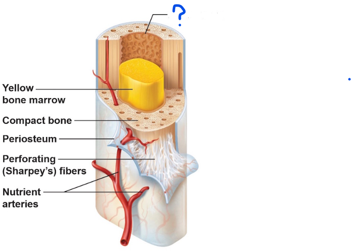
Yellow marrow - adipose cells
leverage - gives muscles a platform to exert force on to cause movement
Protection - every important organ is protected ( ribs protect the heart and lungs, skull protects the brain, ect)
Red marrow -
all the formed elements of blood are produced
Yellow marrow
- adipose cells
histological organization of mature bone
Osseous tissue is a type of connective tissue
Specialized cells called osteocytes make up 2 % of the mass of this tissue
has a solid matrix
made of collagen fibers surrounded by calcium salts (which gives strength to the bones)
Periosteum
outlining continues with the deep fascia

Endosteum
inner lining
Osteocytes
similar to fibrocytes, maintain the protein and mineral content of the matrix
Mature bone cells
Mature = maintain
they oversee everything !!!
Osteoblast
responsible for building bone
immature bone cells
found in the periosteum and endosteum
Produced osteoid, which is involved in making the matrix
make new bone, but the process is called osteogenesis
Osteoprogenitor cells
Produced osteoblasts
differentiate to form new osteoblasts
found in the periosteum and endosteum
Really important for repairing bones after they break
Osteoclast
found in the periosteum and endosteum
performs osteolysis
The dissolving of bone tissue, thereby causing the release of stored calcium ions and phosphate ions into the blood
It makes bones weak, but you’re supposed to replenish them through your diet
Used in muscles, movement
really important in muscle movement
Ground bone slide
What is this slide ?
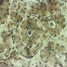
osteon
What is this whole structure called ?

Canaliculi
What is number 3 ?
( strings that connect osteocytes)
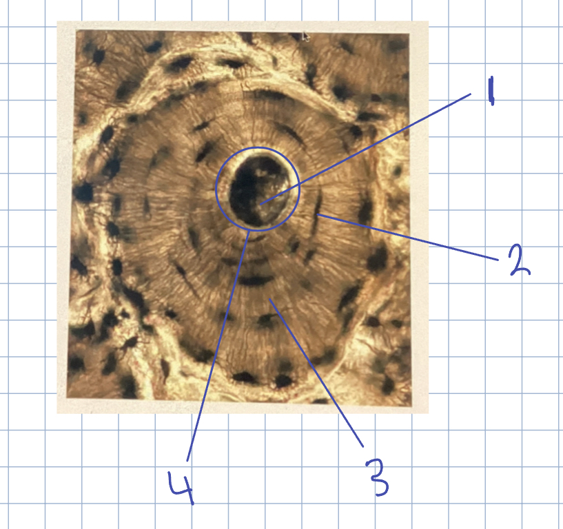
Central canal
What is 1 ?
( blood vessels run through )

Lamella
What is 4?
( similar to rings of a tree)

Osteocytes in lacunae
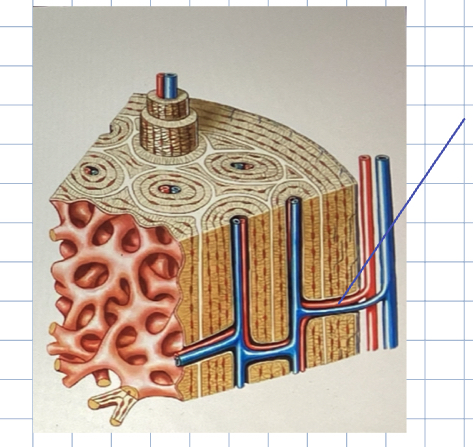
volkmann’s canals
perpendicular to the surface of the blood vessels
designed to give blood to deeper osteons
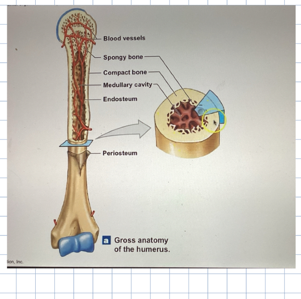
Epiphysis
ends

Diaphysis
Shaft
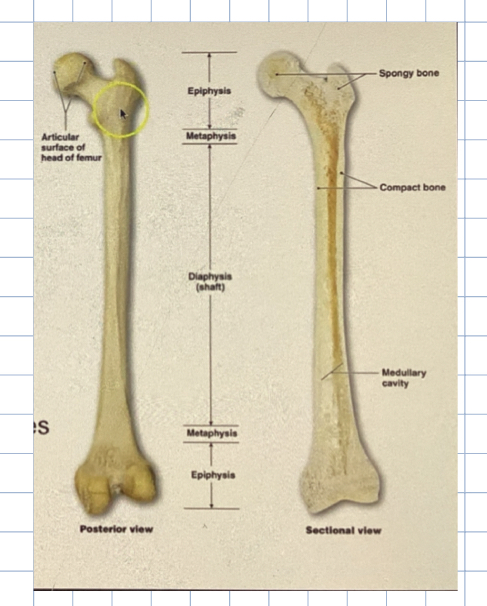
Metaphysis
at the neck of the bone, it’s a narrow growth zone between the epiphysis and diaphysis
found on the distal and proximal portions of the bone ( so not just the neck )

Medullary cavity
where the bone marrow is held

Articular cartilage
usually found on the epiphysis
thin layer of hyaline cartilage over the epiphysis
reduces friction and absorbs shock
Compact bone
aka dense bone
More compact, thicker compact bone in the diaphysis bc its where you find the medullary cavity
Compact bone outlines the medullary cavity
Also covers spongy bone
Function:
gerentes tremendous strength from end to end, so when force is applied, the compactness is what prevents it from breaking.
conducts stress from one area of the body to another area of the body
Spongy bone
trabecular bone
no true osteon
Made of an open network of plates (called trabeculae or spicules)
irregular latticework
makes the lightweight nature of bones
larger spaces filled with red marrow - especially within epiphyses and irregularly shaped bone
Osteocytes and lacunae are found in trabeculae
Not in true osteons, in structures that look like osteons bc they don’t have a central canal
In this type of bone, they get their nutrients from blood
don’t need central or perforating canal bc they are really close to red bone marrow, making them bathed in blood
function:
to deal with stress from the side
Periosteum
outer surface of the bone
covering over bone not covered by articular cartilage, or where tendons and ligaments
Structure :
fibrous layer - outer
connective tissue with blood vessels, lymphatic vessels, and nerves that pass into the bone
Osteogenic Layer - inner
elastic fibers, blood vessels, osteoprogenitor cells, osteoclasts, and osteoblasts
The main job is cell growth, making new bone

Endosteum
inner surface of bone
lines the medullary cavity
consists of osteoprogenitor cells
actively involved in repair & growth
Osteogenesis
the process of bone formation
included
ossification
calcification
Ossification
takes place during calcification, cartilage is replaced by bone cells
Calacififcation
Taking the cartilages jelly like matrix and turning it solid
The deposit of calcium ion salts into the bone tissue makes it solid
Intramembranous ossification
formation of bone directly on or within the fibrous connective tissue membranes
Cells of compact connective tissues undergo differentiation to form osteoblasts, which divide to form bone
involved in the development of the clavicle, mandible, skull, and facial bones
have softer connective tissue within them, which is why they are formed in these membranes
Endochondral ossification
How most of the bones in the body are actually formed
formation of bone from hyaline cartilage
Hyaline cartilage makes a template for the bone
Cartilage is eroded away and eventually replaced with bone
Epiphyseal plate
where endochondral ossification occurs
cartilage in the metaphysis, converted to bone, increasing bone length
A layer of cartilage that slowly turns to bone
As we age, the width of this zone narrows. until at a point it just becomes a line called the epiphyseal line - which is a bony remnant of the epiphyseal plate

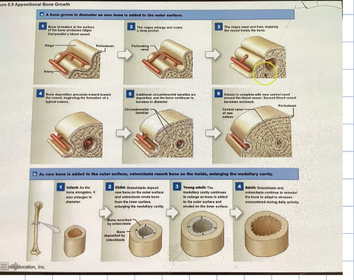
Appositional growth
increasing the diameter of the bone
Blood vessels that run parallel to the bone become surrounded by bone cells
which creates “ tunnels.”
Each “tunnel” has a blood vessel in it, technically making it an osteon
Osteoblasts surrounding the bone begin to produce a matrix, thus creating concentric rings, making it larger
Osteoclasts are dissolving the inner bone, thus creating the marrow cavity
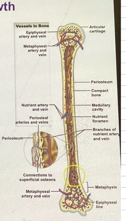
Major blood vessels associated with the long bone
nutrient vessels
metaphyseal vessels
epiphyseal vessels
periosteal vessels
nutrient vessels
enter the diaphysis and branch towards the epiphysis ( both ends, splits )
Re-enter the compound bone leading to the central canal of the osteons

Metaphyseal vessels
supply nutrients to the diaphyseal edge of the epiphysis
region of the metaphysis

Epiphyseal vessels
supply nutrients to the medullary cavities of the epiphysis

Periosteal vessels
supply nutrients to the superficial osteons - outer part of compact bone
found in the periosteum
nutrient muscles are named after where they go, they enter the bone and split
Nutrition
A factor regulating bone growth
mineral: calcium, phosphate, magnesium, citrate, carbonate, and sodium
Vitamins: A, C, and D
calcitriol : vitamin D3 - helps in Ca absorption
If taking vitamins, take calcium citrate instead of calcium carbonate, as it absorbs better; also why milks are fortified
Hormones
A factor regulating bone growth
thyroxine - growth hormone
maintain normal activity in the epiphyseal region
sex hormones - stimulate osteoblast activity
You get a higher influx of sex hormones during adolescence, which is where puberty happens, which is why growth and puberty happen during this time
development of secondary sexual characteristics
growing pains, mostly at night, because growing pains happen at night
Fracture
injury and repair
When a bone is broken or cracked
The healed area is stronger and thicker than normal bone
sometimes susceptible to atmospheric pressure, aches
Aging in the skeletal system
When osteoclast activity is faster than osteoblast activity, bones become more porous - osteoporosis
You dissolve bone faster than you make it
easy to break as you get older
Start bulking up on calcium before you get older bone density exams, as you get older
classification of bone shapes
sutural bones
irregular bone
short bone
pneumatize bones
flat bone
long bone
sesamoid bones
sutural bones
small bones between the joints of certain cranial bones
Irregular bone
Irregular amount of spongy vs compact bone
vertebral column, certain facial bones
Short bones
cube shaped
spongy with a thin layer of compact bone
wrist and ankle
Pneumatized bones
Hollow or contains numerous air pockets
Flat bones
thin; composed of 2 more or less parallel plates of compact bones over spongy bone
protection for soft tissue
extensive areas for muscle attachment
cranial bone, sternum, ribs, scapula
spongy bone sandwich
compact bone, spongy bone, compact bone
really big area important for muscles attachments
important for the protection of soft tissue ( like the brain )
long bones
greater length than width
diaphysis, metaphyses, epiphyses, and medullary cavity
slightly curved for strength- body stress
thighs, legs, toes, arms, forearms. fingers
Processes
any projections or bump
Ramus
an extension of a bone that forms an angle that forms an angle with the rest of the structure
Sinus
a chamber within a bone, normally filled with air
canal
a passageway for blood vessels and or nerves
Fissure
a deep furrow, cleft, or slit
Foramen
a rounded passage way ( hole ) for blood vessels and or nerves
Trochanter
a large, rough projection. A runner that rotates
crest
a prominent ridge
Spine
a pointed process
Line
a low ridge
Tubercle
a small, rounded projection
Tuberosity
a large rough projection
Sulcus
a narrow groove
fossa
a shallow depression
Head
the expanded articular end of an epiphysis, often separated from the shaft by a narrower neck
Neck
a narrower connection between the epiphysis and diaphysis
Facet
a small, flat articular surface
Condyle
a smooth, rounded articular process
a knuckle
Trochlea
a smooth, grooved articular process shaped like a pulley