Vision: Central visual pathways
1/55
Earn XP
Name | Mastery | Learn | Test | Matching | Spaced | Call with Kai |
|---|
No analytics yet
Send a link to your students to track their progress
56 Terms
optic nerve
Ganglion cell axons exit the retina through the optic disk and form the ___ ___
Optic Chiasm
In the ___ ___ ~60% of fibers cross over to the contralateral side
Optic Tract
The ___ ___ contains information from both eyes
Dorsal lateral geniculate nucleus of thalamus
primary visual (striate) cortex
A major target for projections of retinal ganglion cells
Suprachiasmatic nucleus of hypothalamus
Regulation of circadian rhythms
Another for projections of retinal ganglion cells
Pretectum
Reflex control of pupil and lens
Another for projections of retinal ganglion cells
Superior colliculus (midbrain)
Orienting the movements of head and eyes
Another for projections of retinal ganglion cells
Pupillary light reflex
Ganglion cells (bilaterally) → Pretectum → Edinger-Westphal nucleus (both) → Oculomotor nerve (III) → Ciliary ganglion
should be identical for both eyes → diagnostic tool
Ciliary ganglion neurons regulate constriction of the iris (decrease diameter of pupil when activated)
Automatic adjustment of the pupil’s size in response to light
Edinger-Westphal nucleus
A man is brought to the emergency room after a car crash. A doctor shines a light in his right eye and only the right pupil constricts. Which of the following regions is most likely damaged?
visual field
Each eye sees a part of visual space that defines its ___ ___

Nasal, temporal
Vertical line divides retina into ___ and ___ divisions
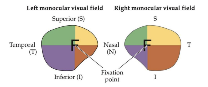
superior, inferior
Horizontal line divides retina into ___ and ___ divisions
B
A binocular portion of the left visual field
→ nasal retina of the left eye and the temporal retina of the right eye
C
A binocular portion of the right visual field
→ nasal retina of the right eye and the temporal retina of the left eye
A and D
Monocular portions of the left and right visual fields (what each eye sees)
→ left (_) and right (_) nasal retinas
left, right
The (left/right?) half of the visual world (coming from either the left or right eye) is represented in the (left/right?) half of the brain
Nasal diviision
Ganglion cells in the ___ ___ of each retina cross in the optic chiasm.
temporal division
Projections of cells that lie in the ___ ___ stay on the same side
opposite
The optic tract carries information from the (same/opposite?) side visual field
fovea
The ___ is represented disproportionally large in the posterior striate cortex, while peripheral stimuli are represented further anterior
upper, below, lower, above
The (upper/lower?) visual field is represented (above/below?) the calcarine sulcus, the (upper/lower?) visual field is represented (above/below?) the calcarine sulcus.
Meyer’s loop
(temporal cortex) → carries info about contralateral superior visual field (inferior retinal quadrants)
Course of the optic radiation to the striate cortex
Baum’s loop
(parietal cortex) → carries info about contralateral inferior visual field (superior retinal quadrants)
Course of the optic radiation to the striate cortex
Scotomas
small visual field deficit
Anopsias
large visual field deficits
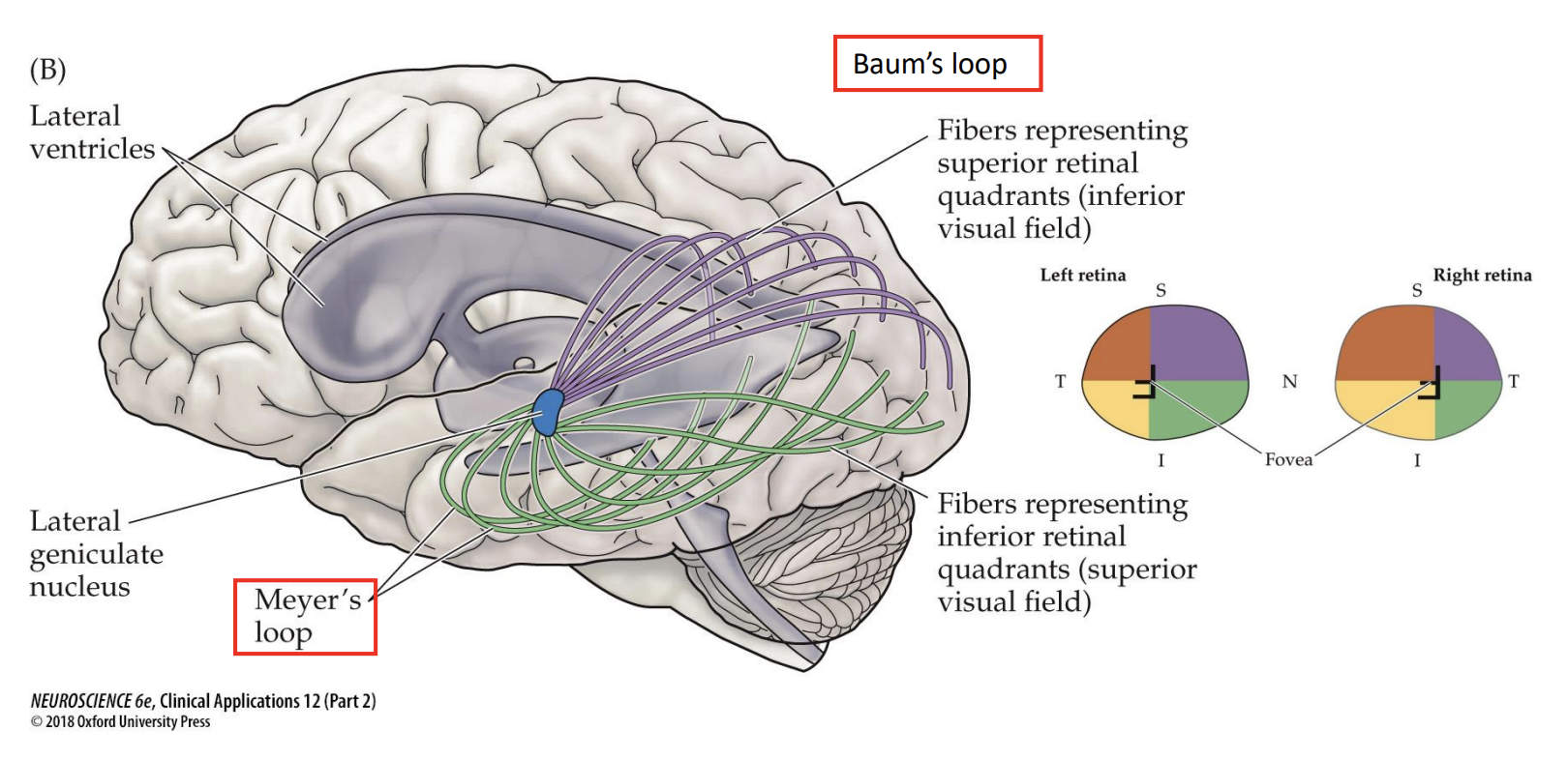
Optic Radiation
Paths of fibers and axons that represent the superior and inferior visual fields
Bitemporal (heteronomous) hemianopsia
• Often the result of pituitary tumors
• Vision loss confined to the temporal visual field of each eye
• The parts of the visual field that are lost in each eye do not overlap
• Peripheral vision is lost
lesion in chiasm → nasal component
Left homonymous hemianopsia
• Loss of sight in the left visual field
• Blindness in the temporal visual field of the left eye & the nasal visual field of the right eye
lesion after chiasm → left (right) visual field
Visual field deficits
Spatial relationships in the retinas are maintained in the central projections → helps to pinpoint the site of neurological damage
Left superior quadrantanopsia
lesion along Meyer’s loop
Loss of visual information from the superior visual field
Left optic tract
A researcher interested in studying how the brain adapts to complete loss of the right visual field should lesion which region to create an animal model of this anopsia?
Magnocellular layers
(layers 1-2, large neurons)
Target in primary visual cortex → layer 4Cα
Parvocellular layers
(layers 3-6, small neurons)
Target in primary visual cortex → layer 4Cβ
Koniocellular
layers in between
Target in primary visual cortex → patchy 2/3
Parvocelluar Pathway
Important for spatial resolution
Detailed analysis of shape, size and color of object
P ganglion cells
Have small receptive fields and are slow → sustained responses
Magnocellular pathway
Critical for high temporal resolution
→ Evaluate location, speed, and direction of a fast-moving object
M ganglion cells
Have large receptive fields and are fast → respond only transiently (short time)
Koniocellular pathway
Also transmits some color information
primary visual cortex
Cells in this area respond selectively to oriented bars/edges → The “preferred” orientation is the orientation to which a cell is most responsive
Layer 4C
Axons from LGN terminate primarily on spiny stellate cells of ___ ___
→ axons convey LGN activity to other cortical layers
Layers 2/3
Pyramidal cells in these layers project to higher order (visual) cortices (red in E)
Layers 5/6
Pyramidal cells in these layers project to subcortical areas, including LGN and superior colliculus
Columns
feature-selective (e.g. orientation)
Microelectrode penetrations perpendicular to the cortical surface encounter ___ of neurons that have similar receptive field properties
• Receptive fields are centered on the same region of visual space (upper right)
• Exhibit similar orientation preferences
Combining inputs from two eyes
→ In the LGN the inputs from the two eyes are separated in different layers.
→ Distinction is maintained in visual cortex in ocular dominance columns (layer 4 inputs).
→ Neurons outside layer 4 integrate inputs from both eyes
far cells
discharge to retinal disparities beyond fixation point (point C)
Type of binocular neuron in the primary visual cortex
near cells
retinal disparities that arise from points in front of the plane of fixation (point A)
Type of binocular neuron in the primary visual cortex
tuned zero
respond selectively to points that lie on the plane of fixation (point B)
Type of binocular neuron in the primary visual cortex
Binocular Neurons
Relative activity in these classes of neurons mediates the sensation of stereoscopic depth
MT (middle temporal area)
Cells in this area respond selectively to direction of a moving edge
V4
Cells in this area respond selectively to color without regard to direction of movement
Cerebral akinetopsia
• Unable to appreciate the motion of objects
• Ex: difficulty pouring tea into a cup because the fluid seemed to be “frozen”
• Problems crossing street – can’t judge movement of approaching cars
Cerebral achromatopsia
• Lose the ability to see the world in color
• Shades of gray
• Normal cone functioning
Dorsal Pathway
Spatial awareness and guidance of actions (e.g., reaching) → Selectivity for direction and speed of movement.
Spatial vision “where” pathway
Located in the parietal lobe
Ventral Pathway
Object recognition and form representation → Selectivity for shape, color, and texture → Preferential response to faces and objects.
Does not merely provide a description of the elements in the visual world → it also plays a crucial role in judging the significance of these elements.
Objection recognition “what” pathway
Located in the temporal lobe