medial epicondyle of humerus
forearm flexors origin
lateral epicondyle of humerus
forearm extensor origin
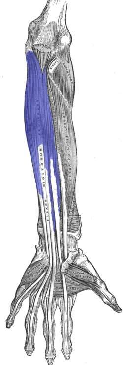
flexor carpi radialis
flexor carpi ulnaris
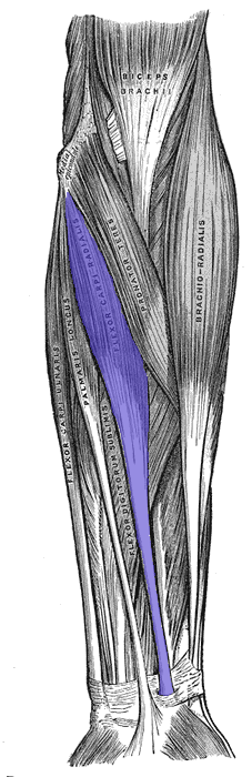
flexor digitorum superficialis
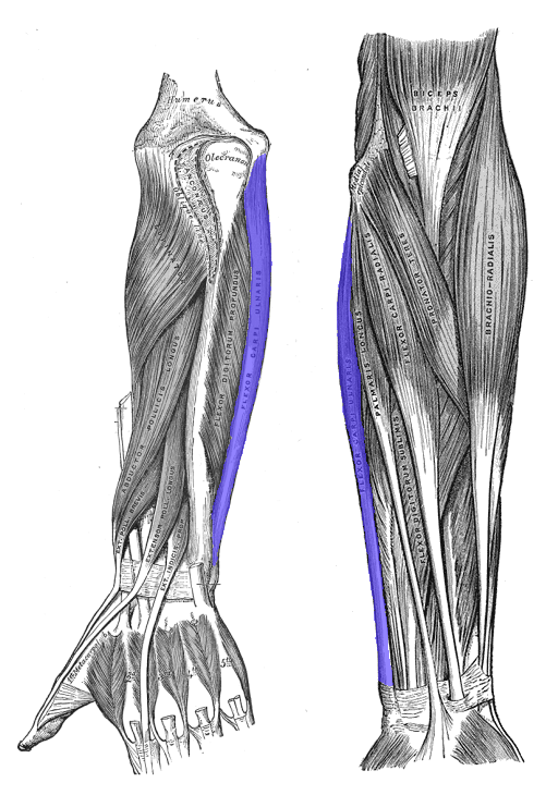
flexor digitorum profundus
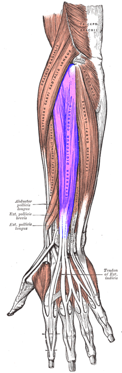
extensor digitorum communis
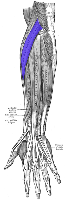
extensor carpi radialis longus
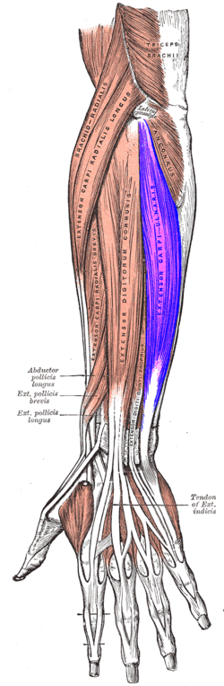
extensor carpi ulnaris
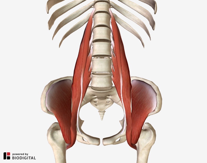
iliopsoas
major flexor of hip joint
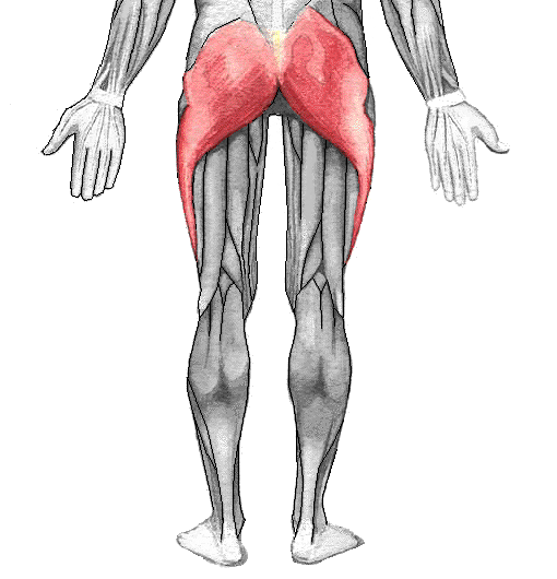
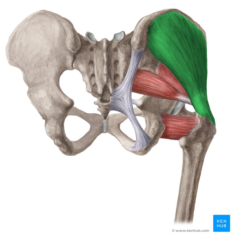
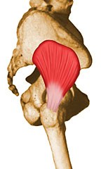
gluteus maximus
O: gluteal surface of ilium
I: gluteal tuberosity
F: extension, external rotation, abduction and adduction of the thigh
ulna
pinky side
radius
thumb side
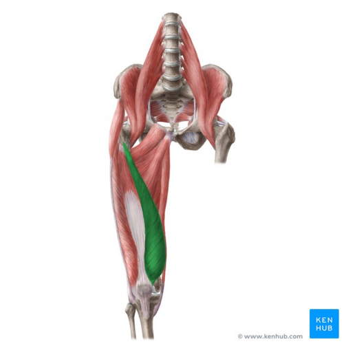
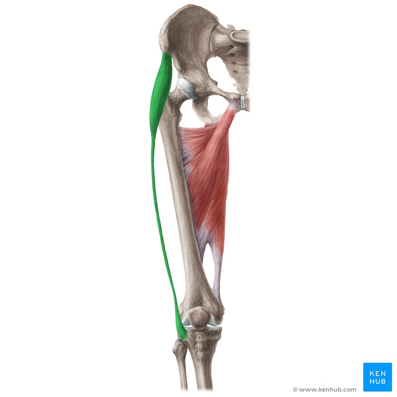
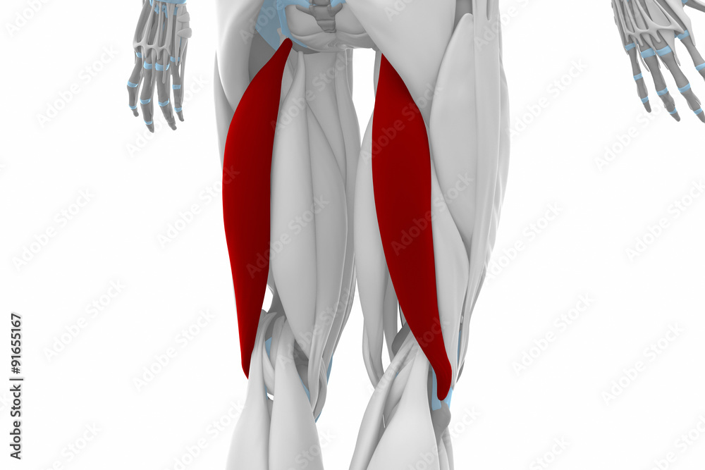
rectus femoris
O: the anterior inferior iliac spine and the other from the ridge of the acetabulum
I: quad tendon at patella
F: only muscle of the quadriceps to cross both the hip and knee joints; flexes the thigh at the hip joint, and extends at the knee joint
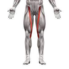
sartorius
O: ASIS
I: tibia
F: flexes hip and knee while laterally rotating leg
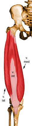
vastus lateralis
O: femur
I: QT (quad tendon)
F: stabilize knee joint
vastus medialis
O: top of femur
I: QT
F: stabilize knee joint
vastus intermedius
under rectus femoris
F: stabilize knee joint
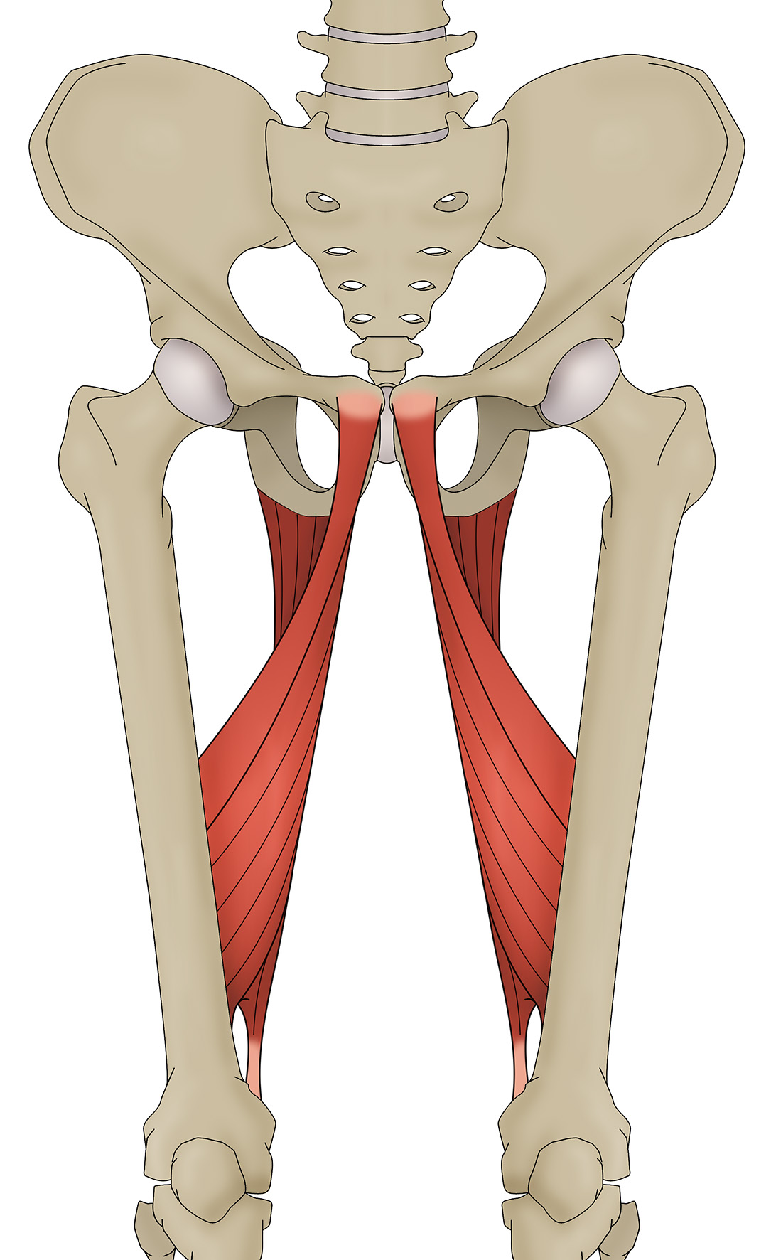
adductor longus
I: Middle third of linea aspera of femur
F: adduct the thigh at the hip joint
adductor magnus
I: Linea aspera and adductor tubercle of femur
F: dynamic stabilizer of the pelvis and femur as well as a prime mover of the femur into adduction
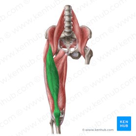
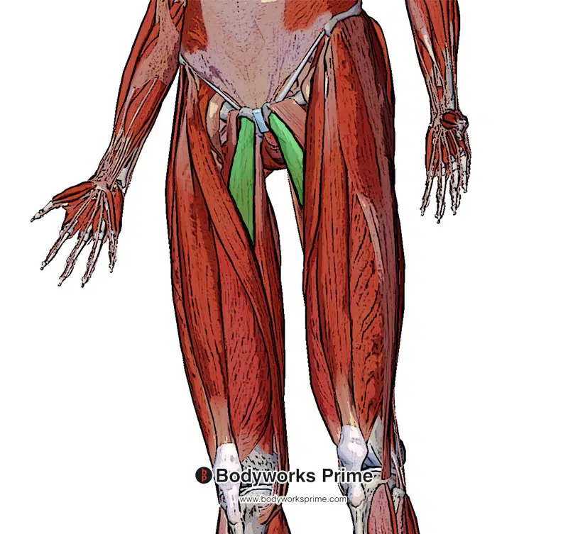
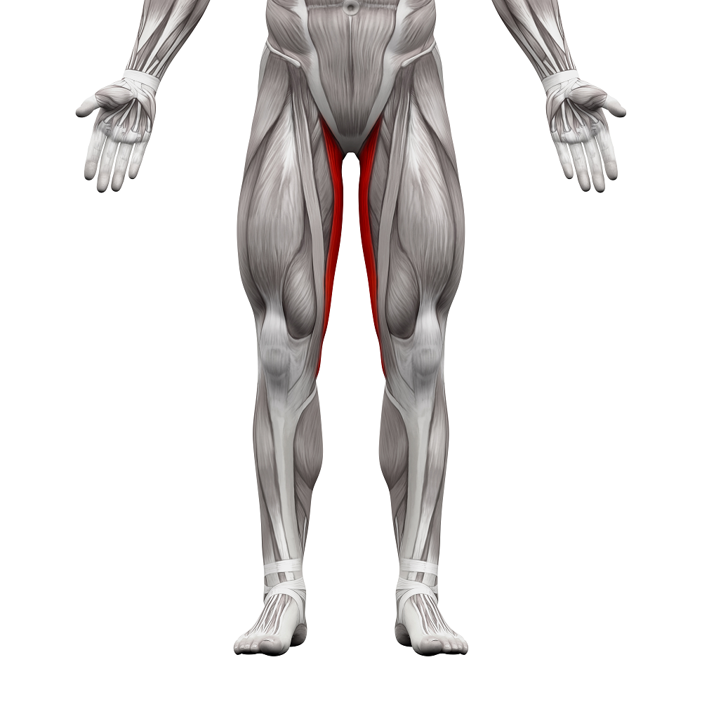
gracilis
I: inserts on the medial tibia
F: hip adduction, knee flexion, and knee internal rotation
gluteus medius
antagonists to adductor group
tensor fascia latae
O: anterior superior iliac spine
I: lateral condyle of tibia
F: thigh internal rotation, weak abduction, external rotation, weak flection and extension, stabilizes hip & knee joints
gluteus medius
O: gluteal surface of ilium
I: greater trochanter
F: abductor
gluteus minimus
O: gluteal surface of ilium
I: greater trochanter
F: abductor
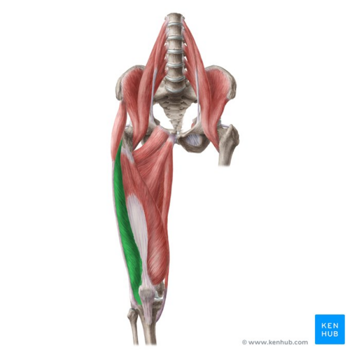
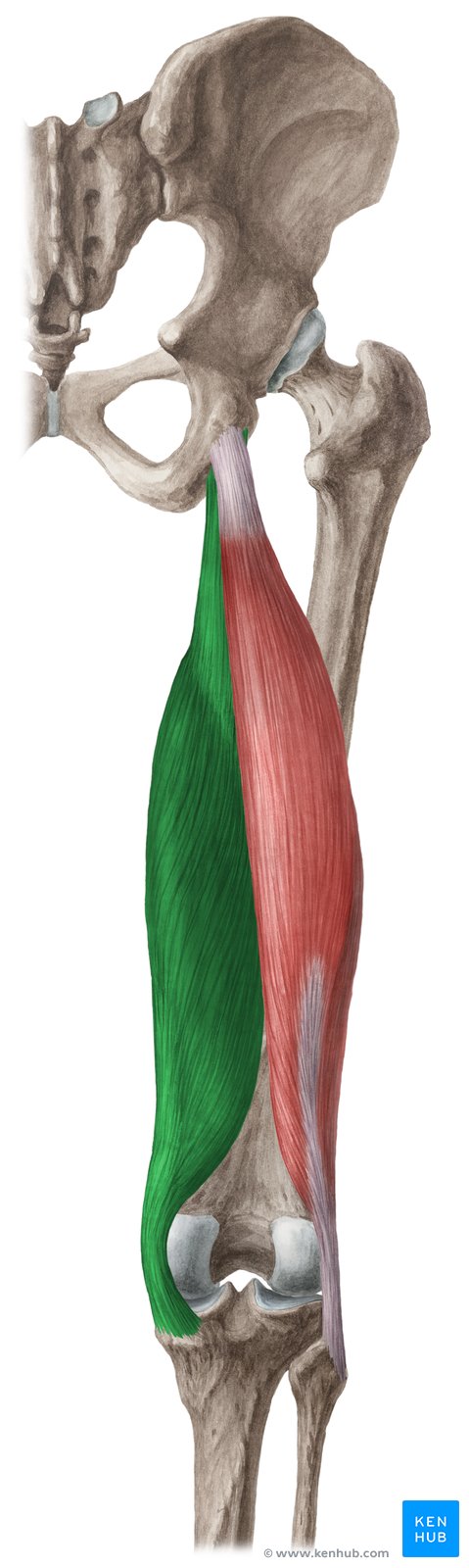
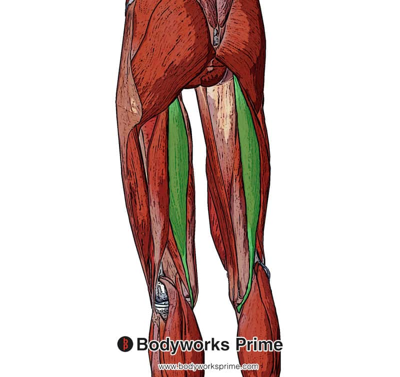
biceps femoris
O: ischial tuberosity
I: tibia and fibula
F: movement at both the hip joint and knee joint
semimembranosus
O: ischial tuberosity
I: tibia
F: flexion and internal rotation of the knee joint
semitendinosus
O: ischial tuberosity
I: tibia
F: extend at the hip and flex at the knee
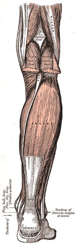
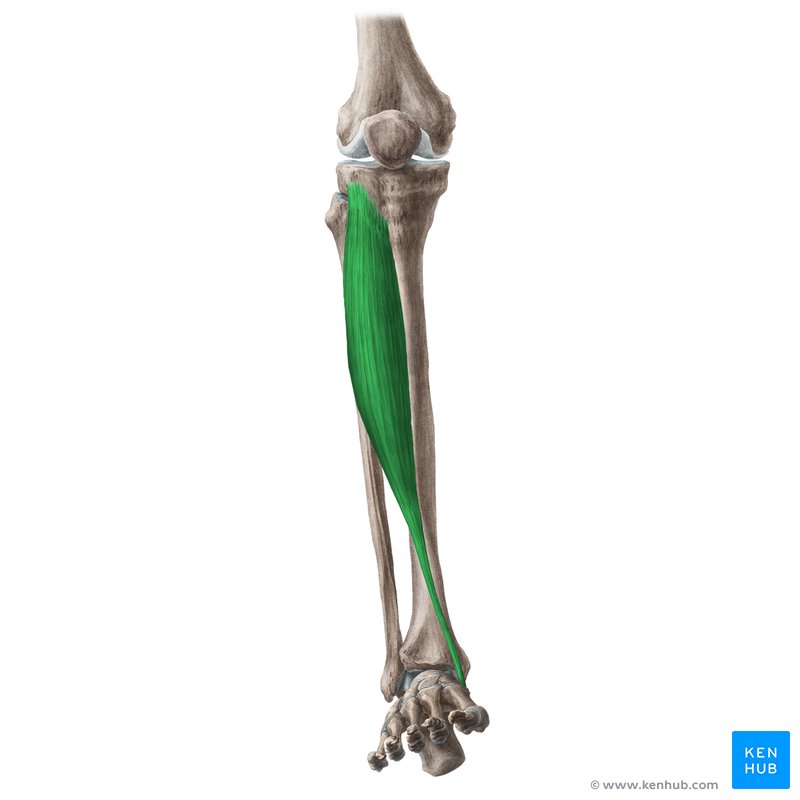
gastrocnemius
O: femoral condyles
I: achilles tendon
F: plantar flexor
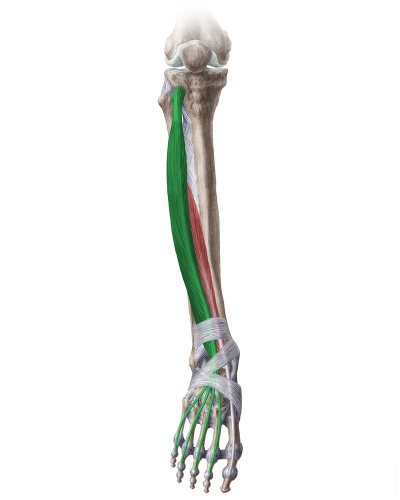
soleus
O: head of fibula
I: posterior surface of calcaneu
F: plantar flexors
tibialis anterior
F: dorsiflexes the foot at the talocrural joint and inverts it at the subtalar joint
extensor digitorum
F: extend the foot at the ankle, along with the four smallest toes
fibularis longus
I: medial cuneiform bone
F: produce the plantarflexion and eversion of the foot on the ankle joint
fibularis brevis
I: tuberosity of the 5th metatarsal bone
F: plantar flexion and eversion
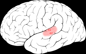
wernickes area
region of the brain that contains motor neurons involved in the comprehension of speech
brocas area
contains neurons involved in speech function
primary motor cortex
the area that provides the most important signal for the production of skilled movements
primary sensory cortex
processing afferent somatosensory input and contributes to the integration of sensory and motor signals necessary for skilled movement
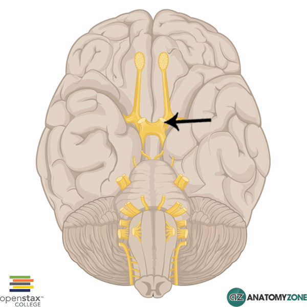
optic nerve
gray matter
brain - superficial tissue
spinal cord - deep tissue
white matter
brain - deep tissue
spinal cord - superficial tissue
CN I (olfactory)
Modality: Sensory
Function: S-smell
Exit from Skull: olfactory fossa from cribiform plate
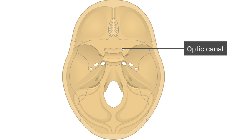
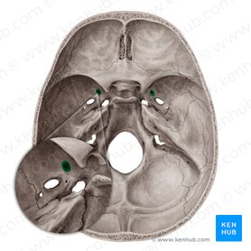
CN II (Optic)
Modality: Sensory
Function: S-vision and visual field
Exit from Skull: optic foramen
CN III (Oculomotor)
Modality: motor
Function: M- eye muscles and pupil
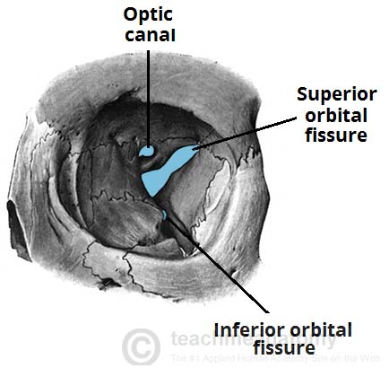
Exit from Skull: superior orbital fissure
CN IV (trochlear)
Modality: motor
Function: eye muscle (superior oblique)
Exit from Skull: superior orbital fissure
CN V (trigeminal)
3 branches
V1 (ophthalmic)
Modality: sensory
Function: S- forehead, around eyes, cornea
Exit from Skull: superior orbital fissure
V2 (Maxillary)
Modality: sensory
Function: S- teeth, skin over maxilla
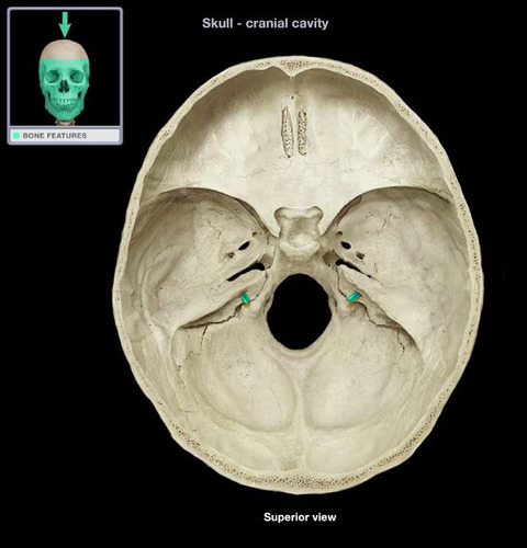
Exit from Skull: foramen rotundum
V3 (mandibular)
Modality: sensory and motor
Function: S-teeth of lower jaw and tongue M- mastication
Exit from Skull: foramen ovale
CN VI (Abducens)
Modality: motor
Function: M-eye muscle (lateral rectus)
Exit from Skull: superior orbital fissure
CN VII (Facial)
Modality: sensory and motor
Function: S-taste in anterior part of tongue, external ear M- facial expression
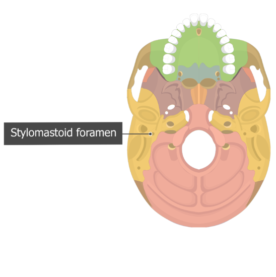
Exit from Skull: internal auditory meatus and stylomastoid foramen
CN VIII (Vestibulocochlear)
Modality: sensory
Function: S-hearing and balance
Exit from Skull: internal auditory meatus
CN IX (Glossopharangeal)
Modality: sensory and motor
Function: S- taste in posterior part of tongue, pharynx M- pharyngeal muscles
Exit from Skull: jugular foramen
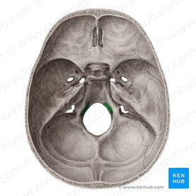
CN X (vagus)
Modality: sensory and motor
Function: S-thoracic and abdominal viscera
M- parasympathetic to abdominal organs
Exit from Skull: jugular foramen
(only cranial nerve to exit the head and neck region)
CN XI (Spinal Accessory)
Modality: motor
Function: M- SCM and trapezius
Exit from Skull: jugular foramen
CN XII (Hypoglossal)
Modality: motor
Function: M- tongue and throat muscles
Exit from Skull: hypoglossal canal
CN III Oculomotor
Innervates 4 of 6 eye muscles
Superior rectus, inferior rectus, medial rectus, and inferior oblique muscle
Controls pupil dilation (sympathetic) and constriction (parasympathetic)
CN IV Trochlear
innervates superior oblique muscle
CN VI Abducens
innervates lateral rectus (abducts eye)
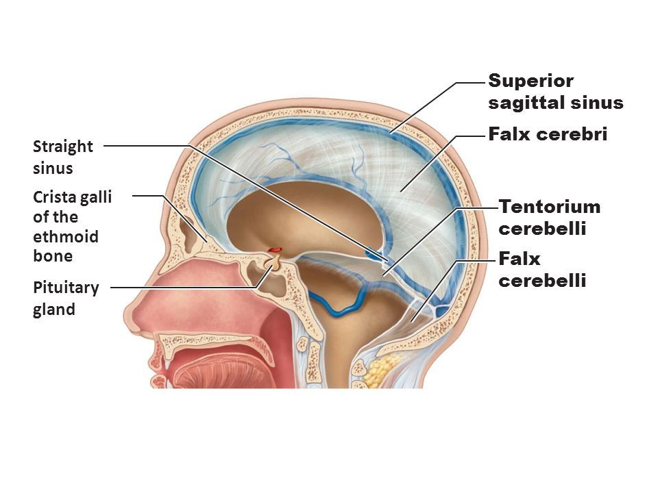
falx cerebri
a large, crescent-shaped fold of dura mater that descends vertically into the longitudinal fissure between the cerebral hemispheres of the human brain
tentorium cerebelli
an invagination of the meningeal layer of the dura mater that separates the occipital and temporal lobes of the cerebral hemispheres from the cerebellum and brainstem
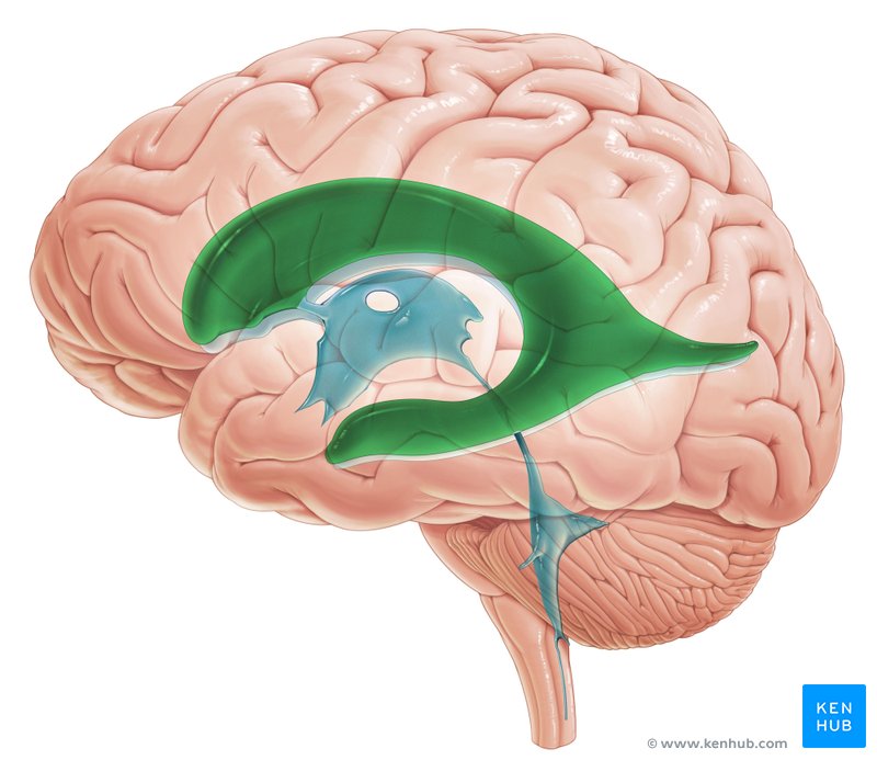
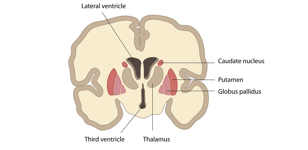
lateral ventricle
Two of them
Go from frontal to occipital lobe
Separated by septum pellucidum
At the bottom there are interventricular foramen that led to third ventricle
third ventricle
found at level of thalamus and hypothalamus
cerebral aqueduct
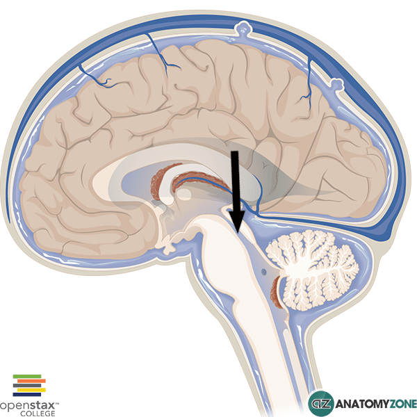
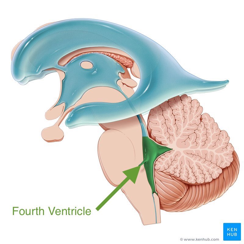
fourth ventricle
In between cerebellum and backside of brainstem
Has opening for CSF (medial and lateral apertures) to flow around the arachnoid layer
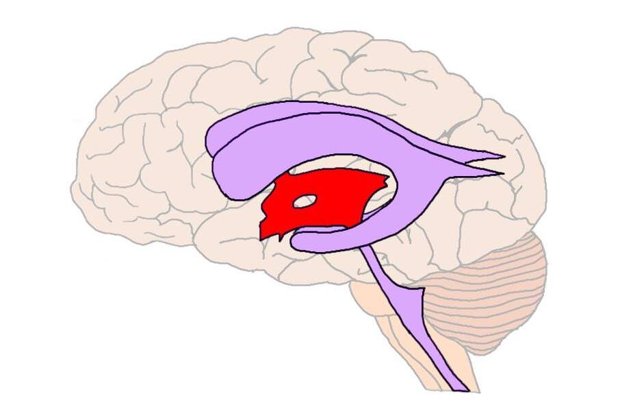
choroid plexus
Surrounded by ependymal cells
Blood vessels inside each ventricle
Forms CSF from blood
Chemical stability
Keeps brain afloat
Impaired drainage or overproduction of CSF
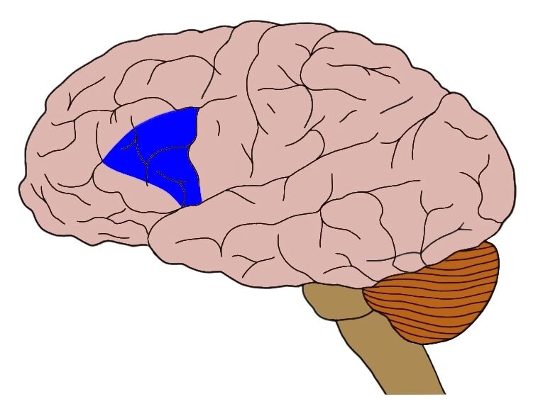
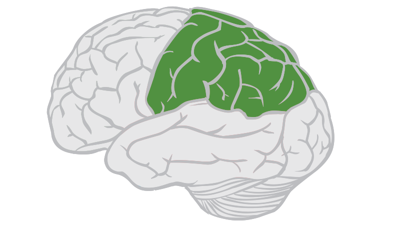
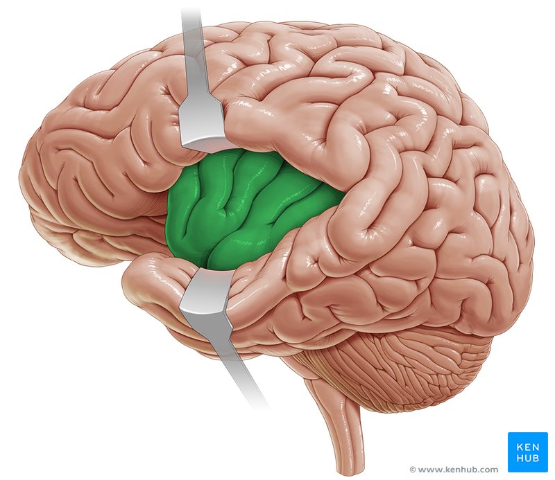
parietal lobe
Major centers for sensory input other than hearing, balance, smell, vision
Touch, temperature, pain, two-point discrimination
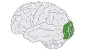
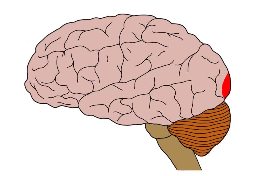
occipital lobe
Coordinates with parietal and temporal lobes
Vision center
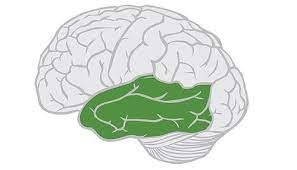
temporal lobe
Memory storage and recall
insula
Behind temporal lobe
4-5 gyri wide
Deepest lobe of cerebrum
Plays a role in addiction and compulsive behavior
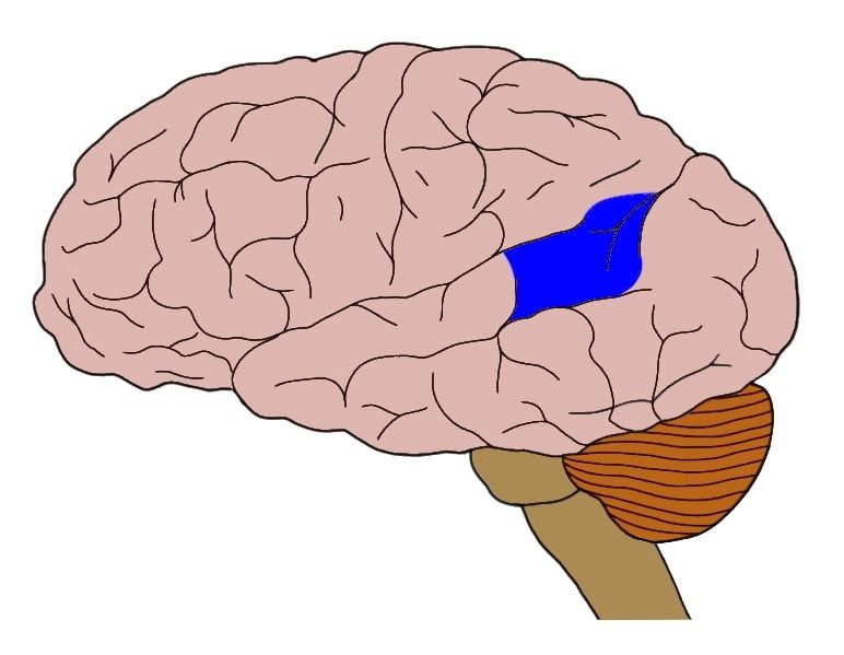
premotor area
very small region; controls learned motor skills (especially those of patterned nature), coordinates multiple muscle groups to work at the same time
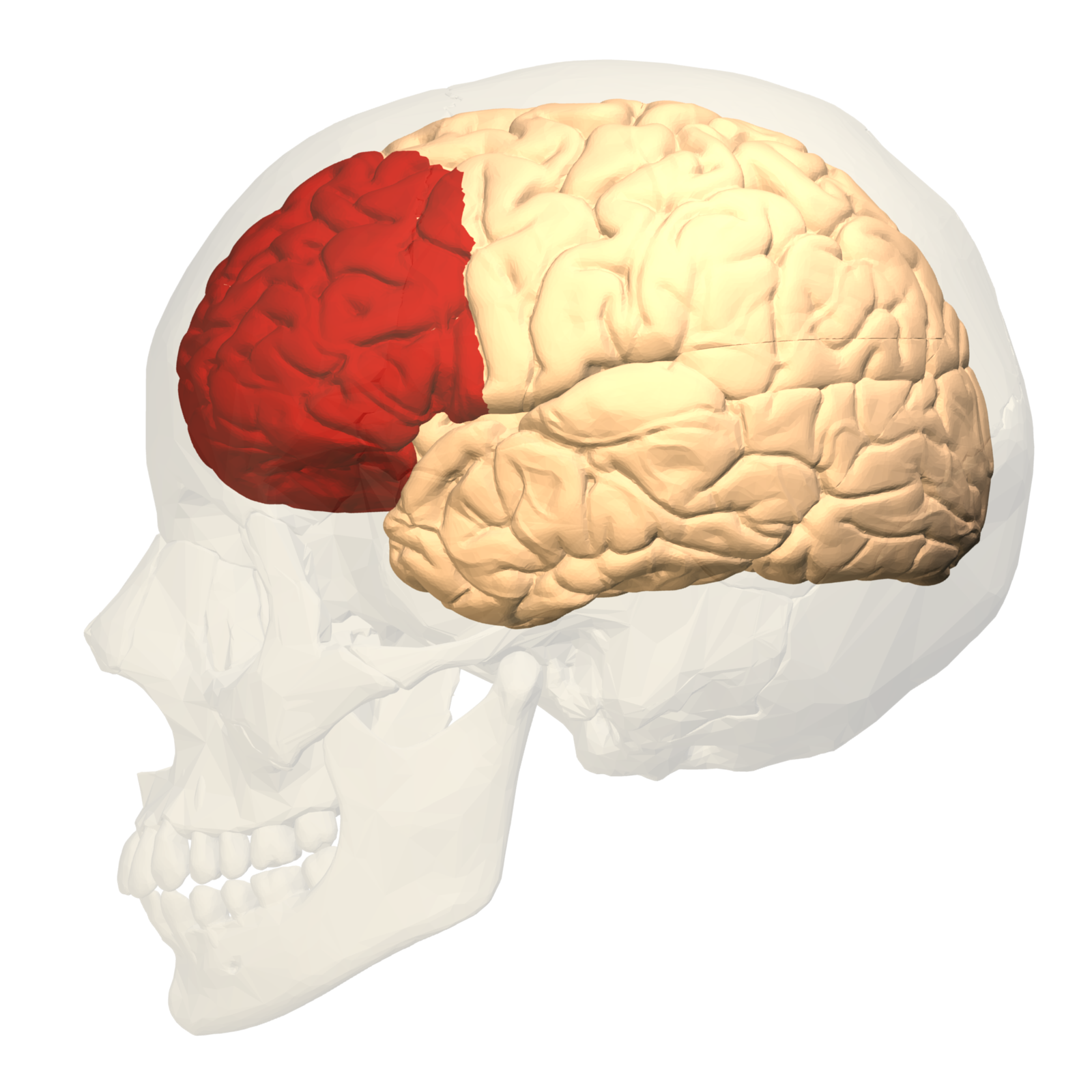
prefrontal cortex
decision making, logic, conscience (right and wrong)
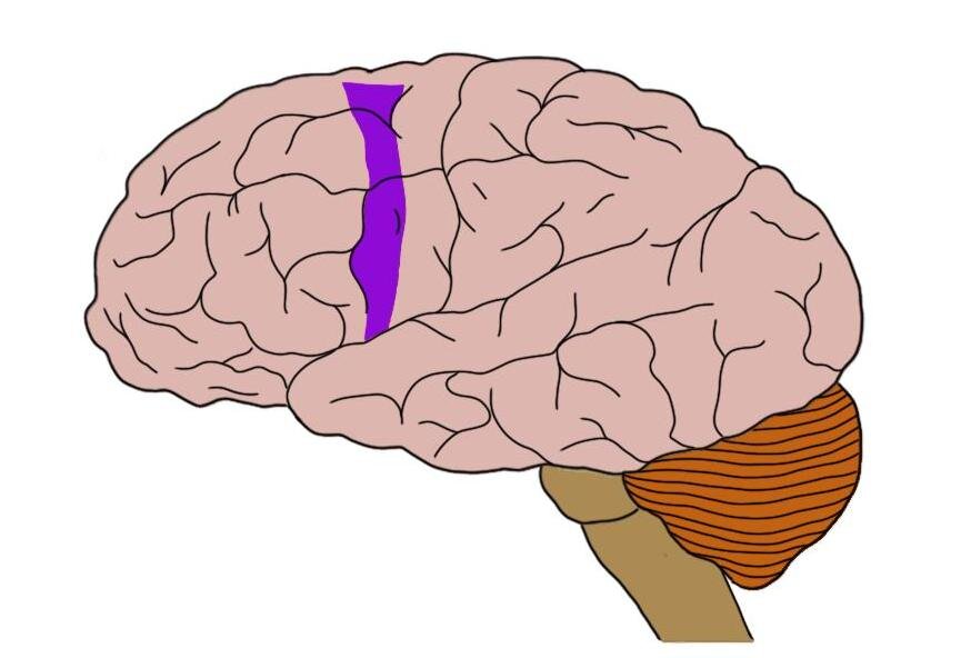
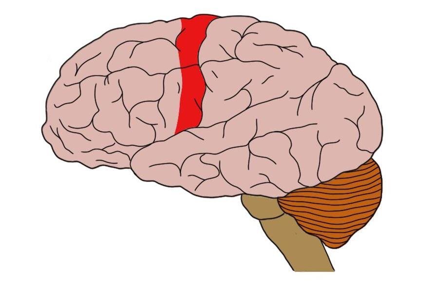
primary motor cortex
precentral gyrus on both sides
Bundle of axons that go through the brain and brainstem that become white matter motor pathways in spinal cord
Areas that have fine tune control over have large surface area on cortex
Face, hands, arm
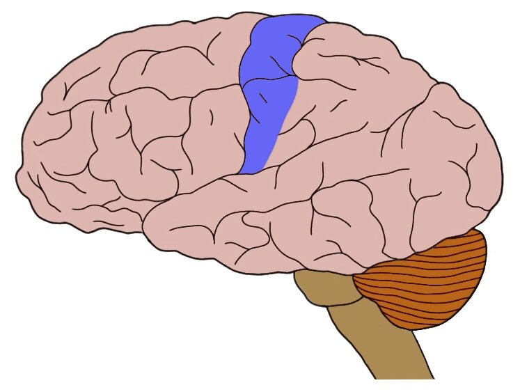
primary sensory cortex
postcentral gyrus on both sides
Areas that have fine tune sensory ability have large surface area on cortex
Hands, face, teeth
sensory association area
can perceive and differentiate emotional aspects of touch and sensations
primary occipital cortex
visual processing, through optic nerve
Visual association area right above it
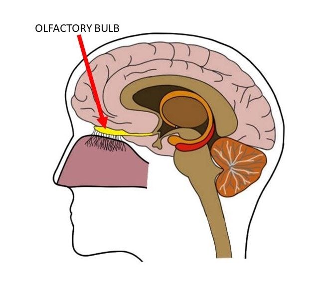
primary olfactory cortex
inside temporal lobe near frontal lobe
Sense of smell is only sense that does not have mandatory stop-over in thalamus
Continuations of olfactory fibers into hypothalamus
auditory association area
top part of temporal lobe
primary gustatory cortex
inferior part of parietal lobe, sense goes through thalamus into here for processing
primary vestibular cortex
buried inside on the posterior border of the insula, parietal, and temporal lobe, conscious perception of balance
left brain
Verbal memory
Speech (motor) area
Right hand control
Feeling shapes w right hand
Superior language and mathematic comprehension (Wernicke's area)
Right visual field
right brain
Memory for shapes (limited language comprehension)
Left hand motor control
Feeling shapes w left hand
Musical ability
Recognition of faces and spatial relationships
Left visual field
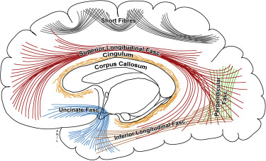
association fibers
Fibers that run back and forth in the same hemisphere
Short and long
commissural fibers
Travel between hemispheres
Make up corpus callosum
projection fibers
travel down from brain to spinal cord
- Most carry motor signals
Travel up from spinal cord
- Most carry sensory signals
Many cross or decussate
- In medulla where they cross before they reach the spinal cord
- 90% of outgoing motor signals cross (left side controls right side motor skills)
basal nuclei
Deep gray matter
Send and receive input regarding movements between brainstem, cerebrum, and cerebellum
Helps inhibit unwanted muscle contractions of skeletal muscles
Disorders of basal nuclei can suffer from resting tremors
CNS
Central
Brain and spinal cord
Integration and control centers
PNS
peripheral
ANS
Autonomic
Somatic and autonomic
Parasympathetic and sympathetic (divides autonomic more)
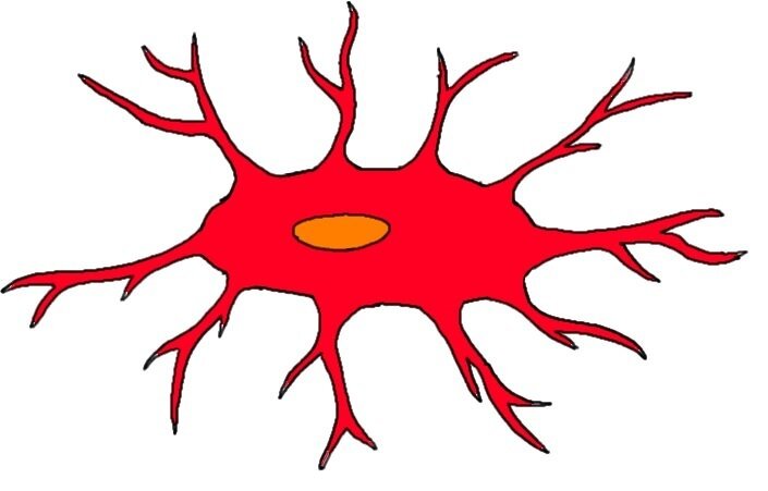
neuroglial cells
Support and protect neurons
Can undergo mitosis
Cells that cause brain cancer through unregulated mitosis
ogliodendrocyte
Myelinates and insulates CNS axons
Allows faster action potential propagation along axons in the CNS
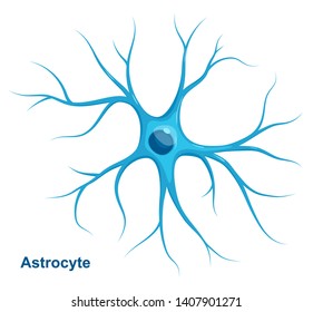
astrocyte
Most abundant
Form blood-brain barrier
Regulate interstitial fluid composition
Structural support and organization to CNS
Assists w neural development
Replicates to occupy space and dying neurons
ependymal cell
Lines ventricles of the brain and central canal of spinal cord
Assists in production and circulation of CSF (cerebrospinal fluid)
microglial cells
Phagocytic cells that move through the CNS
Protects CNS by engulfing infections agents and other potential harmful substances
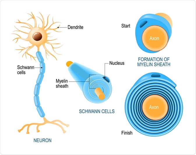
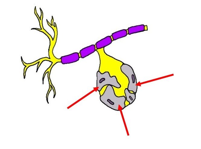
Schwann cell
Found in PNS
Myelinates and insulates PNS axons
Allows for faster action potential propagation along an axon in the PNS
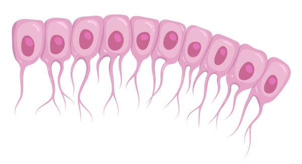
satellite cells
Dorsal root ganglia of spinal nerves
Found in PNS
Electrically insulates PNS cell bodies
Regulates nutrient and waste exchange for cell bodies in ganglia
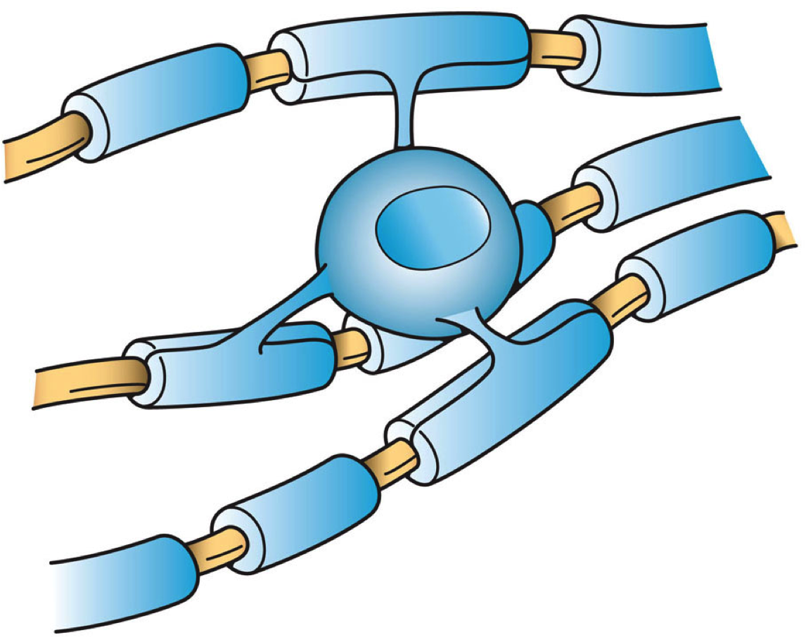
saltatory conduction
Allows action potentials to travel very quickly
Myelin is most important factor in increasing conduction velocity
Allows action potentials to jump over myelinated cells into each node of Ranvier down the axon
myelinated axon
Axons have lipid sheath around them
Created by oligodendrocyte or Schwann cells
Nodes of Ranvier, saltatory conduction
Speed of generation is much greater 5-100m/sec
unmyelinated axon
No myelin sheaths
Conduction is slower at 0.5-2m/sec
Tend to be shorter axons than myelinated ones
axon hillock
Electrical signal is generated down axon