Lecture 9 Nervous System Organization
1/33
There's no tags or description
Looks like no tags are added yet.
Name | Mastery | Learn | Test | Matching | Spaced |
|---|
No study sessions yet.
34 Terms
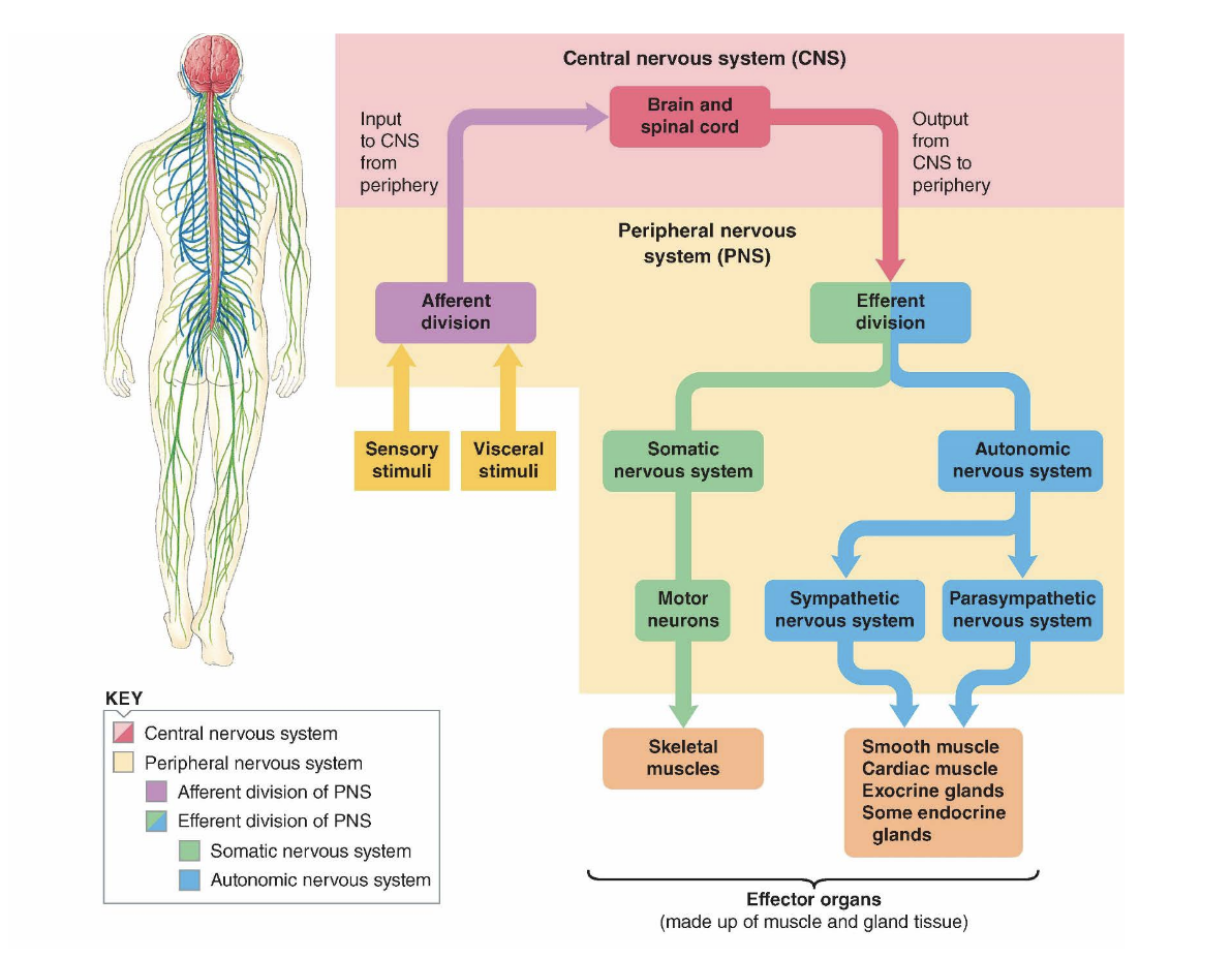
The CNS and the PNS
• Central Nervous System (CNS) – brain and
spinal cord
• Peripheral Nervous System (PNS) – nerve
fibres that carry information between the CNS
and other parts of the body
Three Functional Classes of Neurons
1. Afferent neurons
2. Efferent neurons
3. Interneurons
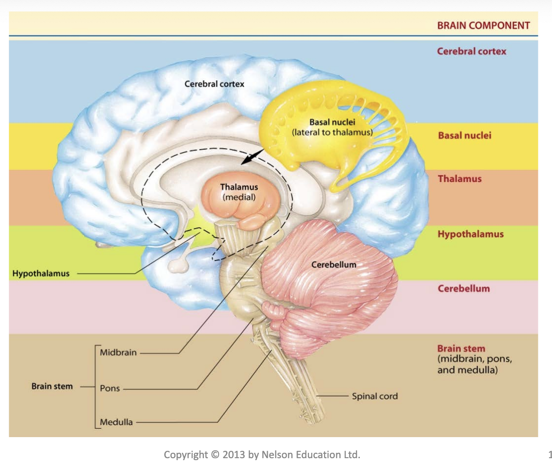
Structure and Function of the CNS
• Consists of the brain and spinal cord
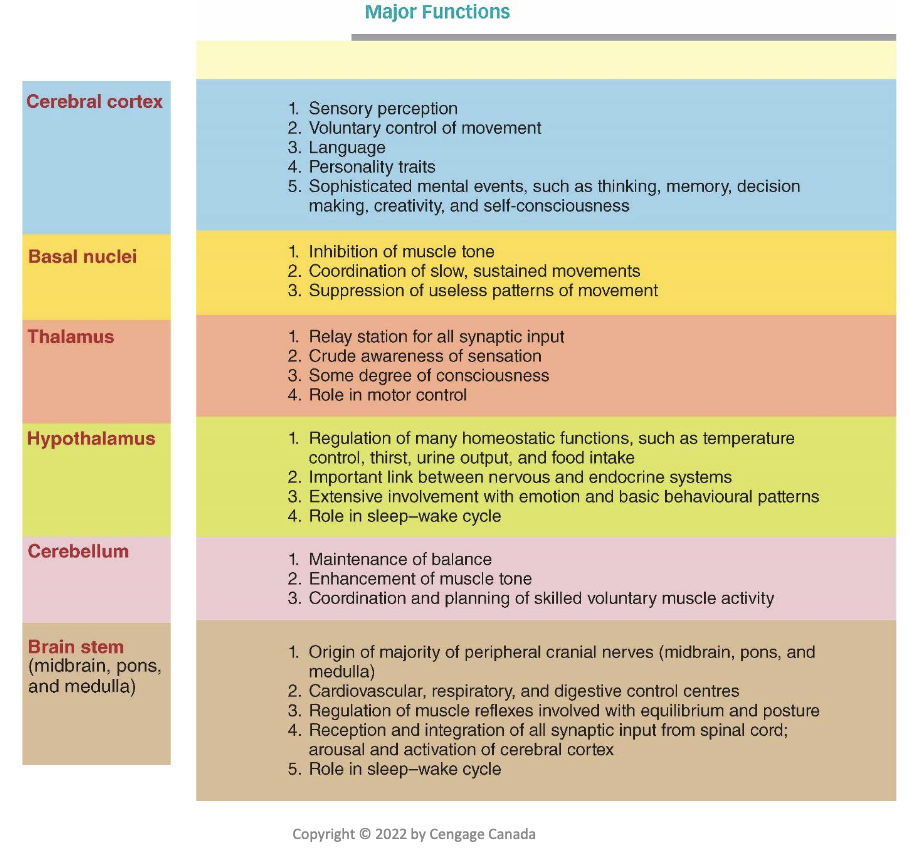
• Cognition refers to the act or process of “knowing,” including both awareness and judgment
• No part of the brain works in isolation from other brain regions
• Brain stem: oldest region of the brain and is continuous with the spinal cord
– Midbrain, pons, and medulla
– Controls many of life-sustaining functions, which are often referred to as “vegetative functions”
• Cerebellum: maintains proper position of the body in space and subconscious coordination
of motor activity; key role in learning skilled motor tasks
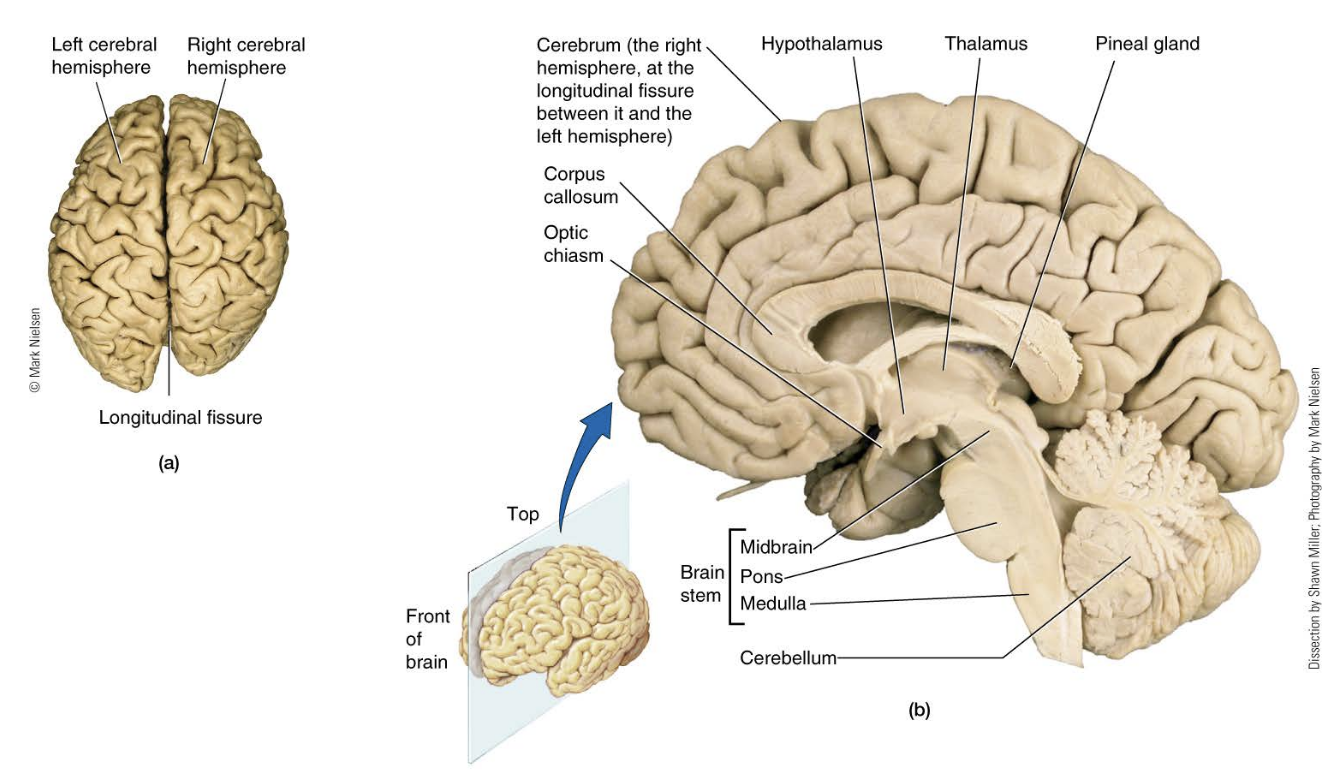
• Cerebrum: most highly developed in humans
and contains 80 percent of brain weight
– Right and left cerebral hemispheres
– Each hemisphere divided into lobes
– Outer layer is the convoluted cerebral cortex
• Covers inner core that houses the basal ganglia
• Most complex integrating area of the brain
Major Functions of diff parts of the brain
Protection of the CNS
• Glial Cells – 90 percent of the cells within CNS, also called neuroglia cells
– Do not conduct nerve impulses
• Astrocytes – most abundant glial cell and fill a number of critical functions
• Microglia – immune cells of the CNS
• Ependymal – line cavities and contribute to formation of CSF

Protection of the Delicate Central Nervous Tissue
• Enclosed in hard, bony structures
• Three protective and nourishing membranes
– Dura mater
– Arachnoid mater
– Pia mater
• Cerebral spinal fluid
– Surrounds the brain, provides shock absorption and exchange of materials
• Blood-brain barrier
Blood-Brain Barrier
• Shields brain from harmful changes in the blood
• Consists of endothelial cells
• Tight junctions prevent exchange across the capillary wall
• Lipid soluble substances such as oxygen and alcohol can penetrate cells
Role of Oxygen and Glucose
• Brain is highly dependent on constant blood supply
• Brain can not produce ATP in absence of oxygen
• Brain does not store glucose as well
• Damage occurs if oxygen is cut off for ~ 5 minutes or glucose for more than 15 minutes
Cerebral Cortex
• Largest portion of the human brain
• Two halves (right and left cerebral hemispheres joined by corpus callosum)
• Corpus callosum allows two hemispheres to communicate and cooperate with each other
• Thin outer shell of gray matter on each hemisphere
• Gray matter is predominately densely packaged neuronal cell bodies
• Bundles or tracts of myelinated nerve fibres constitute the white matter
– Transmit signals from one part of the cerebral cortex to another and to other regions of the CNS
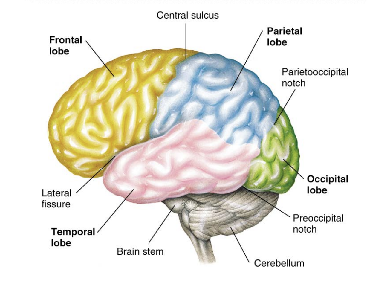
Lobes in the Cerebral Cortex
• Each hemisphere is divided into four lobes:
– Occipital lobes: house visual cortex
– Temporal lobes: house auditory cortex
– Parietal lobes: receive and process somatosensory input
– Frontal lobes: voluntary motor activity, speaking, and elaboration of thought
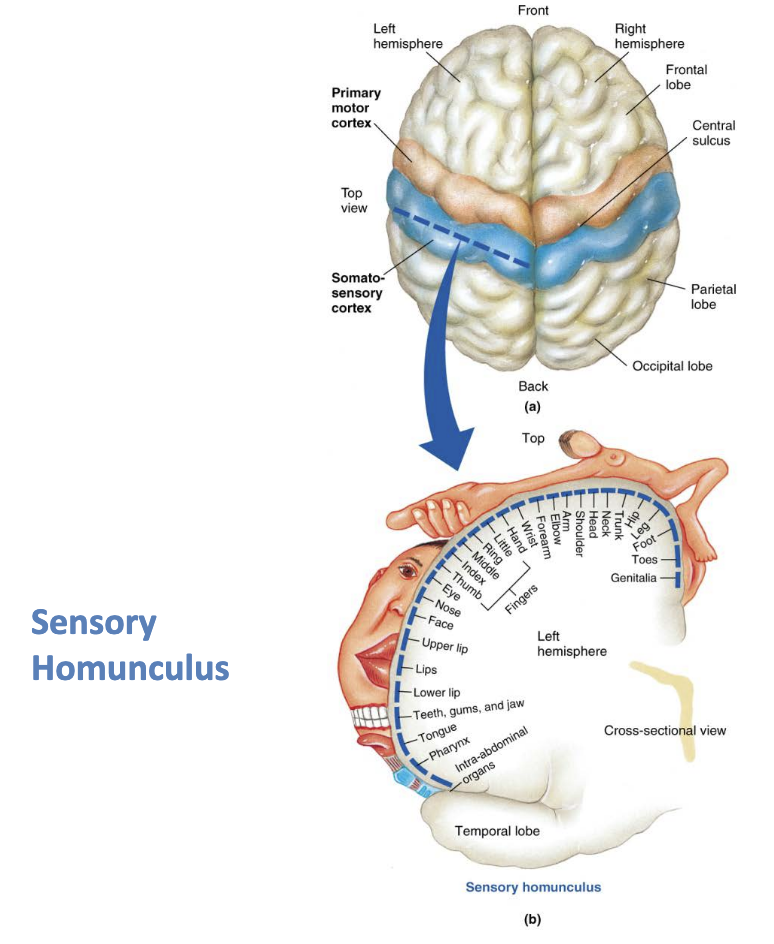
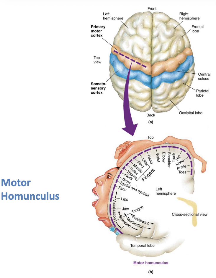
Sensory and Motor Homunculus
Homunculus: proportion of brain real estate does not match proportion of what it affects
space devoted to something is related to complexity of the process not to the size of the system
Plasticity and Neurogenesis
• Ability to change or be functionally remodelled in response to the demands placed on it
– More pronounced in early developmental years.
• When an area of the brain is destroyed, other areas of the brain may gradually assume some or all of the functions of the damaged region
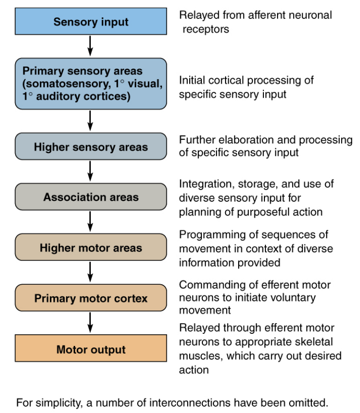
Sensory Input to Motor Output Block Diagram
Cerebral Hemispheres
• Left side is most commonly the dominant hemisphere for fine motor control, so most people are right-handed
– Left side excels in logical, analytic, sequential, and verbal tasks
– Right side excels in nonlanguage skills such as spatial perception and arts and music talents
Basal Ganglia
• Basal ganglia consist of several masses of grey matter located deep within the cerebral white matter
– Complex role in movement
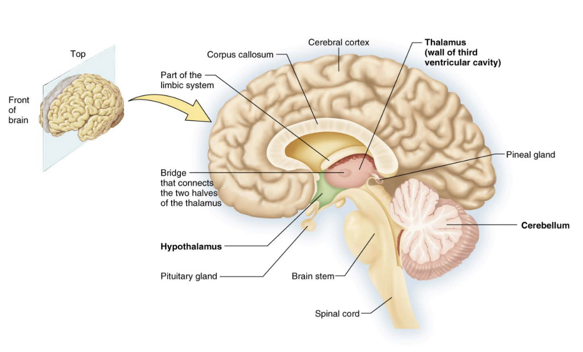
Thalamus
• The thalamus is deep in the brain near the
basal ganglia
– Serves as a relay station and integrating centre
Hypothalamus
• The hypothalamus is an integrating centre that lies beneath the thalamus
– Multifunctional – regulating the internal environment
Cross Section of Brain (Middle)
The Limbic System
• Limbic system surrounds the brain stem and is not a separate structure
• It is a interconnected ring of forebrain structures
– Emotion – amygdala
– Basic behavioural patterns
– Reward and punishment centres
Brain Stem parts
• Pons
• Medulla oblongata
• Midbrain
Five Functions of the Brain Stem
• Majority of cranial nerves arise from the brain
stem
• Contains centers that control cardiovascular,
respiratory, and digestive function
• Regulates postural muscle reflexes
• Reticular activating system (RAS): controls the
overall degree of cortical alertness
• Plays a role in the sleep–wake cycle
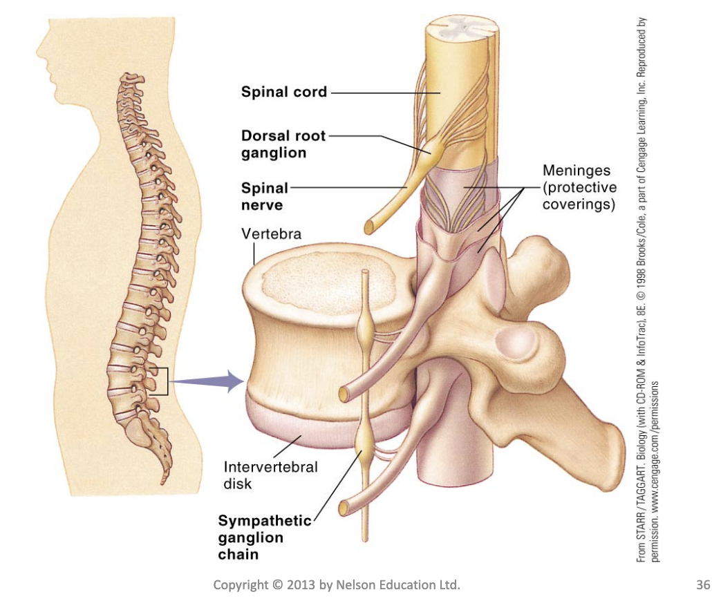
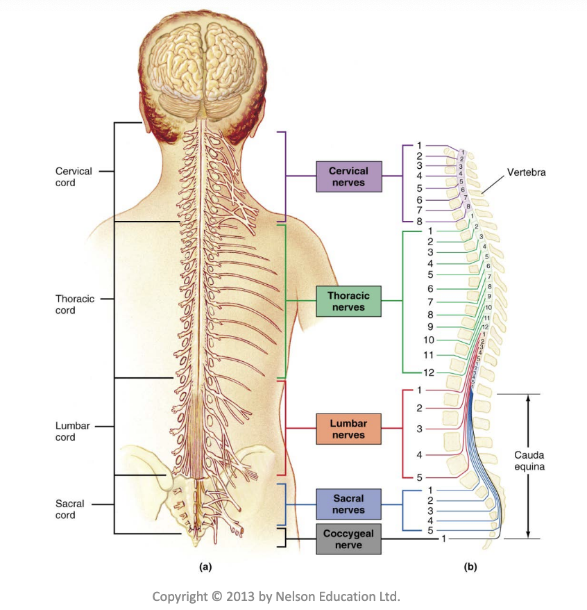
Spinal Cord
• Spinal cord extends from brain stem
• Paired spinal nerves emerge from the spinal cord through spaces in vertebrae
• Thick bundle of elongated nerve roots within the lower vertebral canal is called the cauda equina
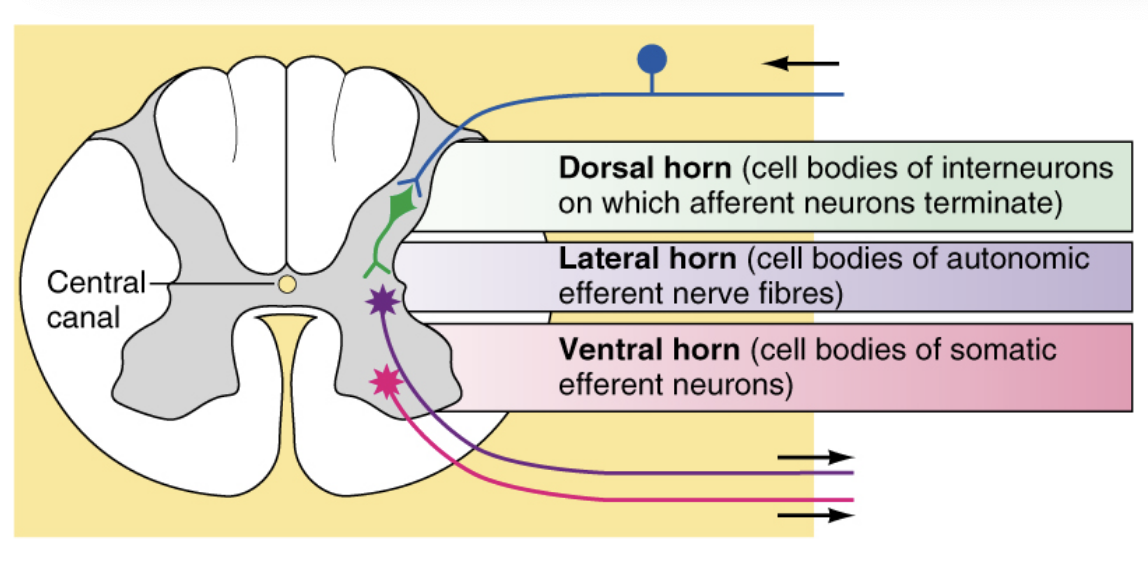
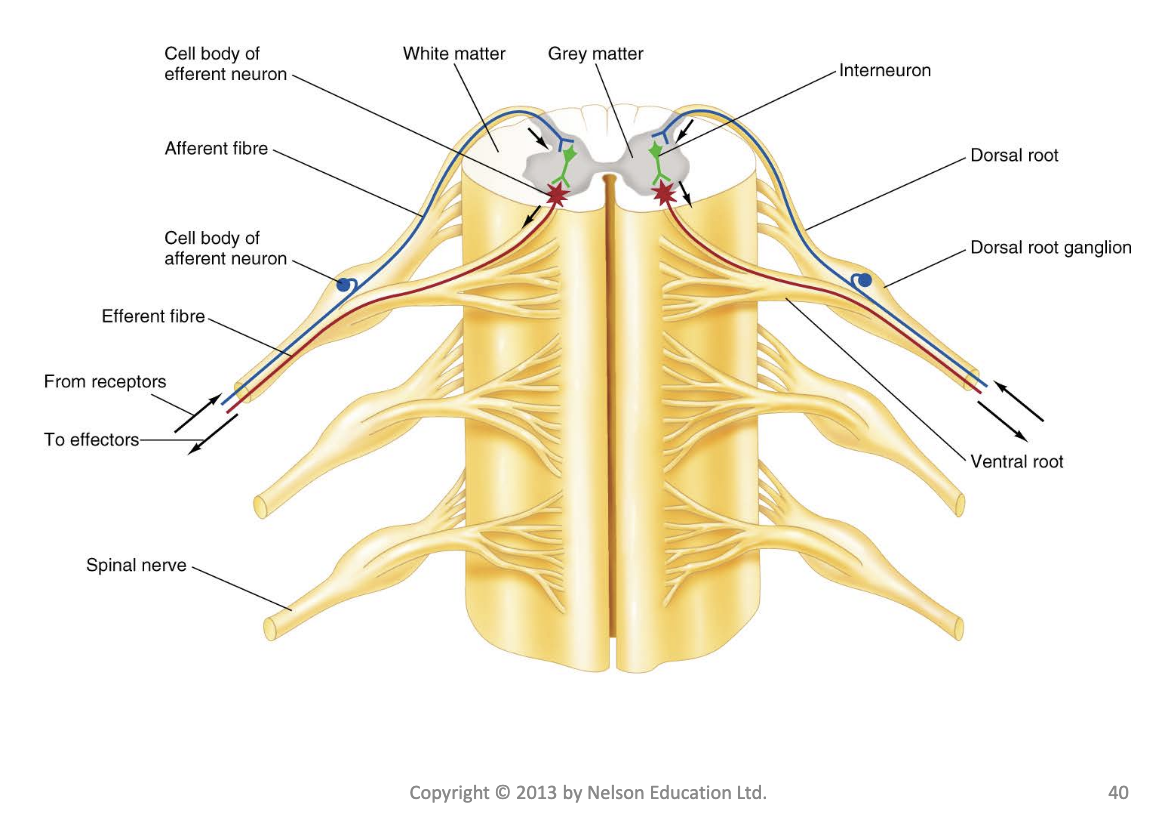
Organization of Neural Tissue in the Spinal Cord
white matter outside to grey matter inside - opposite to the brain
• Spinal Cord White Matter
– Organized into tracts (ascending and descending tracts)
– Located in the outer section of the spinal cord
• Spinal Cord Grey Matter
– Dorsal (posterior) horn
– Ventral (anterior) horn
– Lateral horn
– Located in inner section of the spinal cord
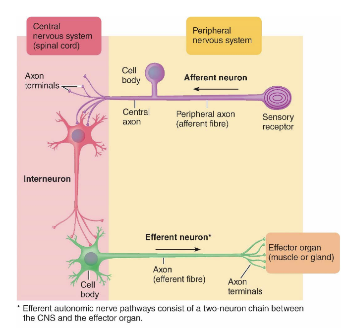
Spinal Nerves
• Spinal nerves connect with each side of the spinal cord by a dorsal root and a ventral root
• A spinal nerve consists of both afferent and efferent fibres
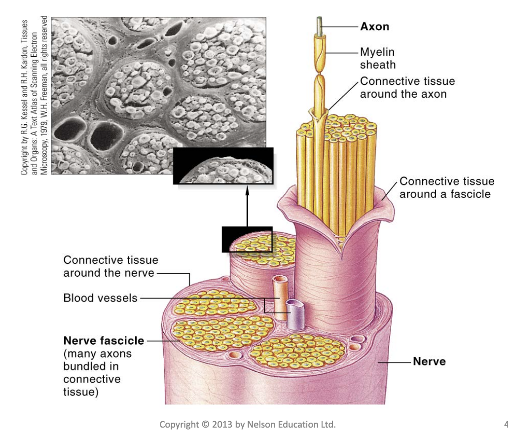
– A nerve is a bundle of peripheral neuronal axons enclosed by a connective tissue covering and following the same pathway
The Peripheral Nervous System
The PNS consists of nerve fibres that carry information between the CNS and other parts of the body
• Afferent division of the PNS: Sends information from internal and external environment to CNS (i.e. carries signals to CNS).
• Efferent division of the PNS: Communication link by which CNS controls activities of muscles and glands (i.e. carries signals from CNS).
Axon-Nerve Structure
some myelated some not
Afferent Division
• Visceral afferent
– Incoming pathway for information from internal viscera (organs in body cavities)
• Sensory afferent
– Somatic (body sense) sensation
• Sensation arising from body surface and proprioception
– Special senses
• Vision, hearing, taste, smell
Receptor Physiology
• Structures (receptors) at peripheral endings of afferent neurons
• Detect stimuli (change detectable by the body)
• Convert forms of energy into electrical signals (action potentials)
– Process is called transduction
Types of Receptors
• Photoreceptors
– Responsive to visible wavelengths of light
• Mechanoreceptors
– Sensitive to mechanical energy (e.g. pressure, stretch, vibration)
• Thermoreceptors
– Sensitive to heat and cold
• Osmoreceptors
– Detect changes in concentration of solutes in body fluids and resultant changes in osmotic activity
• Chemoreceptors
– Sensitive to specific chemicals
– Include receptors for smell and taste and receptors that detect O2 and CO2 concentrations in blood and chemical content of digestive tract
• Nociceptors
– Pain receptors that are sensitive to tissue damage or distortion of tissue
Stimuli and Receptor Permeability
• Receptors may be either
– The specialized ending of an afferent neuron
– The separate cell closely associated with peripheral ending of a neuron
• Stimulus alters receptor’s permeability, which leads to graded receptor potential
• Usually causes nonselective opening of all small ion channels
– This change in membrane permeability can lead to the influx of sodium ions
• This produces receptor (generator) potentials
– The magnitude of the receptor potential represents the intensity of the stimulus
• A receptor potential of sufficient magnitude can produce an action potential
– This action potential is propagated along an afferent fibre to the CNS
*remember: action potential doesnt change but the reaction can get faster
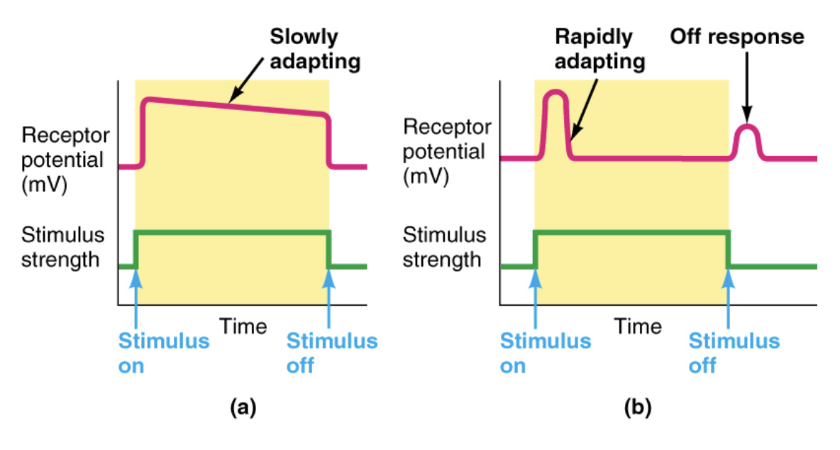
Types of Receptors According to Their Speed of Adaptation
Receptors may adapt slowly or rapidly to sustained stimulation
– Tonic receptors
• Do not adapt at all or adapt slowly
– Muscle stretch receptors, joint proprioceptors
– Phasic receptors
• Rapidly adapting receptors
– Tactile receptors in skin
Efferent Division
• Autonomic nervous system (ANS)
– Involuntary branch of PNS
– Innervates cardiac muscle, smooth muscle, most exocrine glands, some endocrine glands, and adipose tissue
• Somatic nervous system
– Subject to voluntary control
– Innervates skeletal muscle
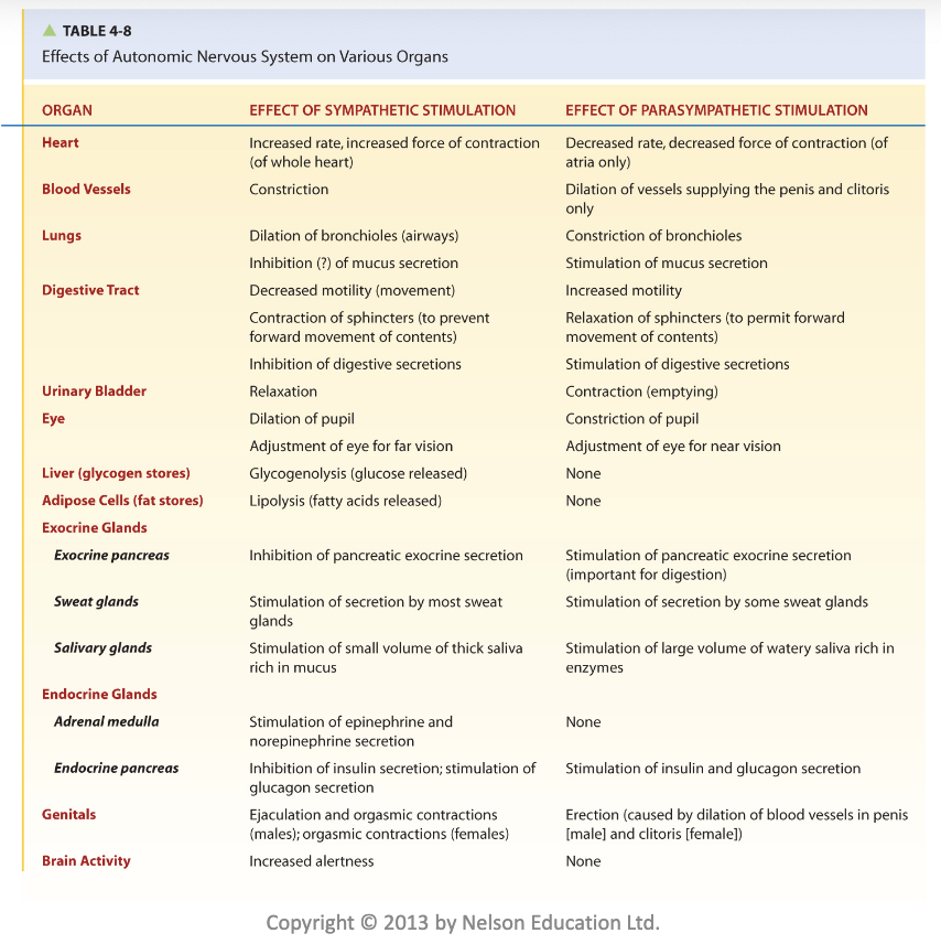
Autonomic Nervous System
• Two subdivisions
– Sympathetic nervous system
– Parasympathetic nervous system
• Sympathetic system dominates in emergency or
stressful (“fight-or-flight”) situations
– Promotes responses that prepare body for strenuous
physical activity
• Parasympathetic system dominates in quiet,
relaxed (“rest-and-digest”) situations
– Promotes body-maintenance activities such as
digestion
Somatic Nervous System
• Consists of axons of motor neurons that originate in spinal cord or brain stem and end
on skeletal muscle
• Motor neurons are the final common pathway by which various regions of CNS exert control over skeletal muscle activity
– These areas of CNS include spinal cord, motor regions of cortex, basal nuclei, cerebellum, and brain stem