EMBRYOLOGY Session 7 – Outflow Tract and Great Arteries
1/65
There's no tags or description
Looks like no tags are added yet.
Name | Mastery | Learn | Test | Matching | Spaced | Call with Kai |
|---|
No analytics yet
Send a link to your students to track their progress
66 Terms
Partitioning of the Truncus begins during the what week?
Partitioning of the Truncus begins during the 5th week
At about 5 weeks of development, the truncus arteriosus starts dividing into the aorta and pulmonary artery.
Partitioning = dividing
Truncus = truncus arteriosus (the early single vessel leaving the heart)
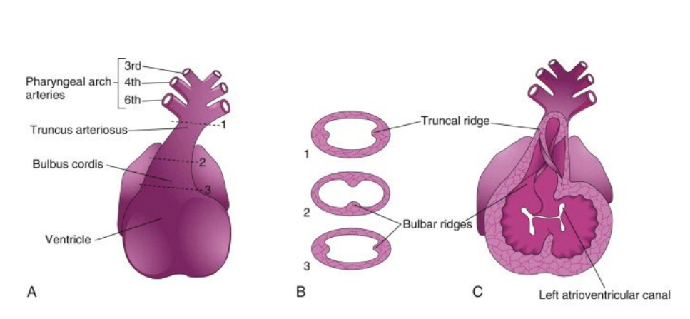
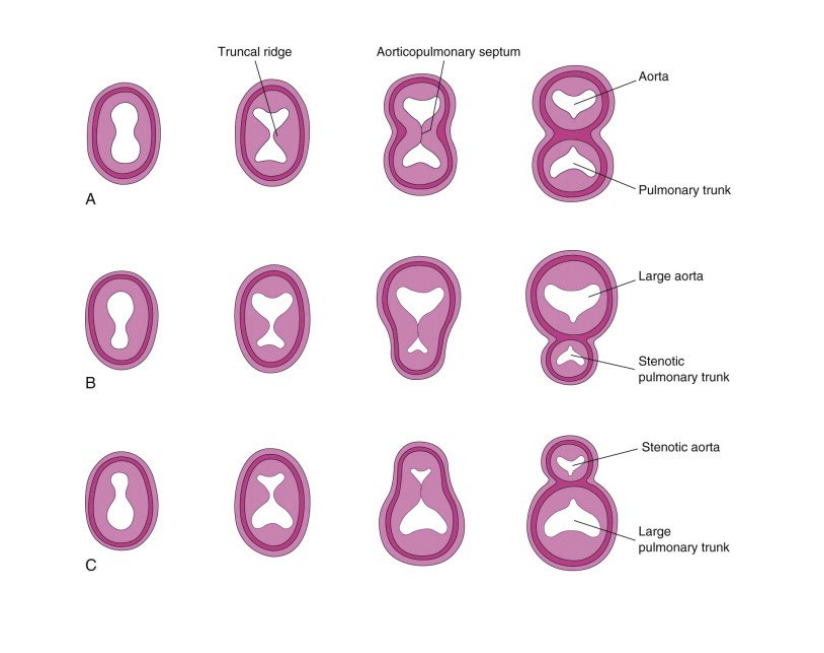
Proliferation, of what cells in the walls of the what creates what?
Proliferation of mesenchymal cells in the walls of the bulbus cordis create bulbar ridges and truncal ridges
The truncal ridges are continuous what?
The truncal ridges are continuous with the bulbar ridges
What are derived from neural crest mesenchymal cells?
Both the bulbar and truncal ridges are derived from neural crest mesenchymal cells
Where are the truncal ridges located?
The truncal ridges are located towards the aortic arch

Where are the bulbar ridges located?
The bulbar ridges are located towards the bulbus cordis
SKIP
SKIP
Neural crest cells continue to migrate through what to reach the what?
Neural crest cells continue to migrate through the pharyngeal arches to reach the ridges
The ridges grow together to septate what into what?
The ridges grow together to septate the truncus arteriosus into two great arteries
Septate = divide something by forming a wall (septum).
A septum is a wall that separates two spaces.
The bulbar and truncal ridges then undergo what? What also helps….?
The bulbar and truncal ridges then undergo a 180-degree spiraling •
Also caused in part by the streaming of blood
Also caused in part by the streaming of blood”
This means:
👉 The direction that blood is flowing helps guide the twist of the septum.
Blood flowing through the heart helps shape how the vessels form.
Very short summary
Neural crest cells form ridges → the ridges fuse and twist (spiral) → this divides the truncus arteriosus into the aorta and pulmonary artery, and blood flow helps guide this twisting.
What does The spiral orientation of the septum creates?
The spiral orientation of the septum creates the criss cross of the great arteries
what does the Spiral form?
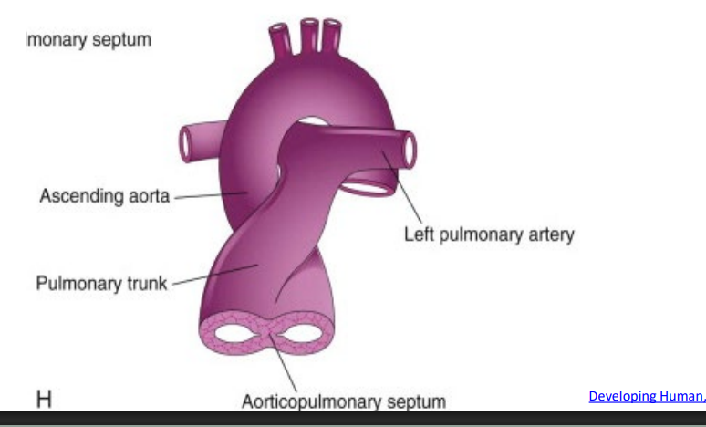
Spiral form of the aorticopulmonary septum
The aorticopulmonary septum divides what, into what?
The aorticopulmonary septum divides the bulbus cordis and truncus arteriosus into two arterial channels, the ascending aorta and pulmonary trunk
The spiraling of the aorticopulmonary septum leads to what?
The spiraling of the aorticopulmonary septum leads to a pulmonary trunk that twists around the ascending aorta (criss cross of the great arteries)
SKIP
Great arteries- which are what? they do what around each other as they leave the heart? and on the echo picture how do we prove this?
Great arteries (ascending aorta and pulmonary trunk) twisting around each other as they leave the heart
In the RVOT view the PA will be elongated and the AO will be transverse and that tells us a true criss croos is happening
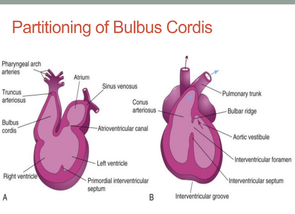
Partitioning of Bulbus Cordis:
The bulbus cordis is incorporated into the walls of the what?
The bulbus cordis is incorporated into the walls of the definitive ventricles, (RV AND LV OUTFLOW TRACTS)
In the RV, the bulbus cordis is represented by the what?
In the RV, the bulbus cordis is represented by the conus arteriosus (infundibulum)
The bulbus cordis develops into the conus arteriosus (infundibulum), which is the smooth outflow tract of the right ventricle leading to the pulmonary artery.
In the LV, the bulbus cordis forms the walls of the what? (talk about the location as well) - the part of the ventricular cavity…
In the LV, the bulbus cordis forms the walls of the aortic vestibule, the part of the ventricular cavity just inferior to the aortic valve
The bulbar ridges extend where to create what?
The bulbar ridges extend down into the bulbus cordis to create the outflow tracts
TRUNCAL DEFECTS:
The conotruncal defects will include? Name 4
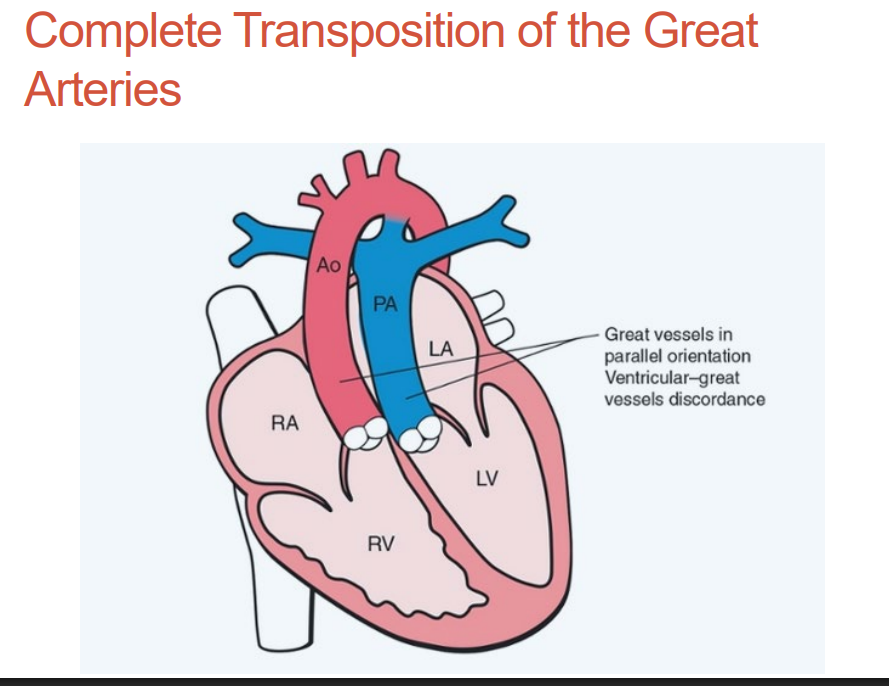
Complete Transposition of the Great Arteries •
Tetralogy of Fallot •
Double Outlet Right Ventricle •
Truncus Arteriosus
Complete transposition of the great arteries • Aka called what?
explain what it is
Aka D-TGA
The great arteries do not spiral and run parallel to each other
Complete transposition of the great arteries
what is the concordance and discordance, tell me whats connected to what?
Atrial – ventricular concordance; ventricular arterial discordance •
Left atrium is connected to the left ventricle, left ventricle is connected to the pulmonary artery
Complete Transposition of the Great Arteries
what is it associated with?
Associated with maternal diabetes
Transposition of the Great Arteries • Deoxygenated systemic venous blood returns to what chambers, and the passes through which great arteries?
Deoxygenated systemic venous blood returns to the right atrium enters the right ventricle and then passes to the body through the aorta
Transposition of the Great Arteries
explain how Oxygenated blood is passes through circulation?
Oxygenated pulmonary venous blood passes through the left ventricle back into the pulmonary circulation
For the Transposition of the Great Arteries, there is no significant what effect on the fetus? Are these often found during a prenatal screnning? what must the pulmonary artery and aorta must do?
Morphological Feature of the PA?
There is no significant hemodynamic effect on the fetus •
This is often missed during a prenatal screening •
Pulmonary artery and aorta must criss cross
Branches/Pants
Complete Transposition of the Great Arteries • Echo Findings:
What is the best view?
Need to perform what interrogation from which views?
Tilting to a what view will show the pulmonary artery with two branches coming off the what?
The aorta and pulmonary artery will run how?
what view will be difficult to see both the pulmonary artery and aorta in the same view?
• Use color flow to look for what?
LVOT
Need to perform outflow tract interrogation from LVOT and RVOT view •
Tilting to a LVOT view will show the pulmonary artery with two branches coming off the LV •
The aorta and pulmonary artery will run parallel •
3vv will be difficult to see both the pulmonary artery and aorta in the same view •
Use color flow to look for the size of the foramen ovale
Complete Transposition of the Great Arteries • what are the Common associations:, name 4
Most are isolated defects •
Membranous VSDs (40%) •
Pulmonary stenosis (30%) •
Maternal diabetes
Complete Transposition of the Great Arteries, name 2 Differentials
Double outlet right ventricle •
Corrected Transposition of the Great Arteries
Complete Transposition of the Great Arteries • Treatment’s
Tolerated well in utero but becomes a what lesion after birth •
Critical need of what to be patent post birth?
Tolerated well in utero but becomes a cyanotic lesion after birth •
Critical need of FO and PDA to be patent post birth
Complete Transposition of the Great Arteries • Treatment
Delivery takes place in a facility that handles urgent cases
what maybe performed emergently while awaiting definitive surgical correction?
A balloon atrial septoplasty maybe performed emergently while awaiting definitive surgical correction
Complete Transposition of the Great Arteries • Treatment
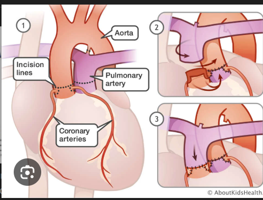
Full surgical correction happens at what weeks of life?
Requires surgical what called a what procedure?
Prognosis post surgery is what result
• Full surgical correction happens at 2 weeks of life •
Requires surgical switch of the great arteries called a Jatene procedure •
Prognosis post surgery is excellent
what is the Most common cyanotic lesion in neonates?
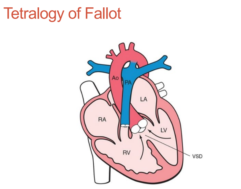
Tetralogy of Fallot
Tetralogy of Fallot • Most common cyanotic lesion in neonates • what are the 4 distinct features?
Overriding aorta •
Malalignment VSD •
Infundibular pulmonary stenosis •
4th (right ventricular hypertrophy) is not seen in utero
Tetralogy of Fallot •
The truncus divides how?
The aorta must override the what by what%?
why would the aorta may be dilated due to what
The truncus divides unequally •
Aorta must override the ventricular septum by less than or equal to 50% •
Aorta may be dilated due to flow from both ventricles
(2 types we will discuss today include: • Stenosis, and absence of the Pulmonary valve)
Tetralogy of Fallot • Echo Findings
what will you see in what chamber views?
^Visualized with what doppler?
which outflow tract view reveals? what?
how will the aorta be slightly shifted? and what?
• Large VSD in 4 chamber and 5 chamber views •
Visualized with color Doppler •
LVOT outflow tract view reveals an overriding aorta and malalignment VSD •
Aorta will be slightly shifted to the right and will be dilated
Tetralogy of Fallot • Echo findings
RVOT will be what but more what than the aorta in the what view?
PA velocity will be what?
Flow across the Ductus Arteriosus may be what?
RVOT will be patent but more narrow than the aorta in the 3VV
PA velocity will be elevated
Flow across the Ductus Arteriosus may be reversed in severe TOF cases
The RVOT is open, but the pulmonary artery is smaller than the aorta on the three-vessel view, which is a sign of outflow obstruction such as in Tetralogy of Fallot.
Tetralogy of Fallot • Common associations: what are they name 5
ASD (83%) •
Right aortic arch (25%) •
Persistent left SVC (11%) •
Trisomies 21, 13 and 18 (30%) •
Alagille syndrome
Tetralogy of Fallot • Differentials what are they name 4
Pulmonary atresia •
Absent pulmonary valve •
Common arterial trunk •
Double outlet right ventricle
The differentials listed
1. Pulmonary atresia
The pulmonary valve is completely closed, so blood cannot leave the RV to the lungs.
2. Absent pulmonary valve
The pulmonary valve is missing or very abnormal, causing severe pulmonary regurgitation and very large pulmonary arteries.
3. Common arterial trunk (Truncus arteriosus)
Instead of two vessels (aorta and pulmonary artery), there is one single vessel leaving the heart.
4. Double outlet right ventricle (DORV)
Both the aorta and pulmonary artery come out of the right ventricle.
Tetralogy of Fallot, what are the Treatment
Routine screening to watch growth of what?
Surgical repair of what?
Routine screening to watch growth of PA and ductus arteriosus flow •
Surgical repair of VSD, repair of obstruction or conduit placement from RV to PA
. Repair of obstruction
In Tetralogy of Fallot there is often narrowing in the RV outflow tract or pulmonary valve.
Doctors widen that area so blood can flow normally to the lungs.
3. Conduit placement from RV to PA
Sometimes the pathway from the right ventricle to the pulmonary artery is too abnormal.
So surgeons place a tube (conduit) that connects:
Right ventricle → Pulmonary artery
This allows blood to reach the lungs normally.
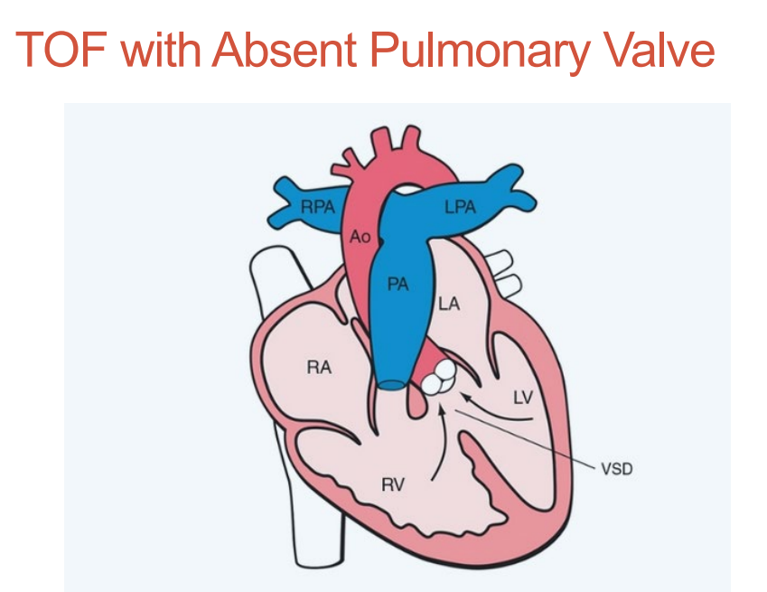
TOF with Absent Pulmonary Valve •
Pulmonary Valve Absence • Absence of the PV • PV Insufficiency
•PV leaflets what wrong and
what does this result in and tell me what cardiac cycle?
Incomplete coaptation of PV leaflets •
Results in backward flow during diastole
TOF with Absent Pulmonary Valve
Massively what? of what
what is the pattern in rudimentary PV?
overriding what?
whats the shunt called?
Massively dilated PA and PA branches •
To and fro pattern in rudimentary PV •
Overriding Aorta •
Malalignment VSD
A rudimentary pulmonary valve is a severely underdeveloped, malformed, or vestigial valve in the heart that fails to function properly
Tetralogy of Fallot with Absent Pulmonary Valve • Common associations are the same: what are they name, whats the 1# common
ASD (83%) • #1
Right aortic arch (25%) •
Persistent left SVC (11%) •
Trisomies 21, 13 and 18 (30%) •
Alagille syndrome
Tetralogy of Fallot with Absent Pulmonary Valve • Differentials what are they name 3
Tetralogy of Fallot •
Common arterial trunk •
Double outlet right ventricle
Tetralogy of Fallot with Absent Pulmonary Valve, what is the Treatment
These tend to be fatal in utero
Double outlet right ventricle, explain what it is?
Both great arteries arise more than 50% or entirely from the RV
Double outlet right ventricle
Complex congenital heart defect that incorporates 3 things, what are they?
what view
Contains an outlet VSD •
Great arteries may be transposed • Transposition of the great arteries (TGA) they run parallel
One of the outflows may be obstructed (typically Pulmonic Valve stenosis)
LVOT view
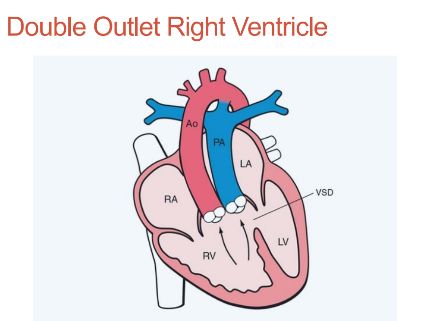
Double Outlet Right Ventricle • Echo Findings
Two great arteries arising form what?
Aorta is over what and how much? %
The goal is to identify how the great arteries lie, what is common? the great arteries may run how?
Location of the VSD in what view? what region?
and what happens to the outflow, and what stenosis ?
Two great arteries arising form the RV •
Aorta is over the ventricular septum more then 50% •
The goal is to identify how the great arteries lie •
Transposed Arteries are common •
The great arteries may run parallel •
Location of the VSD in 5 chamber • Outlet region • O
utflow tract obstruction • Pulmonary stenosis?
Double Outlet Right Ventricle • Common Associations: what are they name 4
Pulmonary stenosis (70%) •
Outlet type VSD •
Right sided aortic arch •
Maternal diabetes
Double Outlet Right Ventricle • Differentials: what are they name 2
Tetralogy of Fallot •
Complete Transposition of the Great Arteries
Double Outlet Right Ventricle, Treatment
what lesion in neonates?
Repair depends on other what?
High pregnancy termination rate due to what?
Cyanotic lesion in neonates •
Repair depends on other anomalies found
• High pregnancy termination rate due to other extracardiac anomalies found
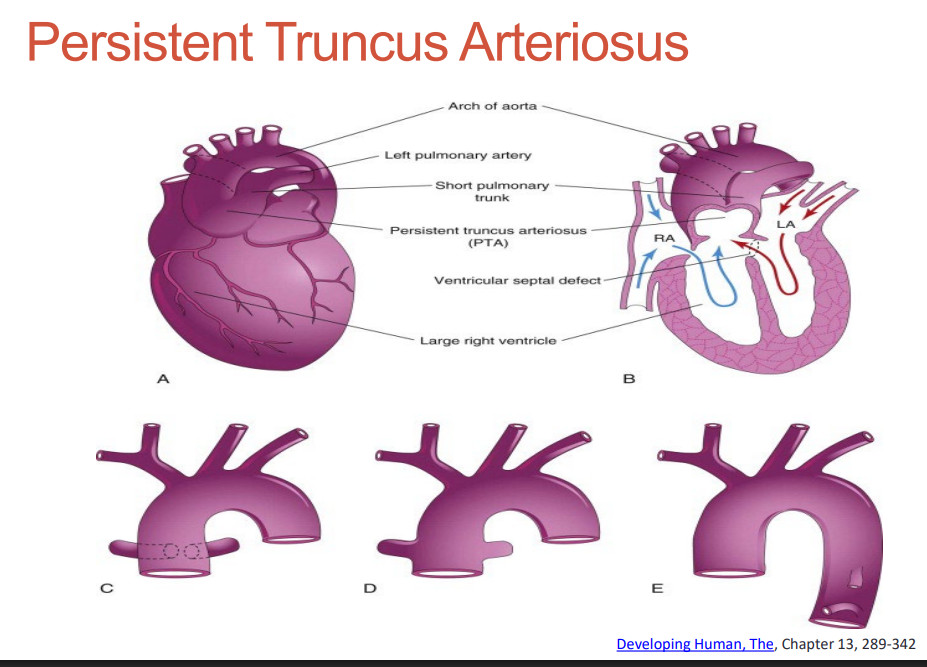
Persistent Truncus Arteriosus- what is this explain?
The truncus arteriosus persists does not divide during cardiac development
Persistent Truncus Arteriosus
A what great vessel arises from the what?
where does it Supplies blood to?
A single great vessel arises from the heart •
Supplies blood to systemic, pulmonic and coronary artery circulations
In Persistent Truncus Arteriosus, what is almost always present?
• A Malalignment VSD is almost always present
SKIP
Persistent Truncus Arteriosus • Echo Findings
The common arterial trunk is what size and arise from where?
Overrides the what?
what shunt is found in what view?
The CAT had how many leaflets in 69% of cases?
how many leaflets in 22% of cases?
describe how leaflets are
what view shows a single vessel?
70% of aortic arches will be what?
The common arterial trunk is large and arise from both ventricles •
Overrides the ventricular septum •
A Large VSD in 5 chamber view •
The CAT had 3 leaflets in 69% of cases •
4 leaflets in 22% of cases •
Leaflets are thick and regurgitant •
3 VV shows a single vessel •
70% of aortic arches will be to the left of the trachea
CAT = Common Arterial Trunk (also called truncus arteriosus)
.
Persistent Truncus Arteriosus • Common Associations what are they? name 4
No ductus arteriosus •
right sided arch •
90% have DiGeorge syndrome •
Maternal diabetes
Persistent Truncus Arteriosus • Differentials name 2
Tetralogy of Fallot •
Pulmonary Atresia with VSD
Persistent Truncus Arteriosus, Treatment
Only what % survive in utero?
what lesion in neonates?
Survival requires corrective surgery…Closure of the what? Separation of the what? what replacement?
And conduit replacement after how many years?
Only 32% survive in utero •
Cyanotic lesion in neonates •
Survival requires corrective surgery •
Closure of the VSD •
Separation of the Pas from aorta and attachment to a conduit •
Valve replacement •
And conduit replacement after 3 years
Surgeons separate the pulmonary arteries from the aorta and connect them to the right ventricle using a conduit so blood can flow normally to the lungs.
Exam ***What view will help diagnosis the complete transpostion of the great arteries? and why
3vv because you only see 2 vessels
What the 3-Vessel View (3VV) normally shows
In a normal fetal heart, the 3VV shows three vessels from left → right:
Pulmonary artery (PA) – largest
Aorta (AO) – medium
Superior vena cava (SVC) – smallest
So normally you see:
PA | AO | SVC
Three circles on ultrasound.
2. What happens in Complete Transposition of the Great Arteries (TGA)
In TGA, the aorta and pulmonary artery do not cross each other.
Instead, they run parallel.
Because of this abnormal orientation, the pulmonary artery moves out of the normal 3VV plane.
3. What you see on the ultrasound
When you scan the 3VV, instead of seeing three vessels, you often see only two:
AO | SVC
The pulmonary artery is missing from the view because it is not crossing in the normal position.
4. The key idea your teacher wants
Normal heart → 3 vessels in 3VV
TGA → only 2 vessels visible
This is a big clue that the great arteries are not crossing normally.
5. Easy memory trick
Normal:
PA – AO – SVC (3 vessels)
TGA:
AO – SVC (PA disappears from view)
The main concept
Your teacher wants you to recognize that TGA causes the great arteries to run parallel instead of crossing, which makes the pulmonary artery disappear from the normal 3-vessel view.
Exam questions ***
Of the complete transpostion of the great arteries what coming off the morphologic RV?
The Aorta
Exam question
For TOF what view will show you the size difference of what?
3VV shows the size difference between the AO - bigger and PA- smaller
Exam Question
what issue is always associated with a conal truncal defect
Maternal Diabetes
Exam Question what is associated with Maternal Diabetes defects
TOF
Double outlet RV
Presistent truncus arteriosus
Com[lete transposition of the great arteries