Second Messenger Signalling
1/19
There's no tags or description
Looks like no tags are added yet.
Name | Mastery | Learn | Test | Matching | Spaced |
|---|
No study sessions yet.
20 Terms
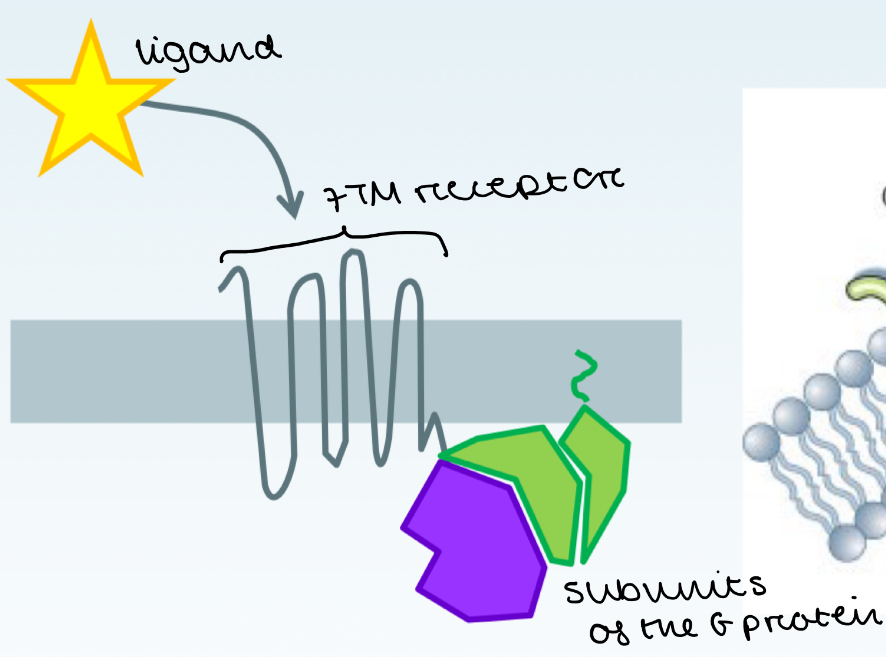
What are G-protein coupled receptors (GPCRs)
Play a crucial role in neuronal signalling, vision, smell and taste
The GPCR binds a ligand and transmits a signal to the inside of the cell
This activates a G protein bound to the inner surface of the GPCR
This triggers an intercellular signalling cascade

Difference between 7TM receptors and GPCRs
7 TransMembrane receptors are a broad class of receptors that has 7 transmembrane segments
They activate different intracellular signaling pathways, but not all of them use G proteins.
GPCRs are specific type of 7TM receptor that always signals through G proteins to trigger intracellular pathways
How many subunits does a G-protein have?
3
Alpha
Beta
Gamma
What are the effects of different GPCRs determined by?
Which G-protein alpha subunit they activate
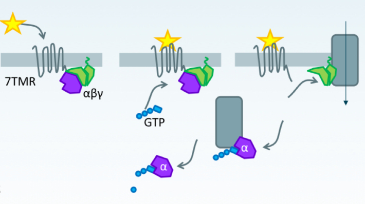
Mechanism of GPCR signalling
Ligand e.g. NE, E binds to 7TMR / GPCR
GPCR undergoes a conformational change
G protein is activated, leading to GTP replacing GDP on the α-subunit of the G protein.
G protein dissociates from the GPCR in 2 parts: Gα-GTP (active form) and Gβγ complex
These components interact with downstream effectors (e.g., proteins, ion channels). One activated, the effectors can activate other proteins leading to an intracellular signalling cascade.
GTP is hydrolysed to GDP and Gα. The Gα rebinds to Gβγ complex, returning the G protein to the inactive state.

3 examples of G-protein coupled receptors used in the sympathetic nervous system
α1 noradrenaline adrenergic receptor
α2 noradrenaline adrenergic receptor
β1/β2 noradrenaline adrenergic receptor
Explain the intracellular signalling mechanism of α1 noradrenaline adrenergic receptor
NE/E binds to receptor coupled with Gq/11 protein
This causes a conformational change in the receptor
Gq/11 protein is activated. GTP replaces GDP on the Gαq subunit. Gαq-GTP dissociates from the Gβγ complex
Gαq-GTP activates Phospholipase C
This produces IP3 and DAG » these are the second messengers
IP3 causes Ca2+ to be released
Ca2+ and DAG activate protein kinase C
Causing smooth muscle contraction
Effect: pupil dilation, constriction of sphincters, contraction of blood vessels
Explain the intracellular signalling mechanism of α2 noradrenaline adrenergic receptor
NE/E binds to receptor coupled with Gi protein
This causes a conformational change in the receptor
Gi protein is activated.
Inhibits adenylate cyclase, decreasing cAMP levels » this is the second messenger
So less activation of protein kinase A = smooth muscle contraction
The Gβγ subunit dissociates and inhibits VG Ca2+ channels, reducing Ca2+ influx = prevents release of neurotransmitter
Effect: decreases release of noradrenaline
Explain the intracellular signalling mechanism of β1/β2 noradrenaline adrenergic receptor
NE/E binds to receptor coupled with Gs protein
This causes a conformational change in the receptor
Gs protein is activated. GTP replaces GDP on the Gαq subunit. Gαq-GTP dissociates from the Gβγ complex.
Gαq-GTP activates adenylate cyclase which converts ATP to cAMP
Increases cAMP levels » this is the second messenger
So more activation of protein kinase A
Effect: increases heart rate, relaxes bronchial smooth muscle, glycogenolysis
β1 adrenergic receptor
After protein kinase A is activated…
In cardiac muscle:
Protein kinase A phosphorylates L-type Ca2+ VG channels
Increased influx of Ca2+ through VG Ca2+ channels
More Ca2+ released from sarcoplasmic reticulum
= stronger contractions
Protein kinase A phosphorylates PLB
So more Ca2+ pumped back into sarcoplasmic reticulum
= relaxation of heart muscle
β2 adrenergic receptor
After protein kinase A is activated…
In bronchial smooth muscle:
Protein kinase A phosphorylates and inhibits MLCK
This prevents actin-myosin cross bridges from forming
= relaxation of bronchial smooth muscle
Protein kinase A also activates phosphorylase kinase
= This increases the breakdown of glycogen to glucose (glycogenolysis)
GPCR vs ligand-gated ion channels
G-protein coupled receptors (metabotropic) are a lot slower than normal ligand-gated channels (ionotropic)
What do we mean by ‘activating’ a GPCR
GPCRs may appear to be active even when no ligand is present » this is called basal activity
This means ligands can either increase or decrease basal activity
NOTE:
Muscarinic receptors are…
A type of GPCR that binds Ach!!
Role of GPCRs in pharmacy
Important drug targets » >30% of drugs target GPCRs
Partial and inverse agonists are being used to regulate signalling e.g. by only partially activating or suppressing the GPCR
Why is it difficult to develop selective drug agonists/antagonists for GPCRs
Many 7TMRs form heterodimers (2 different GPCRs which create a new drug target)
Drug development must consider how GPCRs interact with each other, making drug design more complex.
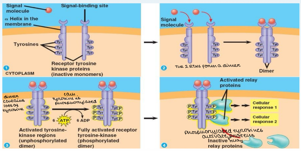
What are Receptor Tyrosine Kinases (RTKs)?
Single transmembrane proteins (have 1 segment that crosses the membrane)
When a ligand binds to the extracellular part of the RTK, it dimerises (2 RTK molecules come together)
Each tyrosine is phosphorylated, forming an activated RTK
The phosphorylated tyrosines activate relay proteins which then produce a response

What does it mean by RTKs have constitutive activity?
Have the capacity to autophosphorylate themselves
What conditions have treatments that target RTKs?
Cancer
Examples of TRKs
TrkA and TrkB which bind nerve growth factors
Ephrins