DHY 207: Introduction to Preliminary Diagnosis of Oral Lesions Vocabulary
DHY 207
Introduction to Preliminary Diagnosis of Oral Lesions Vocabulary
Describing and Recording Clinical Findings
Detailed descriptions
History
Location
Distribution
Definition
Size
Shape
Color
Consistency
Surface texture
Photographs
History
Location
Terminology: Clinical Appearance of Soft Tissue Lesions
Distribution
Localized vs generalized

Margins
Well-defined vs poorly defined
Regular vs irregular


Number multiple configuration
discrete vs coalescing

Size of Lesion
Centimeter (cm)
one hundredth of a meter; equivalent to a less than one half inch
Millimeter (mm)
One thousandth of a meter
the periodontal probe is of great assistance in documenting the size or diameter of a lesion that can be measured in millimeters
Size and Shape
Note general shape, measure size of lesion with a probe
Measure diameter of round lesions
Measure width and length of square, rectangular, and oval lesions
Record measurements- width first then length
Height is listed after the length of the lesion
Relate size of a large lesion to the area it cover
Size and Shape: Flat
- Macule: Flat lesion differentiated from. the surrounding tissue b color alone (less than 1 cm in diameter)
- Patch: May also describe area with a different surface texture with or without a color change (more than 1 cm)

Size and Shape: Elevated Fluid Filled
- Vesicle: A small (0.5 cm or less) elevated lesion filled with clear fluid. It is a small, elevated lesion that contains serous fluid
- Bulla: Larger (larger than 0.5 cm) elevated lesion filled with clear fluid. It is a circumscribed, elevated lesion that is more than 5 mm in diameter. It usually contains serous fluid, and looks like a blister
- Pustule: A raised lesion filled with pus or purulent exudate like an acne pimple It is various sized circumscribed elevations containing pus

Size and Shape: Elevated Solid
- Papule: Solid raised lesion 0.5 cm or less
- Nodule: Solid raised lesion larger than 0.5 cm but less than 2 cm
- Tumor: Solid raised lesion larger than 2 cm

- Lobule: An elevated solid segment or lobe that is part of a whole. These lobes sometimes appear fused together

Size and Shape: Plaque Broad Flat
- The slightly raised and flat configuration of this white lichen planus lesion covering a relatively broad area is indicative of a plaque. Also note the well-defined irregular margin
Size and Shape: Base/Attachment
- Sessile: Describing the base of a lesion that is flat or broad instead of stemlike
- Pedunculated: Attached by a stemlike or stalklike base similar to that of a mushroom

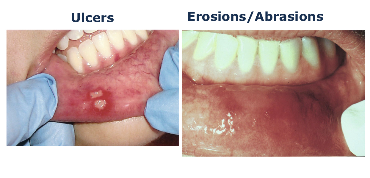
Size and Shape: Depressed
- Ulcers
- Erosions/abrasions
Direction of Growth
- Endophytic: Grow into the surrounding tissues with little or no observable swelling
- Exophytic

Color of Lesion
Most frequent color descriptions:
Red
Pink
Salmon
White
Blue-black
Gray
Brown
Black
Used to identify specific lesions
may be incorporated into general descriptions
Color: White
White lesions usually involve excess keratin in the tissues, making them more opaque
Leukoplakia: A clinical term for a white, plaque-like lesion on the oral mucosa that cannot be rubbed off or diagnosed as a specific disease
Pallor: Paleness of the skin or mucosal tissues


Color: Red
- Erythema is an abnormal redness of the mucosa or gingiva
- Erythroplakia: A clinical term used to describe an oral lesion that appears as a smooth red patch or glandular red and velvety patch
- less common than leukoplakia
- 90% of erythroplakias demonstrate epithelial dysplasia or squamous cell carcinoma

Color: Yellow
- Xanthelasma: A yellowish plaque located around the eyelids, is associated with high levels of cholesterol

Color: Pigmented
- Endogenous: Come from within the body- melanin pigmentations
- Exogenous: Come from outside the body- lead, amalgam, and others
- Black
- Brown
- Blue

Consistency
- The consistency of soft tissue abnormalities is often soft or normal feeling
- Indurated soft tissue lesions, such as an inflamed lymph node, feel quite hard
- Fluctuate is used to describe a fluid-filled lesion that moves fluid from one area to another when the lesion is pressed
Surface Texture
- Corrugated: Wrinkled
- Fissure: A cleft or groove, normal or otherwise, showing prominent depth
- Papillary: Resembling small, nipple-shaped projections or elevations found in clusters
- Smooth
- Rough
- Folded


Describing Radiographic Findings/Lesions in Bone
- History-aware/not aware
- Location and size
Opacity
Radiopaque
Describes the light or white area on a radiograph that results from the inability of radiant energy to pass through the structure
the more dense the structure, the more light or white it appears on the radiograph
Radiolucent
Describes the black or dark areas on a radiograph
Radiant energy can pass through these structures
Less dense tissue, such as pulp, is seen as a radiolucent structure

Radiolucent and Radiopaque
- A mixture of light and dark areas within a lesion
- Denotes a stage in lesion development


Inner Appearance
- Unilocular: Having one compartment or unit that is well defined or outlined as in a simple radicular cyst
- Multilocular: Describes a lesion that extends beyond the confines of one distinct area. Defined as many lobes or parts that are somewhat fused together. A multilocular radiolucency is sometimes described as resembling soap bubbles

- Coalescence: The process by which parts of a whole join together, or fuse, to make one

Radiographic Terms Used to Describe Lesions in Bone
Diffuse
Describes a lesion with borders that are not well defined, making it impossible to detect the exact parameters of the lesion
Can make treatment more difficult and depending on the biopsy results, more radical

Margins
Well-defined vs poorly defined

- Well circumscribed: Used to describe a lesion with borders that are specially defined and in which one can clearly see the exact margins and extent

Surrounding Tissues
Root resorption
Radiographically, the apex of the tooth appears shortened or blunted and irregularly shaped
Occurs as a response to stimuli, which can include a cyst, tumor, or trauma

External root resorption
Arises from tissue outside the tooth, such as the PDL

Internal root resorption
Triggered by pupal tissue reaction from within the tooth
The pulpal area can be seen as a diffuse radiolucency beyond the confines of the normal pulp area

Scalloping around the root
A radiolucent lesion that appears to extend up the PDL and between the roots


Additional Vocabulary Words
- Anomaly: Something that deviates from what is standard or normal
- Dysphasia: Difficulty swallowing
- Dysphonia: Difficulty speaking
- Dyspnea: Difficulty breathing
Reactive Tissue Responses: Hyperplasia, Hypertrophy, and Atrophy
- Hyperplasia: An increase in the number of cells, often in response to chronic irritation or abrasion. May return to normal if the insult subsides, or may persist after removal of the irritant
- Hypertrophy: An increase in the size of cells. May be seen in cardiac muscle as a response to hypertension
- Atrophy: A decrease in size or function of a cell, tissue, organ, or entire body