Imagenes Histologia Ex 1
1/172
There's no tags or description
Looks like no tags are added yet.
Name | Mastery | Learn | Test | Matching | Spaced |
|---|
No study sessions yet.
173 Terms
Connective Tissue
Mesenchyme CT
Mucous CT
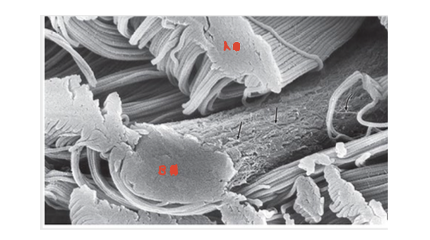
1: Elastic Fibers
2: Collagen Fibers
Collagen Fibers
Collagen Fibrils
Collagen Fibrils
Reticular Fibers
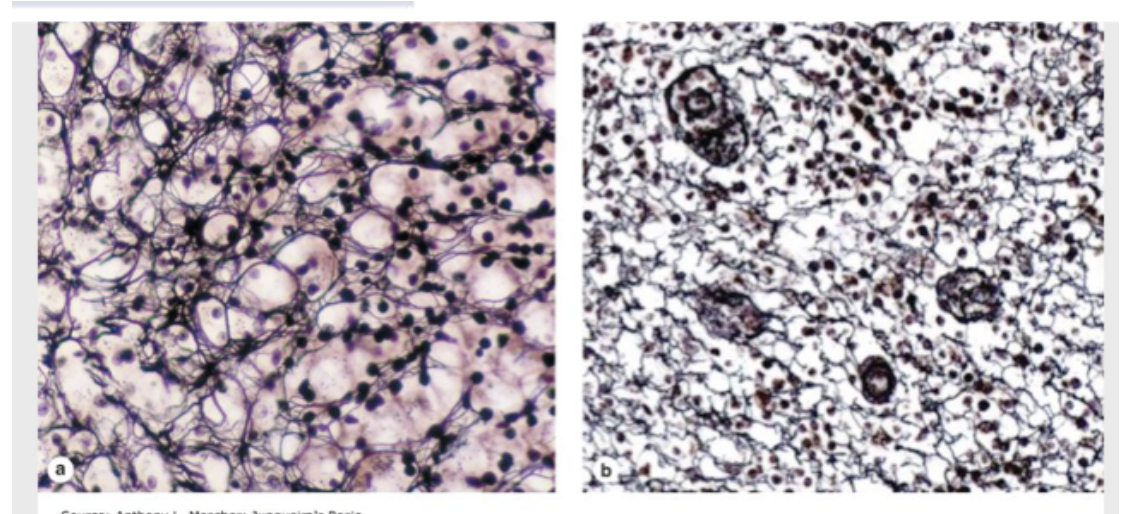
Healthy CT vs Scurvy CT
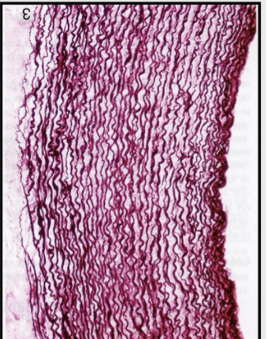
Elastic Fibers in Elastic Artery
Skin
Fibrilin-1
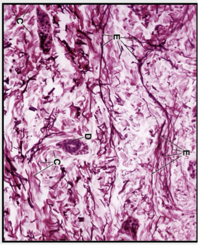
A= Collagen Fibers
B= Elastic Fibers
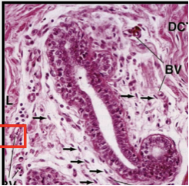
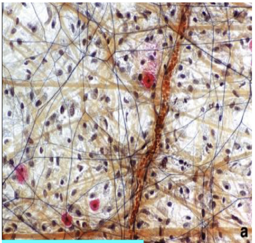
Loose CT(Areolar)
Loose CT(Areolar)
Loose CT(Areolar)
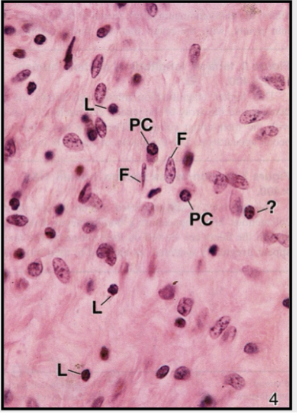
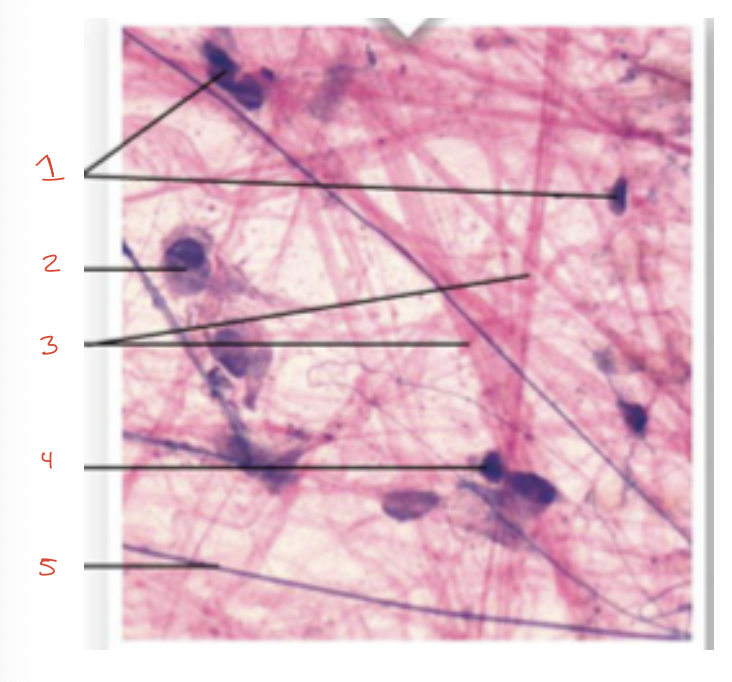
1: Fibrocytes
2: Macrofagos
3: Collagen Fibers
4: Mast Cells
5: Elastic Fibers
Loose Cellular CT
Loose Cellular Ct
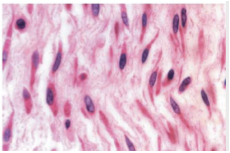
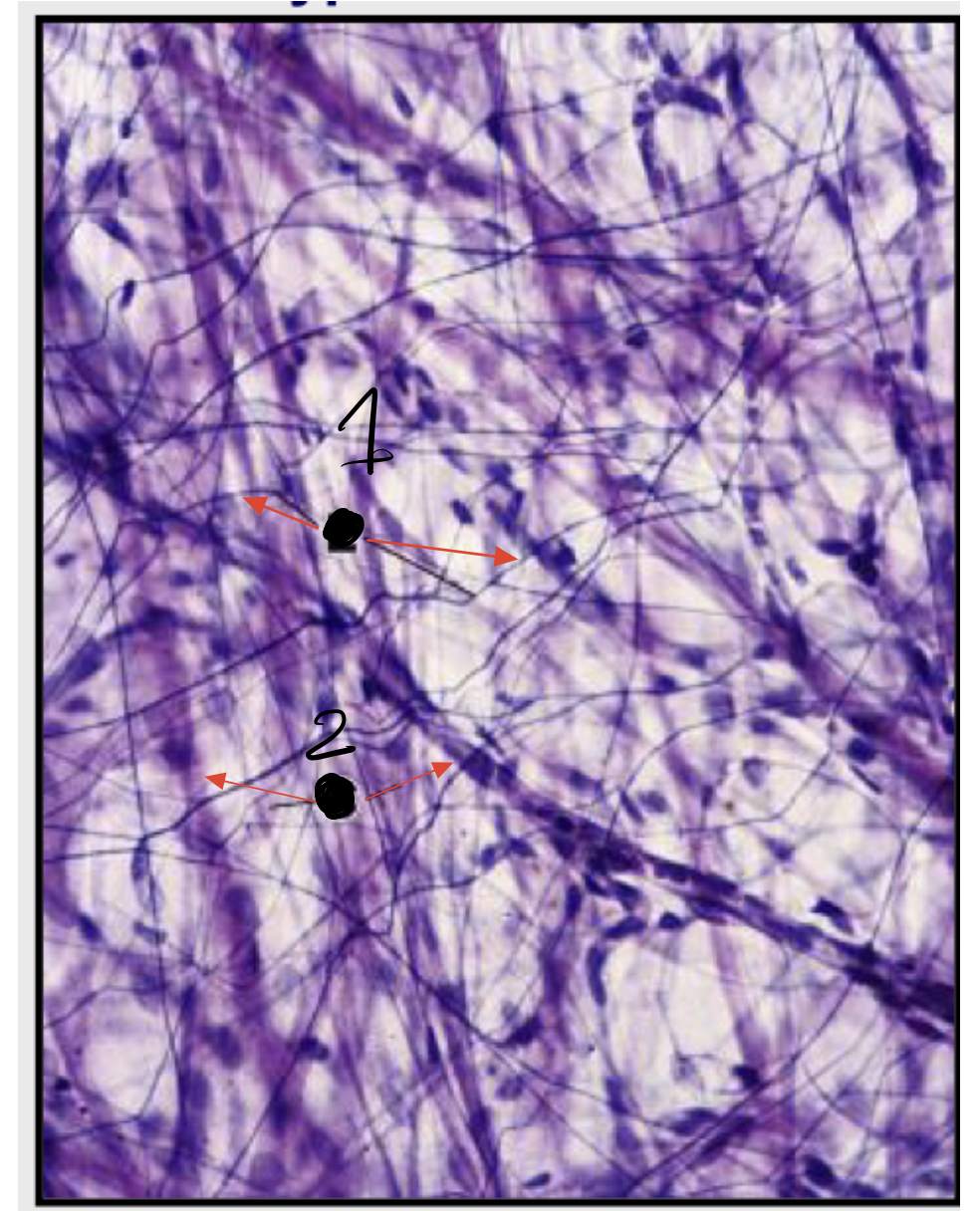
Dense Irregular CT
Dense Irregular CT
Dense Irregular CT
Dense Regular Collagenous CT
Dense Regular Collagenous CT
Dense Regular Elastic CT
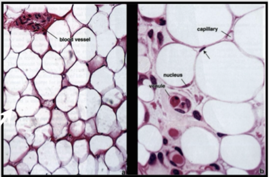
Adipose Tissue
White Fat(Unilocular)
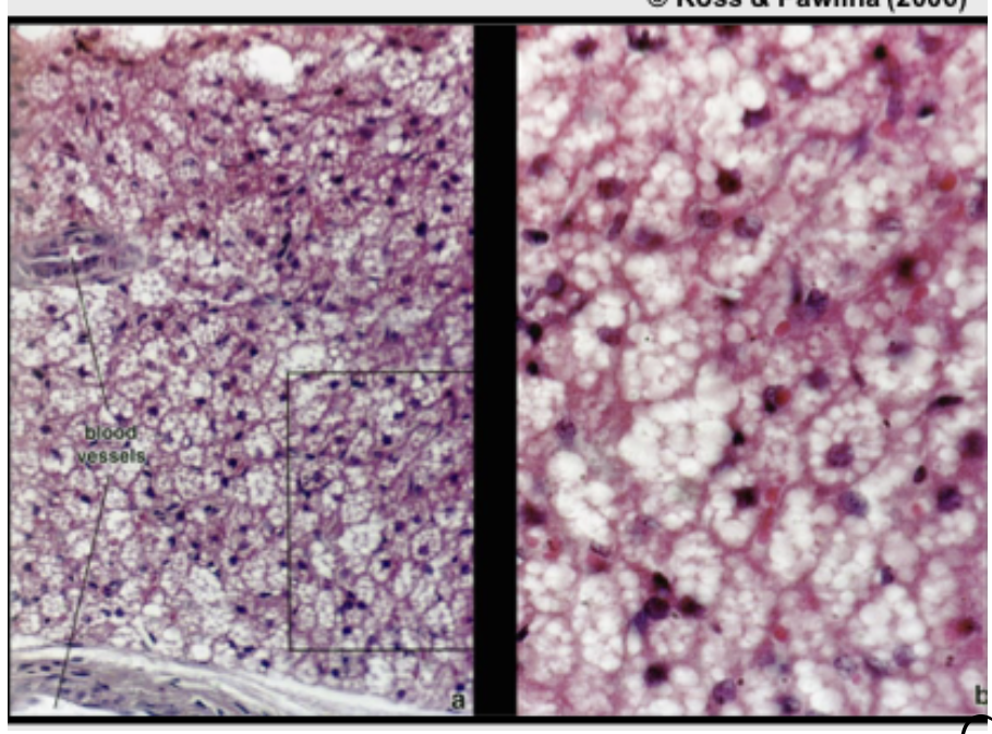
Adipose Tissue
Brown Fat(Multilocular)
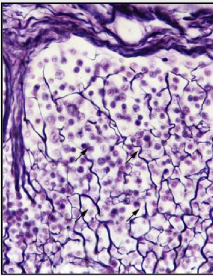
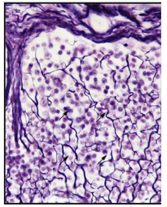
Reticular CT
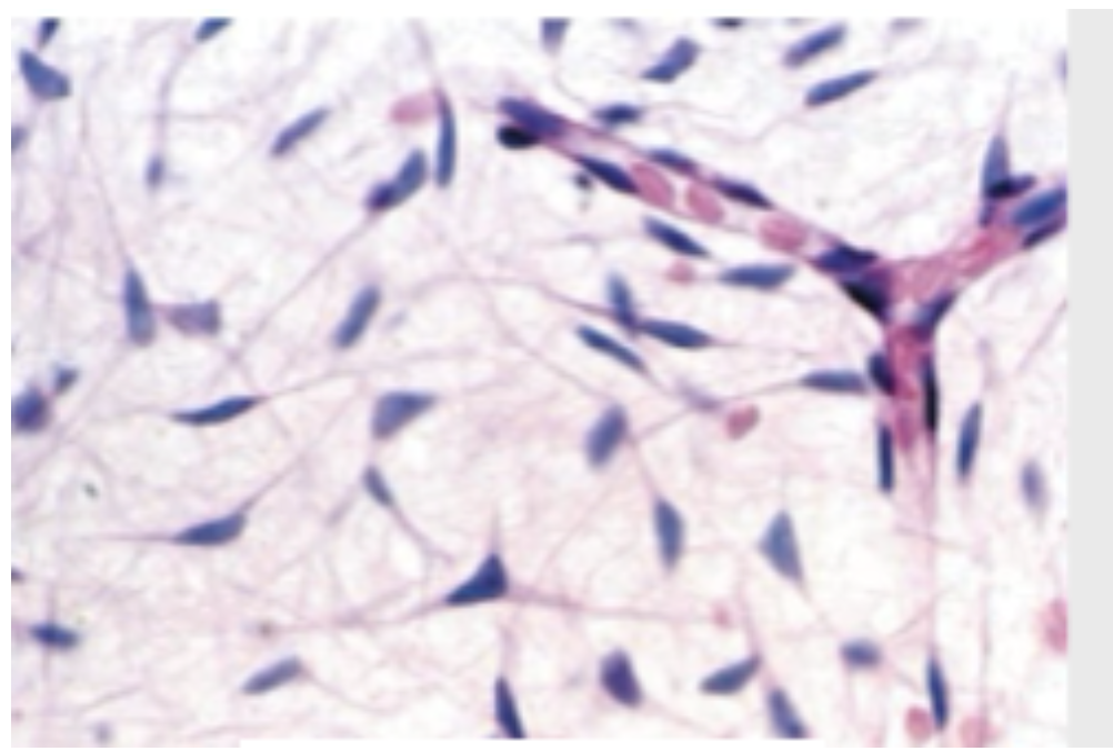
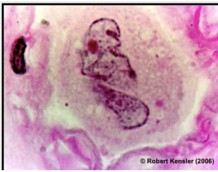
Fibroblast
1: Fibroblasts
2: Long Whip of fibroblasts
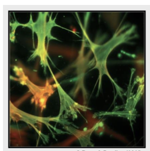
Myofibroblast
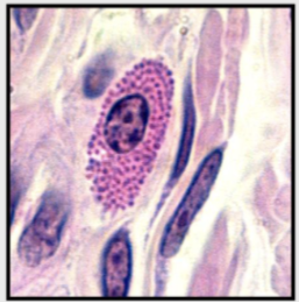
Mast Cell with eosin stain
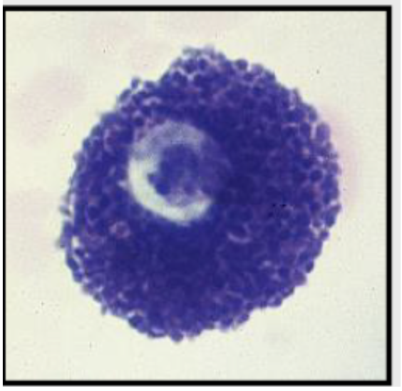
MAst Cells with toluidine Blue
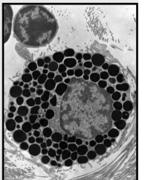
Mast Cell
Mast Cell
Granules of Mast Cells
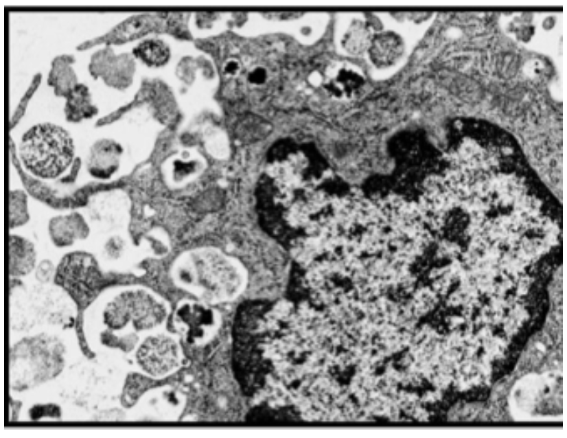
Macrofago
Macrofagos
Macrofagos
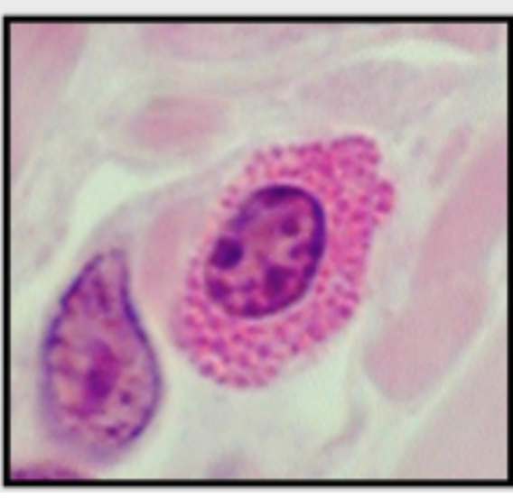
Plasma Cells
Plasma Cells
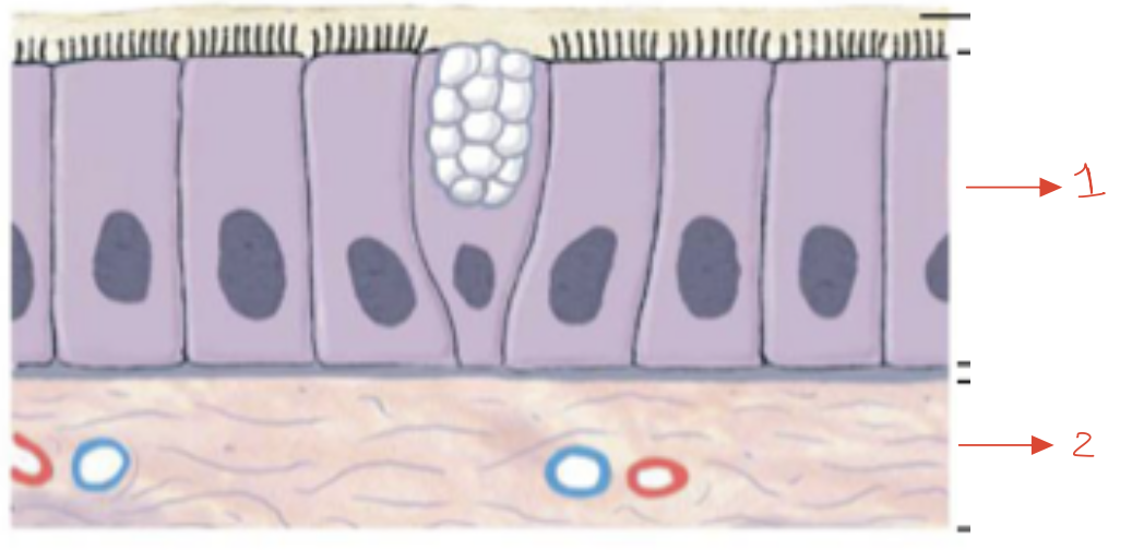
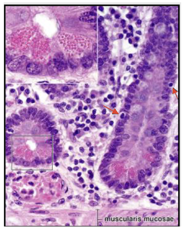
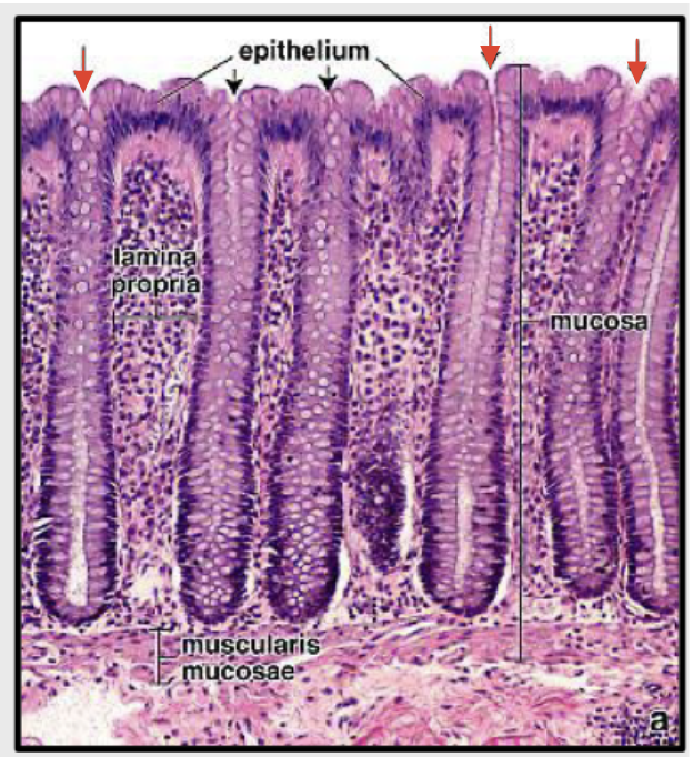
Mucous Membrane
1: Epithelium
2: Lamina Propia
Serous Membrane
Cutaneos Membrane
Synovial Membrane
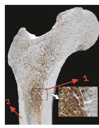
1: Spongy Bone
2: Compact Bone
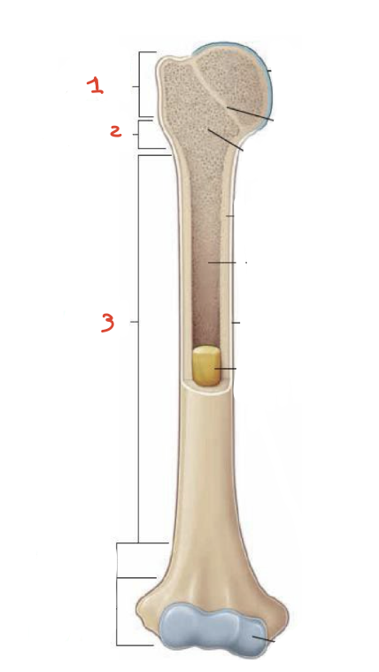
1: Epiphysis
2: Metaphysis
3: Diaphysis
1: Endosteum
2: Marrow Space
Mature Bone
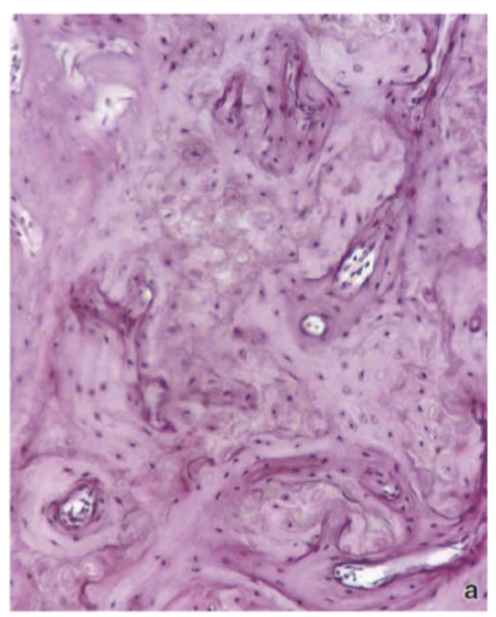
Inmature Bone
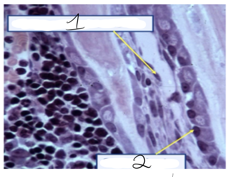
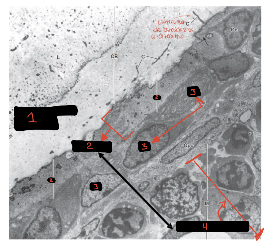
1: Osteoprogenitors Cells
2: Osteoblasts
1: Osteoid
2: Mineralized Bone
3: Osteoblastos
1: Osteoblasts
2: Osteoid
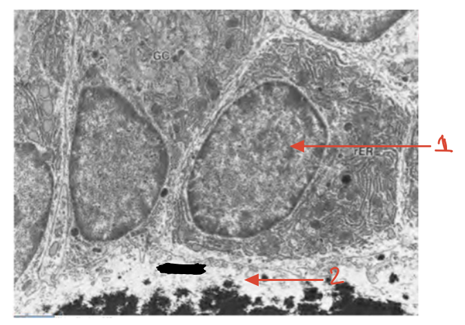
Osteoblast
Osteocytes
Osteocytes
Bone Lining Cells
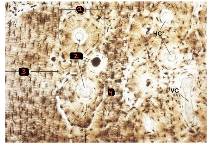
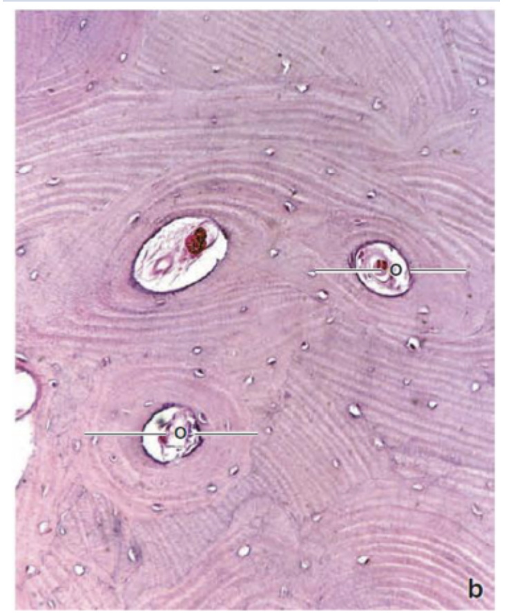
1: Osteon
2: Harversian Canal
3: Circumferential Lamellae
4: Interstitial Lamellae
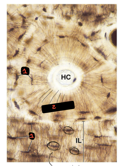
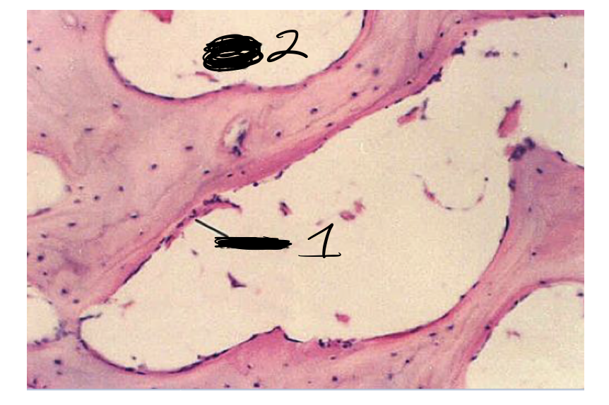
1: Lacuna with osteocyte inside
2: Canaliculos
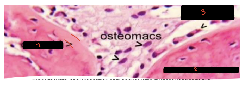
1: Mineralizaded Tissue
2: Osteoid
3: OsteoBlast
4: Bone Marrow
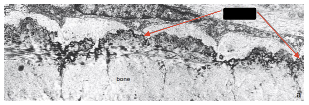
Bone-Lining Cells
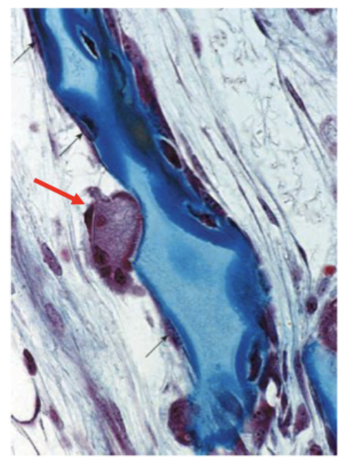
Osteoclast
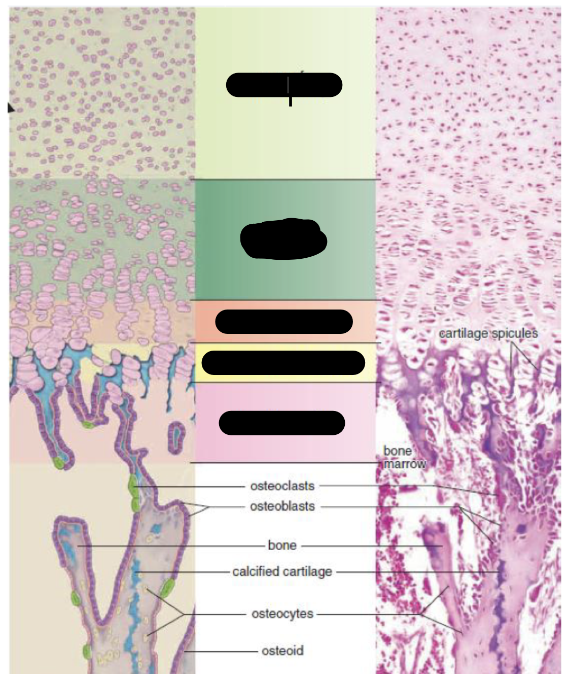
Zone of Reserved Cartilage
Zone of Proliferation
Zone of Hypertrophy
Zone of Calcified Catilage
Zone of Resorption
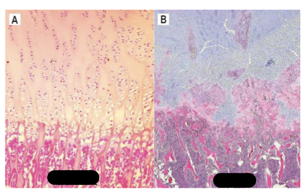
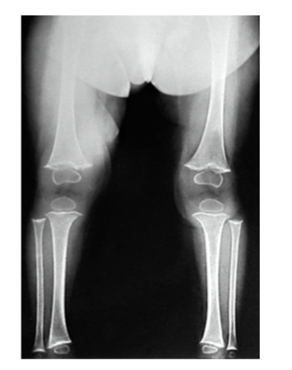
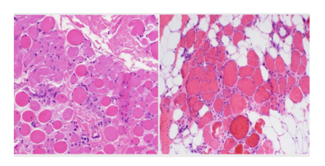
Normal tissue Vs Rickets
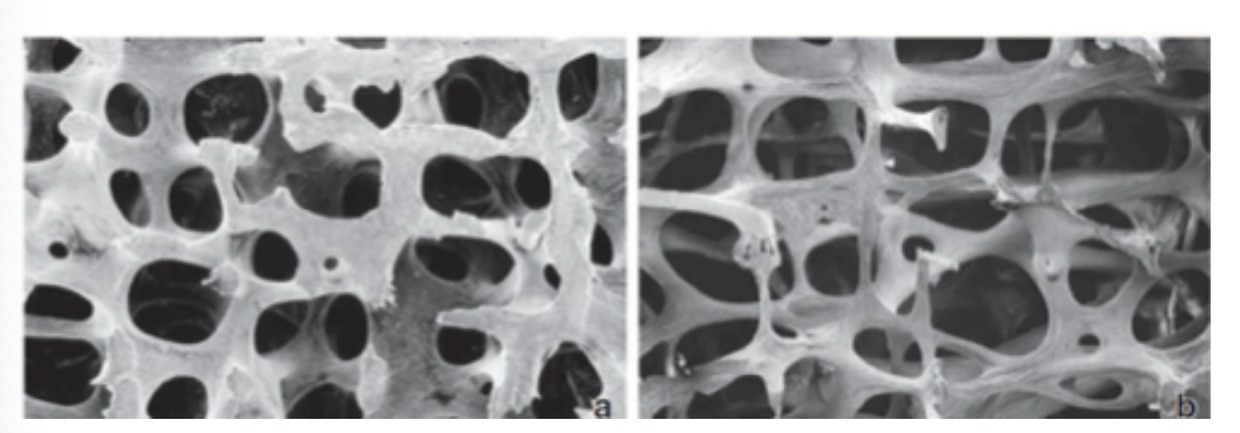
Normal Trabeculae Vs Osteoporosis Trabeculae
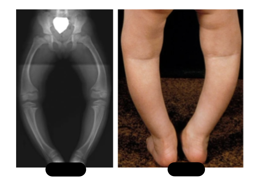
Normal vs Rickets
Scurvy
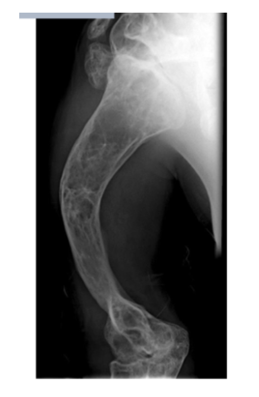
Osteogenesis Imperfecta
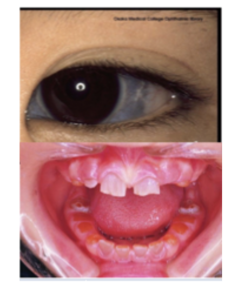
Osteogenesis Imperfecta
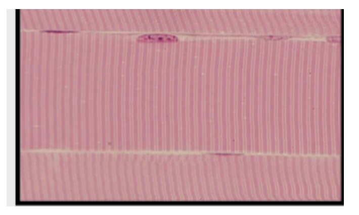
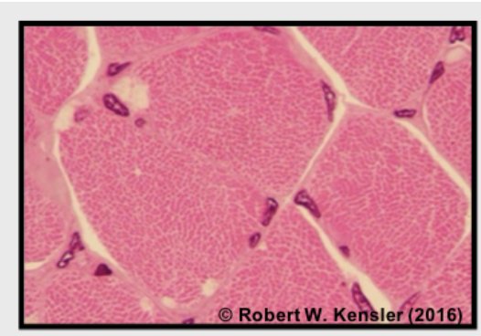
Skeletal Muscle
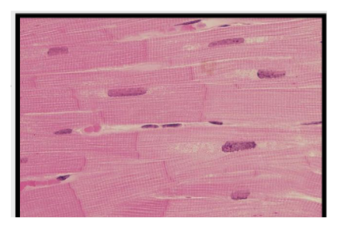
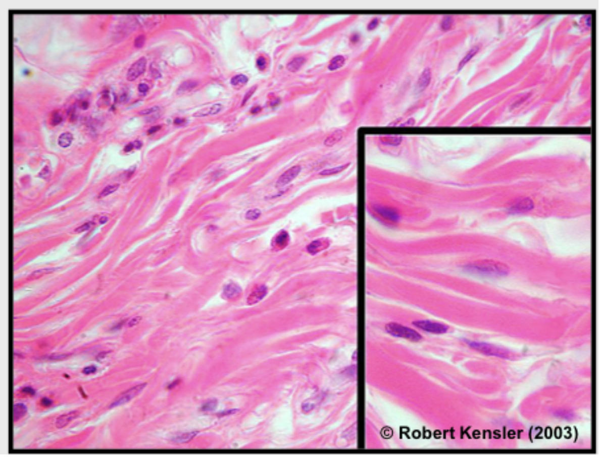
Cardiac Muscle
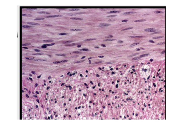
Smooth Muscle
Skeletal Muscle
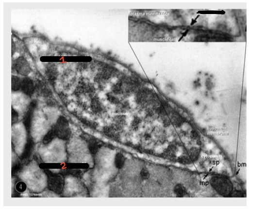
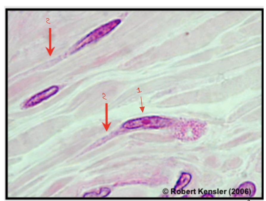
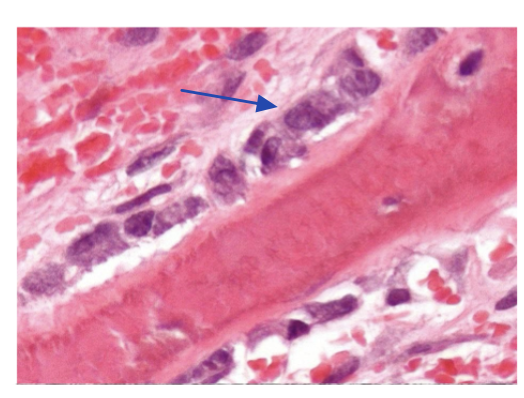
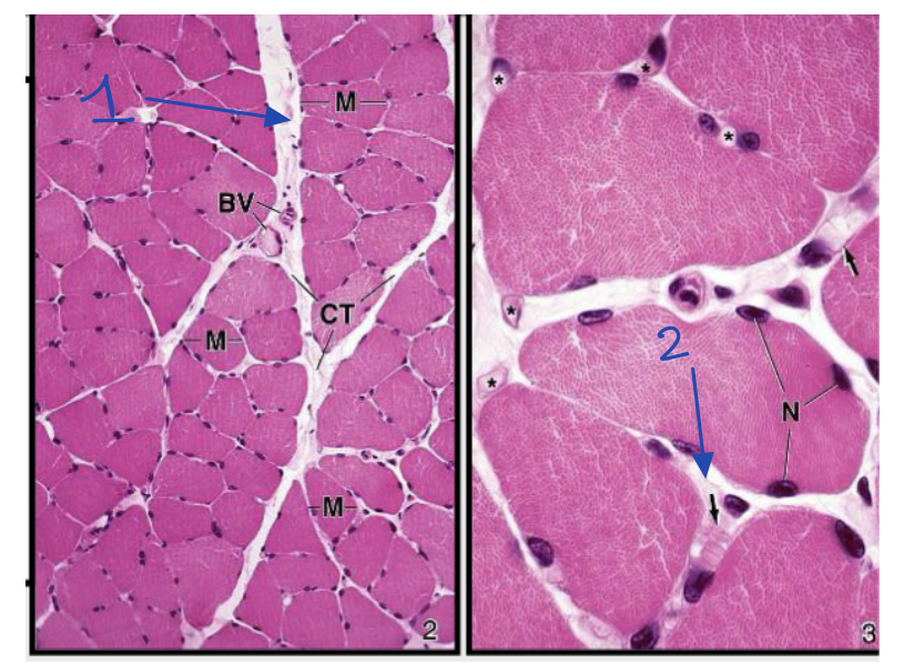
1: Satelital Cell
2: Muscle Cell
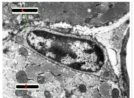
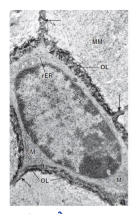
1: Satelital Cell
2: Muscle Cell
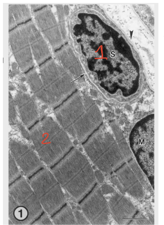
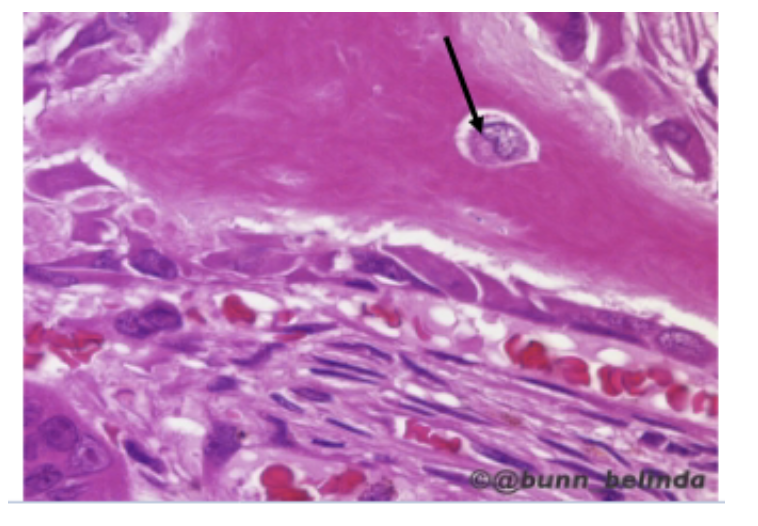
1: Satelital Cell
2: Muscle Cell
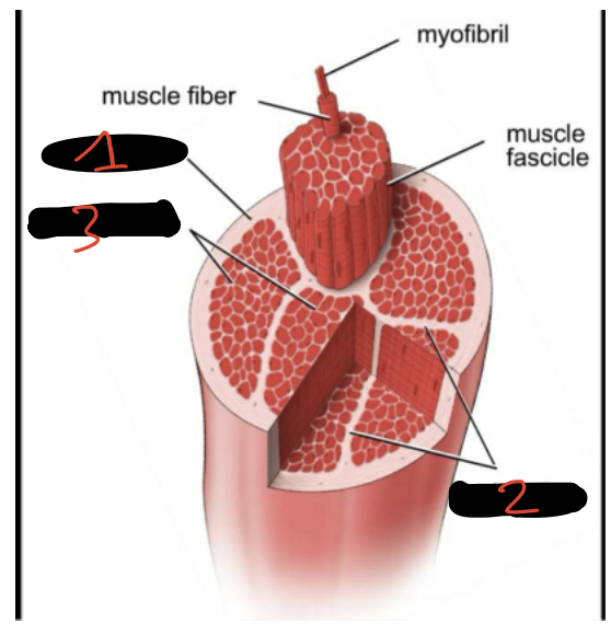
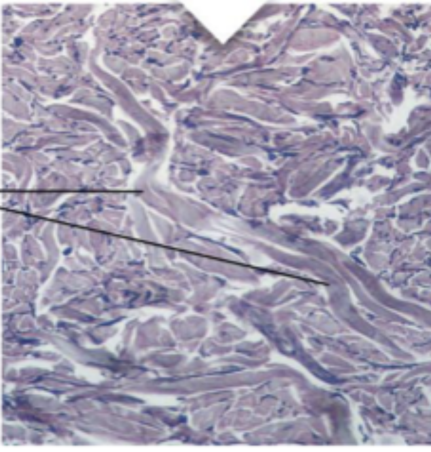
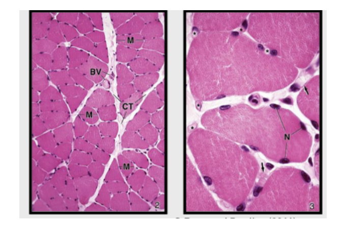
1: Epimysium
2: Perimysium
3: Endomysium
1; Perymysium
2: Endomysium
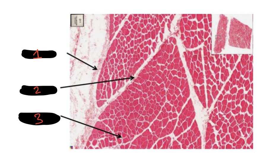
1: Epimysium
2: Perimysium
3: Endomysium
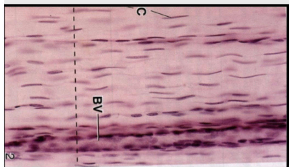
Endomysium
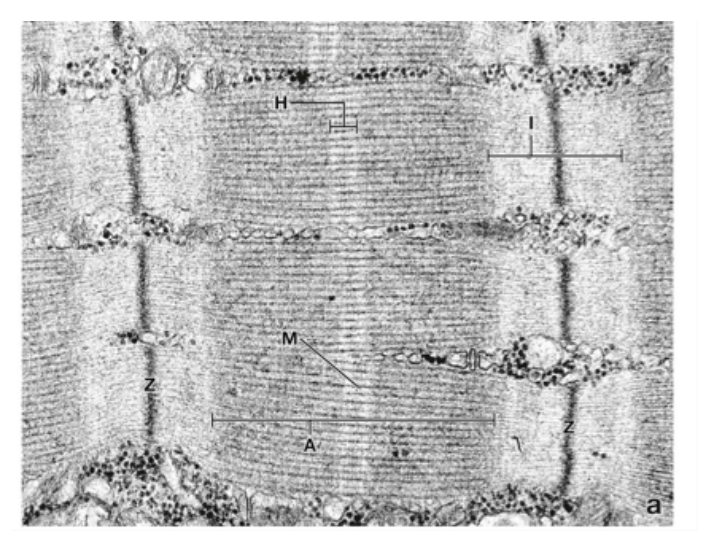
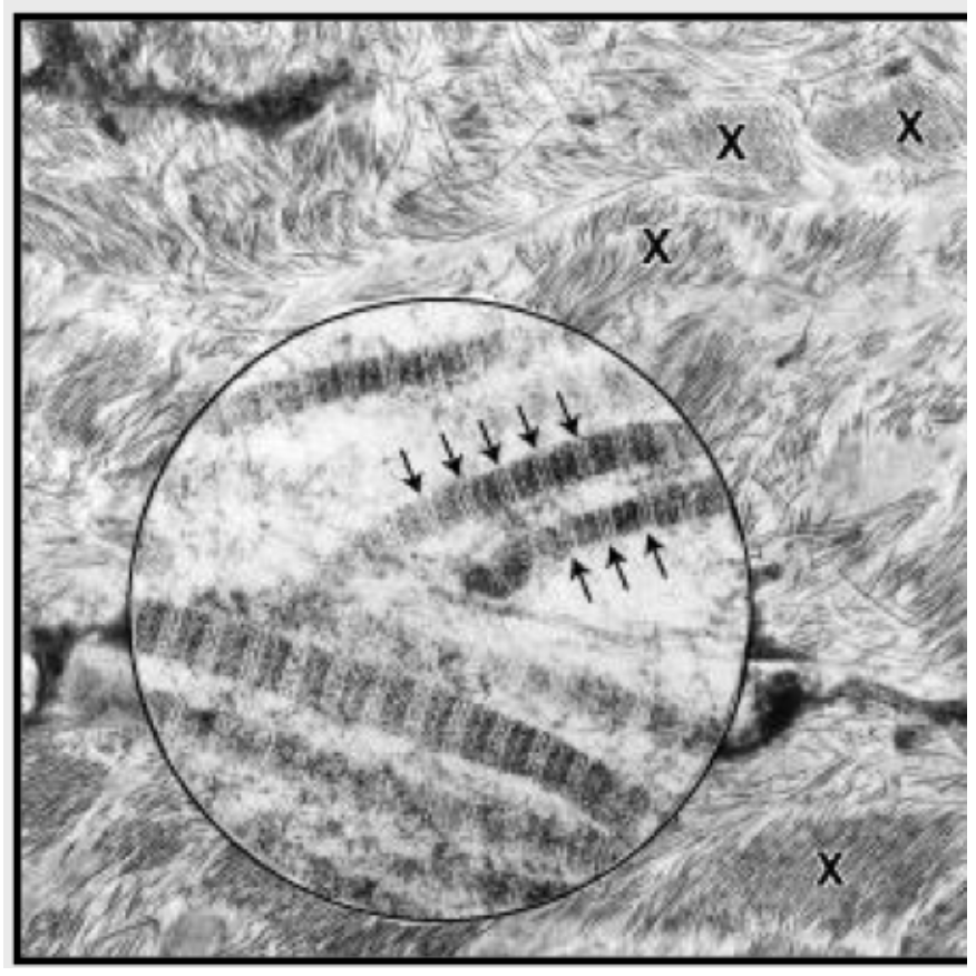
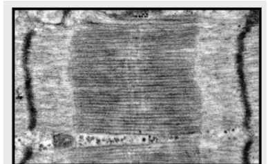
Sarcomero
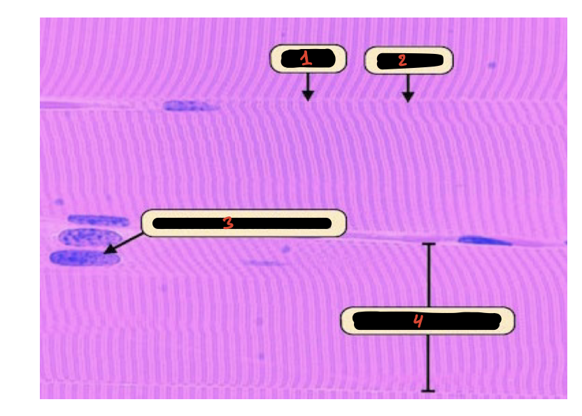
1: I Band
2: A Band
3: Skeletal Muscle Nucleous
4: Skeletal Muscle Cell
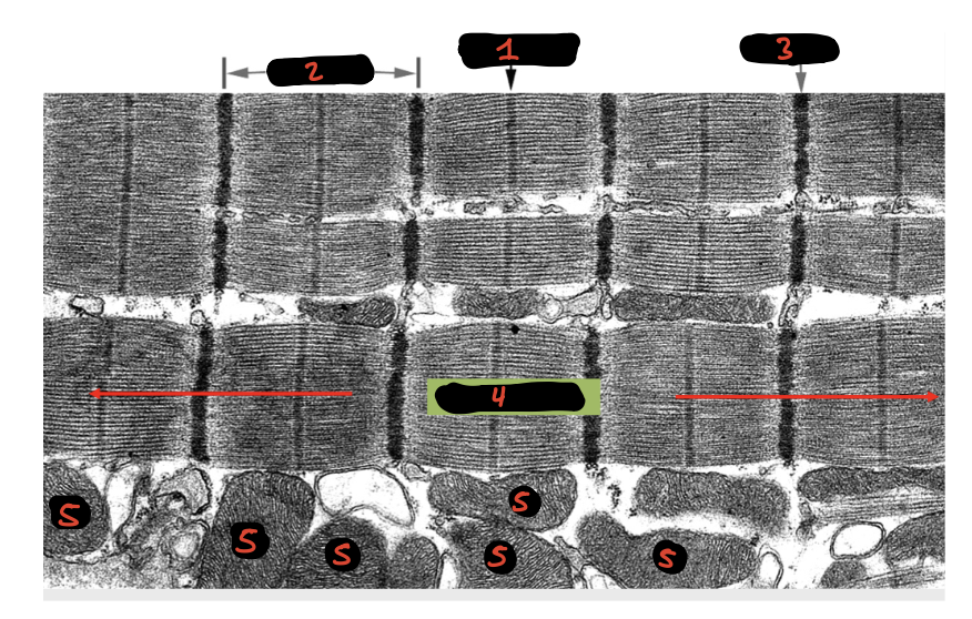
1: M Band
2: Sarcomero
3: Z line
4: Myofibril
5: Mitocondrias
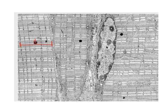
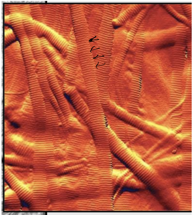
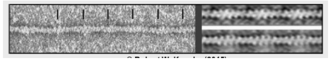
Myofibril
Myofibrils
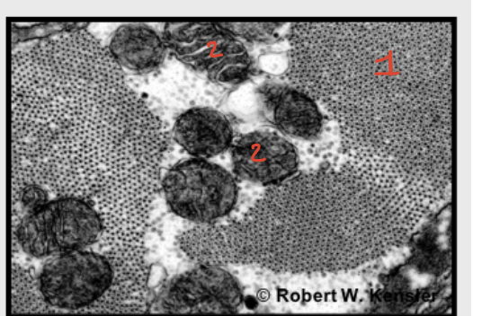
1: Cross Sections of Myofibrils
2: Mitocondrias
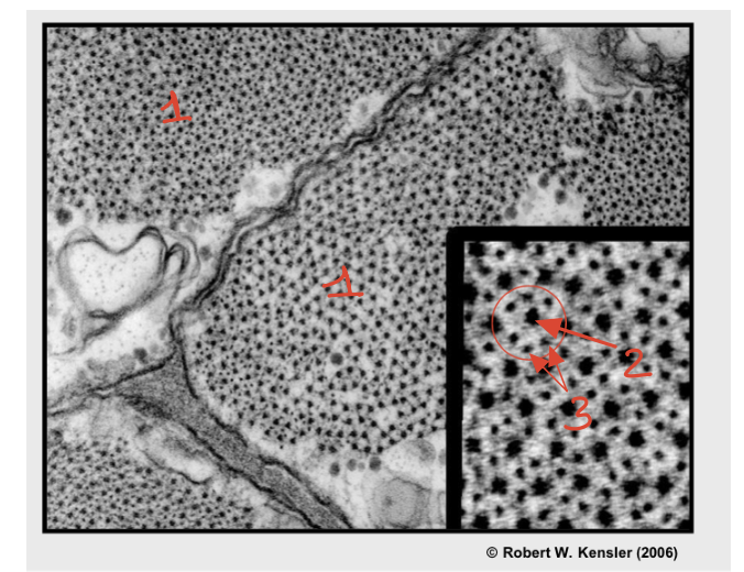
1: Cross Sections of Myofibrils
2: Thick Filament
3: Thin Filaments
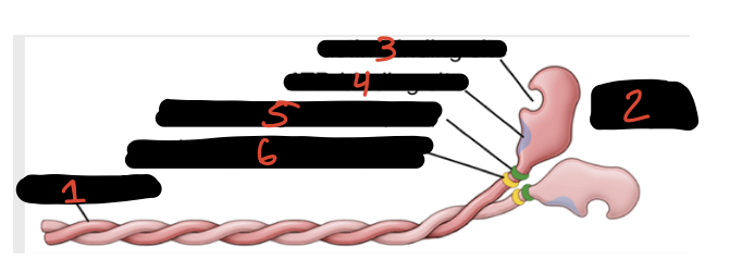
1: Heavy Chains
2: Heads
3: Actin-Binding Site
4: ATP-Binding site
5: essential light Chain
6: Regulatory light Chain
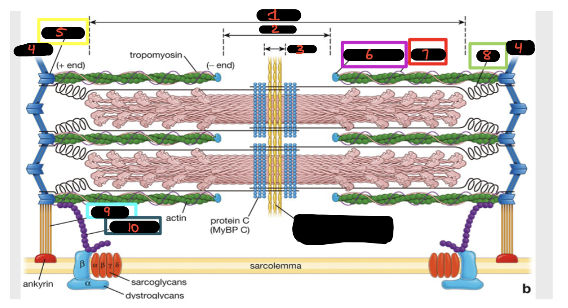
1: A Band
2: H Band
3: M line
4: Z line
5: Alfa-actinin
6: Tropomodulin
7: Nebulin
8: Titin
9: Desmin
10: Dystrophin
Lo oscuro del centro es?
Myofibiril Thick Filaments
Myofibril Thin filaments
Triads
Sarcomeros with Normal Dystrophin vs Sarcomeros with Duchenne Muscular Dystrophy (DMD)
Cells with Normal Dystrophin vs Cells with Duchenne Muscular Dystrophy (DMD)
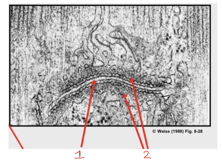
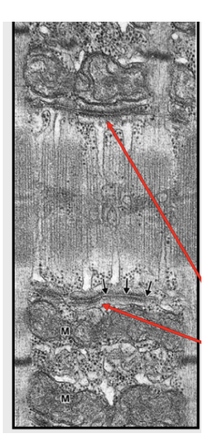
1: T tubule
2: Terminal Cisterna of SR
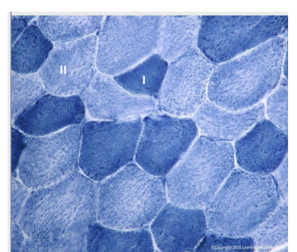
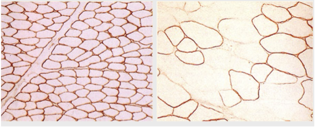
Fibers type I and II of the Skeletal Muscle
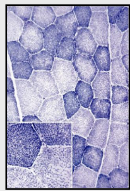
Varios types of Skeletal Muscle Fibers
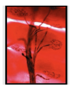
Neuromuscular Junction
Neuromuscular Junction
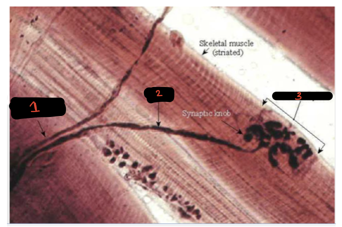
1: Nerve Bundles of Axons
2: Axons
3: Motor end plate
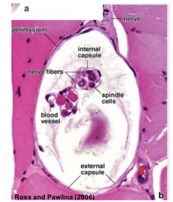
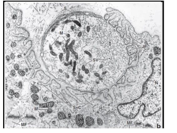
Muscle Spindle