Module 4- Objectives & Questions
1/13
There's no tags or description
Looks like no tags are added yet.
Name | Mastery | Learn | Test | Matching | Spaced | Call with Kai |
|---|
No analytics yet
Send a link to your students to track their progress
14 Terms
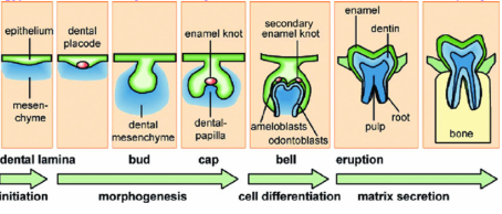
List for each stage of tooth development (up to eruption) the defining characteristics of that stage.
-initiation: placode, ecto-mesenchymal condensation below epithelial thickening (epithelial ingrowth); 4-5 weeks; 4 separate thickenings on maxilla and MNP, 2 on mandible
-bud stage: 10 upper jaw buds (rounded epithelial outgrowth into ectomesenchyme), 10 lower jaw buds, dental lamina, vestibular lamina; 5-6 weeks
-cap stage: enamel knot, dental sac, dental papilla; 8 weeks
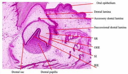
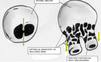
-bell stage: 4 layers of enamel organ- stellate intermedium, inner-enamel epithelium (IEE), outer enamel epithelium (OEE), stellate reticulum (SR)
-appositional stage: ameloblasts and odontoblasts (deposition of enamel and dentin matrix), continued morphodifferentiation, cytodifferentiation begins
-eruptions: HERS = IEE + OEE = epithelial root sheath
-cytodifferentiation starts at the end of bell stage and beginning of appositional stage- BL thickens and mesenchymal cells line up to become pre-odontoblasts
Describe the defining characteristics and future roles of IEE, SI, SR, and OEE. Describe also the differences in cell shape the future role of the condensed mesenchyme in the concavity of the enamel organ and surrounding the enamel organ.
-IEE: single layer of low columnar epithelial cells, lines inner concave surface of enamel organ and is separated by the basal lamina from the dental papilla, will differentiate into ameloblasts
-SI: 1-4 layers of squamous epithelial cells, immediately adjacent and external to IEE, support ameloblasts during amelogenesis
-SR: stellate shaped cells with cell bodies far apart, support ameloblasts during amelogenesis, release proteoglycans and heparin → pressure to maintain the shape of the tooth, transports nutrients between IEE and OEE
-OEE: single layer of low columnar almost squamous epithelial cells, lines the outer convex surface of the enamel organ, separated by a basal lamina from the dental sac, protective, exchange with blood vessels
-dental papilla (condensed mesenchyme): beneath IEE, cells become odontoblasts (dentin)
-dental follicle (condensed mesenchyme): around enamel organ, forms cementum/PDL/alveolar bone
Name the sites where the earliest formed dentin and enamel matrix of a tooth are deposited. Where are the final layers of dentin and enamel deposited?
-earliest dentin: cusp tips (or incisal edges)
-earliest enamel: immediately adjacent, at DEJ
-final dentin: innermost surface of pulp chamber
-final enamel: outermost surface of crown
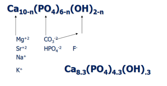
Provide the chemical formula of a unit of crystal of biologic hydroxyapatite.
Ca10(PO4)6(OH)2
Explain the possibility of ion substitutions in biologic hydroxyapatite. Name examples.
-mineral is an imperfect microcrystalline analogue of geologic hydroxyapatite
-F substituting for hydroxyl ion in hydroxyl column- F fits inside plane of triangle → more stable configuration
-carbonate (triangular) substituting for either phosphate (pyramidal) or hydroxyl ions → poor fit and thus strain in atomic arrangement
-Pb substituting for Ca ion- poisonous for mineral and individual (lost to mineral homeostasis)
Explain what intermediate step in biologic mineralization may occur if no mineralized tissues are present.
-formation of amorphous calcium phosphate (lower energy intermediate) that then transforms into crystalline hydroxyapatite
-formed in mitochondria of scleroblasts
-minerals released in matrix vesicles
Name on which side of the enamel organ of a primary tooth the vestibular lamina forms. A successional lamina develops from which side of the enamel organ of the primary tooth?
-vestibular lamina: develops on labial side of enamel organ
-successional lamina: develops on lingual side of enamel organ
Explain how the cervical opening of an enamel organ of a molar tooth is subdivided into 2 or 3 cervical openings, leading to the formation of multiple roots.
-start of root formation coincides with start of eruptive movement of the tooth
-subdivision accomplished by horizontal outgrowth of 2 or 3 epithelial flaps across the cervical opening- first visible in late cap or early bell stages
-usually consist of IEE and OEE, resembling histologic structure of HERS- flaps fuse once they meet in the center of the cervical opening
Name 3 possible mechanisms that may be involved in the interactions between epithelial and mesenchymal cells that may lead to the differentiation of odontoblasts and ameloblasts
-diffusible substances (paracrine growth factors)
-cell-ECM interactions (basement membrane molecules, integrins)
-direct cell-cell contacts (juxtacrine signaling)
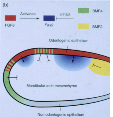
Name the growth factor that is expressed in the oral epithelium at locations of future tooth development. Which growth factor is found in non-tooth forming epithelium? In which locations of the jaw mesenchyme is PAX9 expressed?
-tooth forming epithelium: FGF8
-non-tooth forming epithelium: BMP4
-PAX9 expression in jaw mesenchyme at underlying future tooth sites (PAX9 required for tooth initiation)
Give a possible explanation for the determination of the different morphology of incisors, canines, premolars, and molars
-ectodermal FGF8 and BMP4 signals lead to expression of different homeobox-containing genes in mesenchyme
-determination controlled by epithelial-mesenchymal signaling
-Homeobox code model: incisors- (+) MSX1/2, ALX3, (-) BARX1, DIX1/2; canine/premolar- (+) MSX1/2, DIX1/2, (-) ALX3, BARX1; molars- (+) BARX1, DIX1/2, MSX1/2, (-)ALX3
Explain the role of the epithelial cells of the primary and secondary enamel knots
-primary enamel knot: non-dividing signaling center, regulates crown shape, cusp initiation, induces differentiation
-secondary enamel knot: regulate cusp number and position
-express signaling molecules that guide morphogenesis
Describe the mineralization in the first formed dentin of a tooth, and the mineralization in the adjacent enamel matrix
-mantle dentin first to be deposited
-dentin mineralization occurs by globular mineralization: hydroxyapatite crystal form in spherical foci, globules enlarge and fuse together; collagen fibers act as nucleation sites for crystal deposition
-mineralization of enamel is immediate and partial: newly secreted enamel matrix is ~30% mineralized at secretion, hydroxyapatite crystals are long, thin ribbons aligned parallel; maturation stage follows- crystals grow in width and thickness
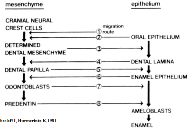
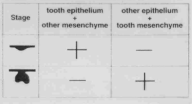
Sequence of developmental interactions in the crown