BIO4 - Exam revision babeyyy
5.0(1)
Card Sorting
1/126
Study Analytics
Name | Mastery | Learn | Test | Matching | Spaced |
|---|
No study sessions yet.
127 Terms
1
New cards
Nucleic acids - Define
Polymer of nucleotides
2
New cards
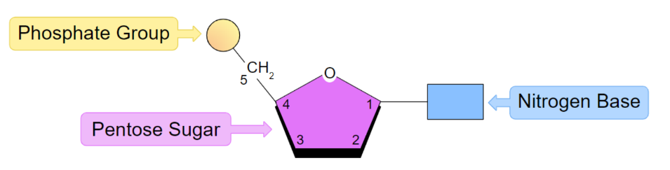
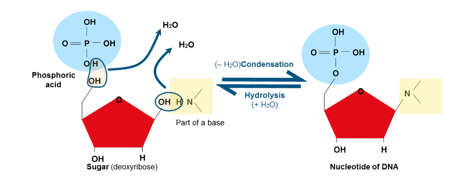
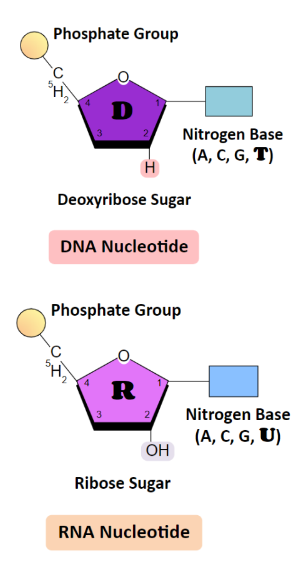
Nucleotide - Define + list structural components
Monomer of nucleic acids, comprised of
A phosphate group
A pentose (five carbon) sugar
A nitrogenous base - either guanine, cytosine,
adenine, thymine or uracil
A phosphate group
A pentose (five carbon) sugar
A nitrogenous base - either guanine, cytosine,
adenine, thymine or uracil
3
New cards
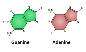
Purines
adenine & guanine
A purine is a heterocyclic aromatic organic
compound containing 4 nitrogen atoms and two
carbon rings.
A purine is a heterocyclic aromatic organic
compound containing 4 nitrogen atoms and two
carbon rings.
4
New cards
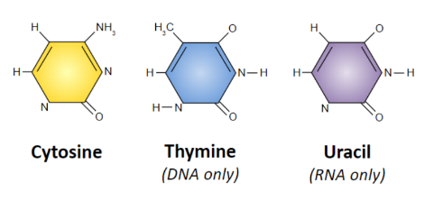
Pyrimidines
cytosine, thymine &
uracil
heterocyclic aromatic organic
compound containing 2 nitrogen atoms and only one
carbon ring.
uracil
heterocyclic aromatic organic
compound containing 2 nitrogen atoms and only one
carbon ring.
5
New cards
Formation of a nucleotide
Condensation reactions
6
New cards
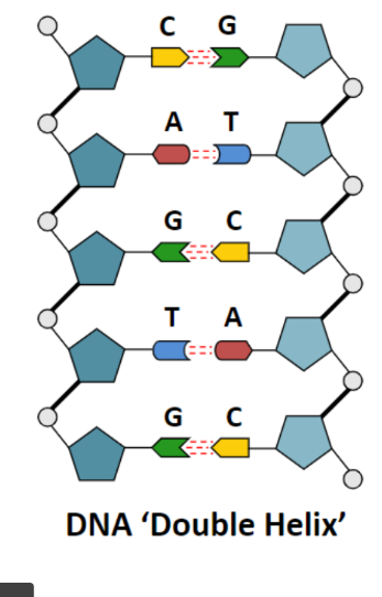
DNA Structure – The Double Helix
\
\
* Purines and pyrimidines are attracted to each other by hydrogen bonds, which cause two nucleic acid chains to join together. (C-G pairs have 3 bonds!)
* The sugars and phosphates are bonded by covalent bonds, making a very strong and rigid ‘back bone’.
* The 5’- phosphate group of one nucleotide attaches to the sugar of another nucleotide (at the 3’-hydroxyl group) IMPORTANT!!
* The sugars and phosphates are bonded by covalent bonds, making a very strong and rigid ‘back bone’.
* The 5’- phosphate group of one nucleotide attaches to the sugar of another nucleotide (at the 3’-hydroxyl group) IMPORTANT!!
7
New cards
Directionality
DNA strands run in opposite directions
* The direction is determined by the carbons in the deoxyribose ring.
* The strand with a free third carbon on the sugar is called the 3’ end.
* The strand with a phosphate bonded to the fifth carbon on the sugar is called the 5’ end.
* The direction is determined by the carbons in the deoxyribose ring.
* The strand with a free third carbon on the sugar is called the 3’ end.
* The strand with a phosphate bonded to the fifth carbon on the sugar is called the 5’ end.
8
New cards
Base - Pair rule
DNA strands are complementary
* Adenine always bonds with Thymine
* Guanince always bonds with cytosine
* Adenine always bonds with Thymine
* Guanince always bonds with cytosine
9
New cards
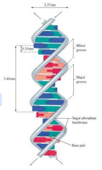
The Helix
* The helix forms due to forces between the bases of adjacent nucleotides.
* The helix forms major & minor grooves.
* In an aqueous environment, the grooves are filled with water molecules.
* The helix forms major & minor grooves.
* In an aqueous environment, the grooves are filled with water molecules.
10
New cards
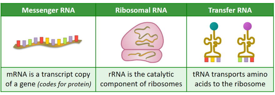
RNA
* similar to DNA but Thymine is replaced with Uracil
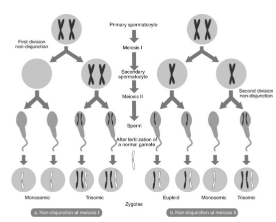
* RNA forms a single strand encoding for portions of the genome - it is much shorter than DNA.
* RNA is usually single-stranded but can form loops via
complementary base pairing
* RNA forms a single strand encoding for portions of the genome - it is much shorter than DNA.
* RNA is usually single-stranded but can form loops via
complementary base pairing
11
New cards
RNA - Name 3 functions
* Messenger RNA (mRNA)
* Ribosomal RNA (rRNA)
* Transfer RNA (tRNA)
* Ribosomal RNA (rRNA)
* Transfer RNA (tRNA)
12
New cards
Chromatin
combination of DNA, histone
protein, and other proteins that makes up
chromosomes. It is found inside the nuclear
envelope of eukaryotic cells.
The functions of chromatin are:
To package DNA into a smaller volume to fit in the cell
and nucleus
To strengthen the DNA to allow mitosis and meiosis to
occur
To control gene expression and DNA replication
13
New cards
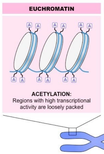
Euchromatin
(extended) is high in gene concentration and
often indicates higher amounts of transcription is
occurring.
often indicates higher amounts of transcription is
occurring.
14
New cards
Heterochromatin
(condensed) is tightly packed and often
indicates the DNA region is transcriptionally inactive.
indicates the DNA region is transcriptionally inactive.

15
New cards
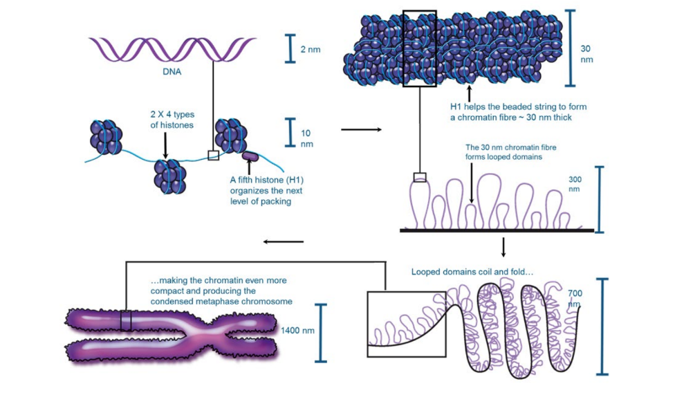
Chromatin → Chromosome
The DNA wraps around a bundle of 8 histone
proteins (2 x 4) to form a nucleosome
An additional histone (H1) helps the chromatin
fibre form looped domains. The looped domains
are attached to a scaffold non-histone protein.
The looped domains themselves fold repeatedly.
Repeated folding produces the condensed
chromosome.
proteins (2 x 4) to form a nucleosome
An additional histone (H1) helps the chromatin
fibre form looped domains. The looped domains
are attached to a scaffold non-histone protein.
The looped domains themselves fold repeatedly.
Repeated folding produces the condensed
chromosome.
16
New cards
Acetylation
Adding an acetyl group to the tail (acetylation) neutralises
the charge, making DNA less tightly coiled and increasing
transcription (euchromatin)
the charge, making DNA less tightly coiled and increasing
transcription (euchromatin)
17
New cards
Methylation
Adding a methyl group to the tail (methylation) maintains
the positive charge, making DNA more coiled and reducing
transcription (heterochromatin)
* As well as the histone tails, DNA bases can also be methylated
* During development, differentiated cells develop a stable and unique DNA methylation pattern that regulates tissue-specific gene expression
the positive charge, making DNA more coiled and reducing
transcription (heterochromatin)
* As well as the histone tails, DNA bases can also be methylated
* During development, differentiated cells develop a stable and unique DNA methylation pattern that regulates tissue-specific gene expression
18
New cards
Epigenetics
Epigenetics is the study of changes in phenotype as a
result of variations in gene expression levels
* DNA methylation patterns may change over a lifetime
* influenced by heritability but is not genetically pre-determined (identical twins may have different DNA
methylation patterns)
* Different cell types in the same organism may have markedly different DNA methylation patterns
* Environmental factors (e.g. diet, pathogen exposure, etc.) may influence the level of DNA methylation within cells
* Direct methylation of DNA (as opposed to the histone tails) can also affect gene expression patterns
* Increased methylation of DNA decreases gene expression (by preventing the binding of transcription factors)
* Consequently, genes that are not transcribed tend to exhibit more DNA methylation than genes that are actively transcribed
19
New cards
Histone
bind to DNA, help give chromosomes their shape, and help control the activity of genes (acetylation and Methylation).
20
New cards
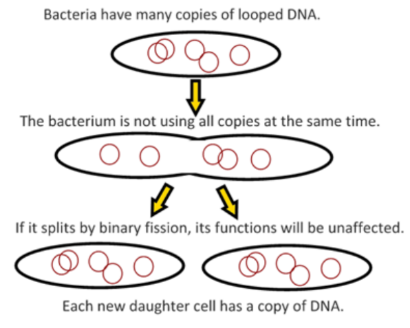
Binary fission
The process where prokaryotic cells divide their genetic material
21
New cards
Mitosis - definition and functions
* Creation of New cells
* Asexual reproduction - certain eukaryotic organisms may reproduce asexually by mitosis (e.g. protozoa, hydra).
* Tissue repair - damaged tissue can recover by replacing dead or damaged cells.
* Embryonic development - a fertilized egg (zygote) will undergo mitosis & differentiation to develop into an embryo
22
New cards
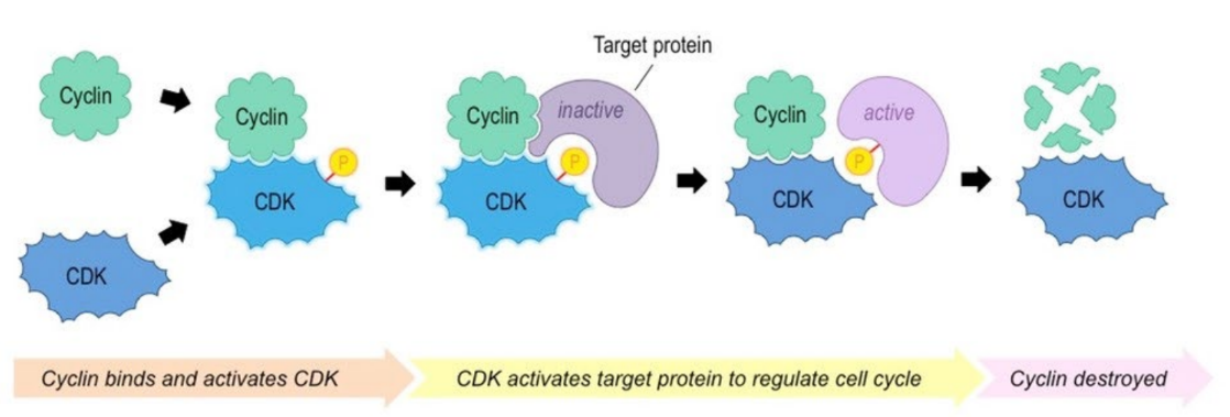
Cyclins
* Regulatory proteins that control the cell cycle
* Cyclins bind to enzymes called cyclin-dependent kinases
(CDKs) that become active and attach phosphate groups to
other proteins in the cell which in turn trigger other proteins to
become active and carry out tasks specific to phases of the cell
cycle.
* Cyclins bind to enzymes called cyclin-dependent kinases
(CDKs) that become active and attach phosphate groups to
other proteins in the cell which in turn trigger other proteins to
become active and carry out tasks specific to phases of the cell
cycle.
23
New cards
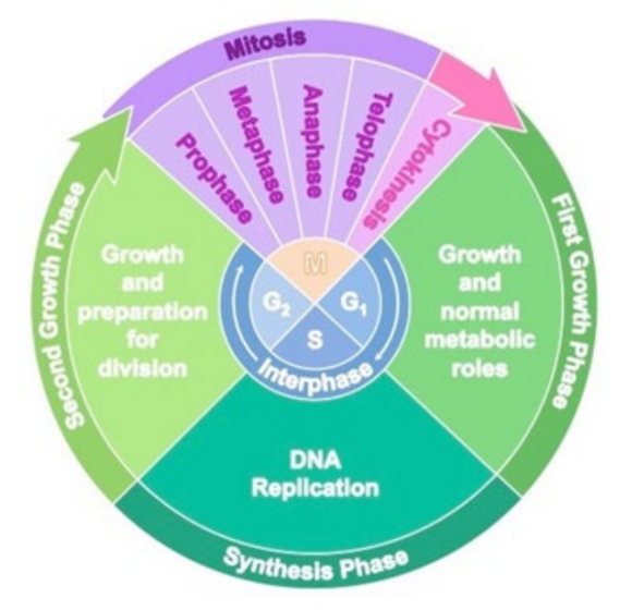
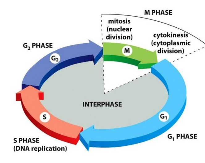
The cell cycle - Recall phases
The cell cycle has several distinct components:
* Interphase
* Mitotic division
* Prophase
* Metaphase
* Anaphase
* Telophase
* Cytokinesis
* Interphase
* Mitotic division
* Prophase
* Metaphase
* Anaphase
* Telophase
* Cytokinesis
24
New cards
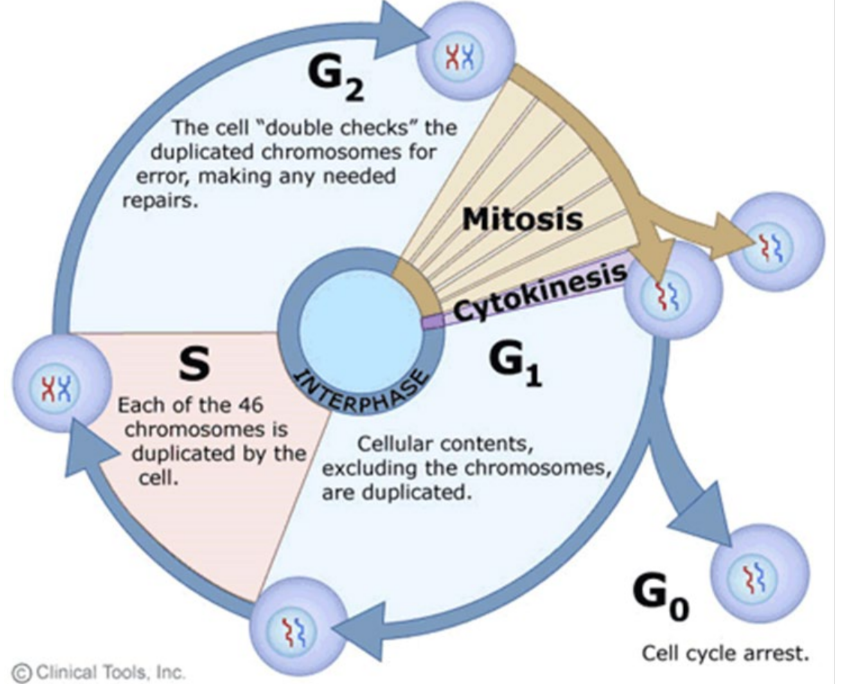
Interphase - Recall phases + processed included
Interphase is a very active phase of the cell cycle with many processes occurring in the nucleus and cytoplasm.
* G1 – Cell growth and metabolism
* S – DNA replication
* G2 – Cell growth and proof-reading
Growth and preparation includes:
* Organelle duplication
* Cell growth / cytoplasmic volume increase
* Protein and enzyme synthesis
* Obtain nutrients
* Respiration / ATP production
* G1 – Cell growth and metabolism
* S – DNA replication
* G2 – Cell growth and proof-reading
Growth and preparation includes:
* Organelle duplication
* Cell growth / cytoplasmic volume increase
* Protein and enzyme synthesis
* Obtain nutrients
* Respiration / ATP production
25
New cards
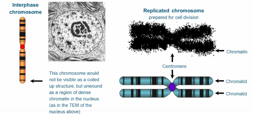
Recall chromosome states during interphase vs dividing cells
* Interphase: Single- armed structures + unwound
* Dividing: Double-armed structure, forming two chromatids
* Dividing: Double-armed structure, forming two chromatids
26
New cards
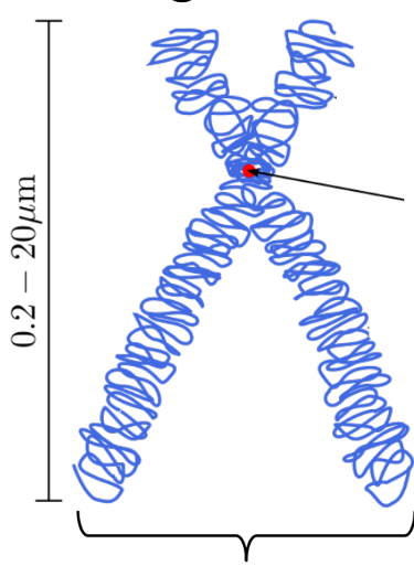
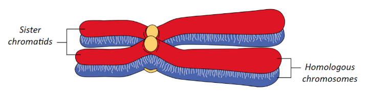
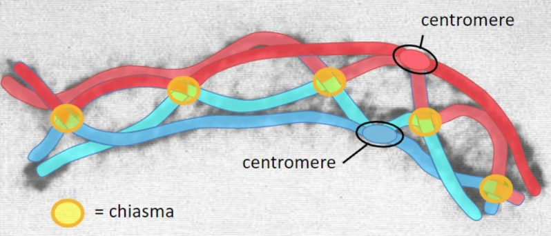
Centromere
The part of a chromosome that links sister chromatids
27
New cards
Sister chromatids
Duplicated chromosomes attached by a centromere
\
Note: Should be referred to as chromosomes after separation
\
Note: Should be referred to as chromosomes after separation
28
New cards
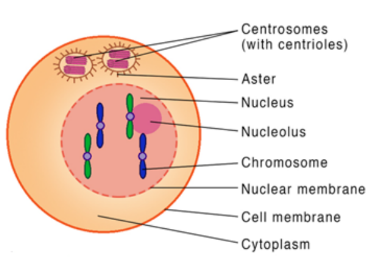
Centrioles
Organise Spindle microtubules

29
New cards
Spindle microtubules/ Spindle fibers
It assembles around the chromosomes and distributes the duplicated genome to the daughter cells during mitosis.

30
New cards
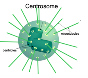
Centrosome
The centrosome is the primary microtubule-organizing centre in animal cells, and so it
* regulates cell motility,
* adhesion and polarity in interphase
* facilitates the organization of the spindle poles during mitosis.
* regulates cell motility,
* adhesion and polarity in interphase
* facilitates the organization of the spindle poles during mitosis.
31
New cards
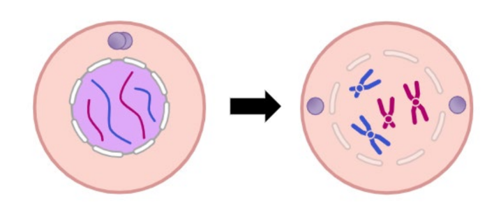
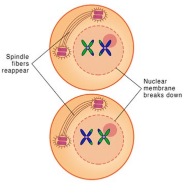
Prophase
* DNA supercoils and chromosomes condense
* Nuclear membrane breaks down
* Paired centrosomes move to poles
* Nuclear membrane breaks down
* Paired centrosomes move to poles
32
New cards
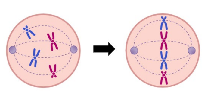
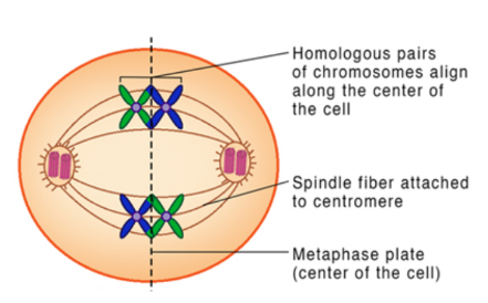
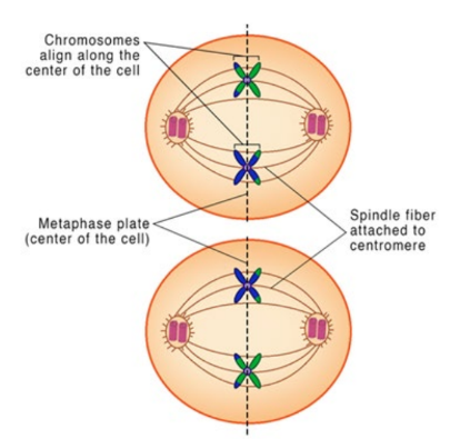
Metaphase
* Microtubule spindle fibres connect from centrosomes to centromeres
* Spindle fibres contract, causing the chromosomes to align at the centre
* Spindle fibres contract, causing the chromosomes to align at the centre
33
New cards
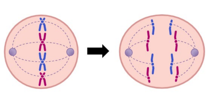
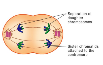
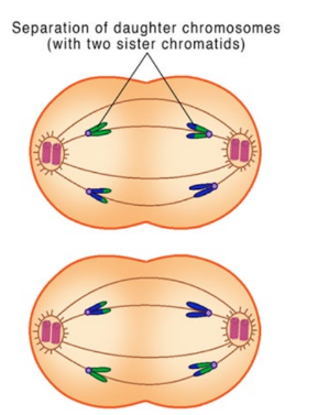
Anaphase
Spindle fibre contraction cause the sister chromatids to separate and become identical chromosomes that move to opposite poles of cell
34
New cards
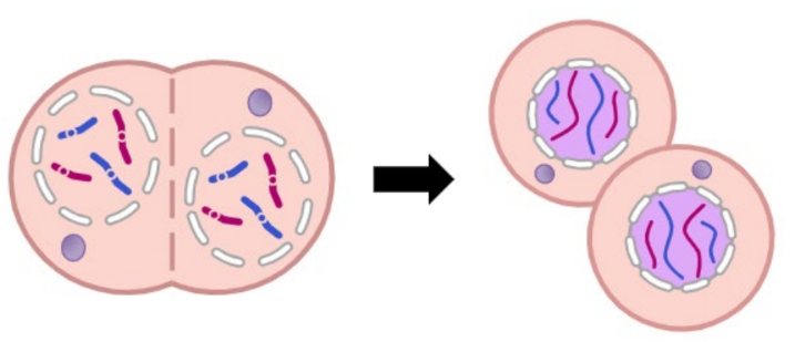
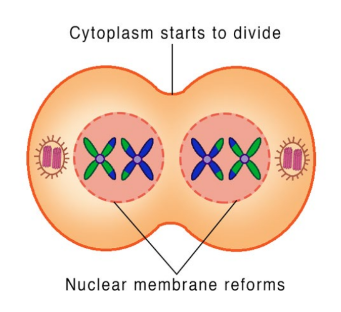
Telophase
* Chromosomes decondense
* Nuclear membranes reform around the two identical chromosome sets
* Nuclear membranes reform around the two identical chromosome sets
35
New cards
Cytokinesis - Plant cells
vesicles are moved to the equator were they
fuse to form a tubular structure which merges with more
vesicles to form the plasma membrane and divide the
cytoplasm. Substances such as pectin and cellulose are
then deposited by exocytosis to form the lamella and cell
wall.
fuse to form a tubular structure which merges with more
vesicles to form the plasma membrane and divide the
cytoplasm. Substances such as pectin and cellulose are
then deposited by exocytosis to form the lamella and cell
wall.

36
New cards
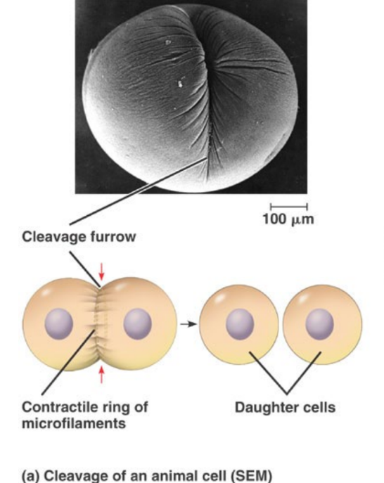
Cytokinesis - Animal cells
the plasma membrane is pulled inwards
around the equator of the cell to form a cleavage furrow
by contractile proteins actin and myosin – the same as in
muscles.
around the equator of the cell to form a cleavage furrow
by contractile proteins actin and myosin – the same as in
muscles.
37
New cards
When does DNA replication occur?
Occurs during S phase -duplicates DNA
38
New cards
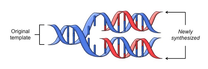
Semi-conservative replication
DNA replication is a semi-conservative process, because when a new double-stranded DNA molecule is formed:
* One strand will be from the original template molecule
* One strand will be newly synthesised
* One strand will be from the original template molecule
* One strand will be newly synthesised
39
New cards
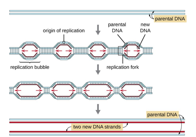
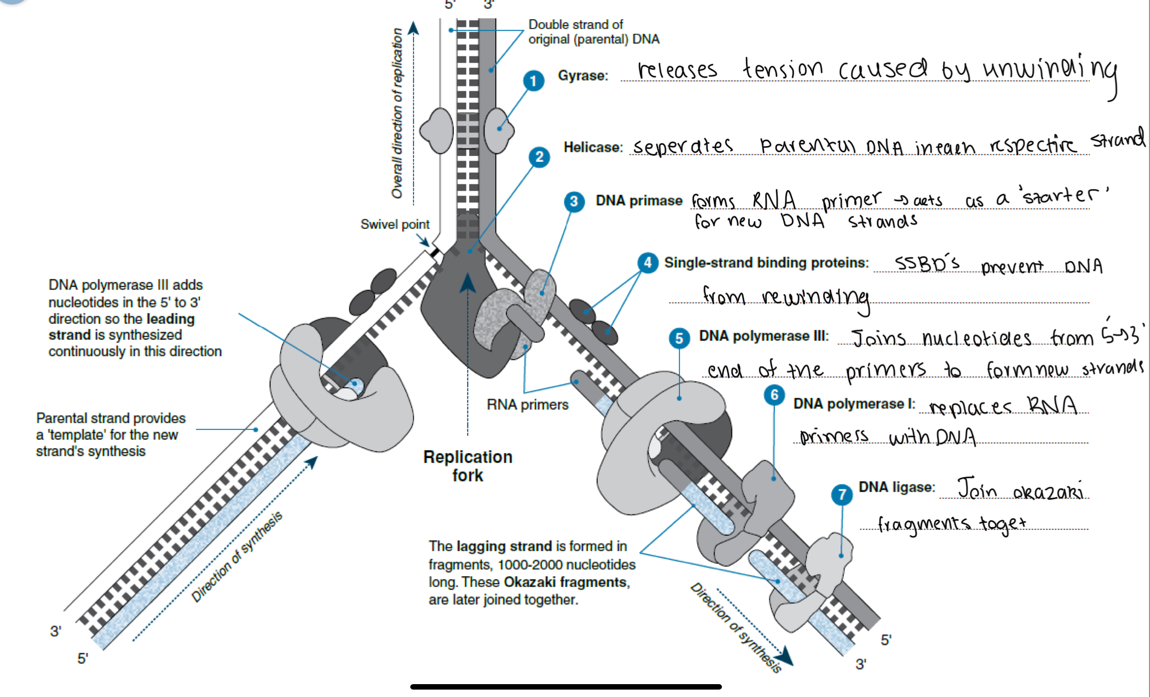
Steps in DNA Replication - 1
1. DNA replication in eukaryotes occurs at
many points along the chromosome, called
origins of replication.
40
New cards
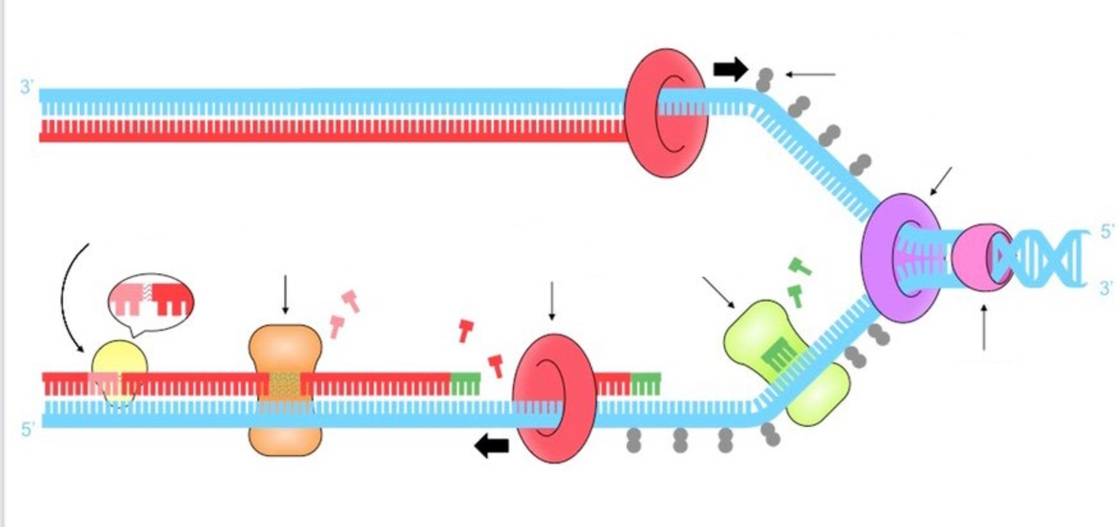
Steps in DNA replication - Label each enzyme and which step they are involved in

41
New cards
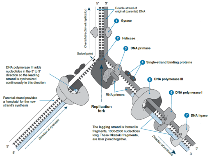
Recall each enzymes function
7) Joins okazaki fragments together
42
New cards
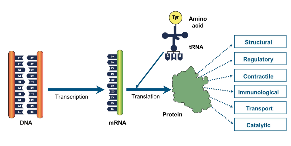
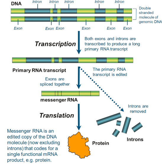
Transcription and translation - Steps
In eukaryotic cells, protein synthesis occurs in
three steps.
1. Transcription of mRNA
2. mRNA processing
3. Translation
three steps.
1. Transcription of mRNA
2. mRNA processing
3. Translation
43
New cards
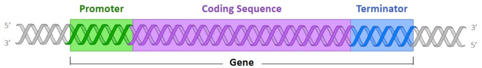
Sections of a gene + Function
A gene is a sequence of DNA that is transcribed into RNA and has 3 main parts:
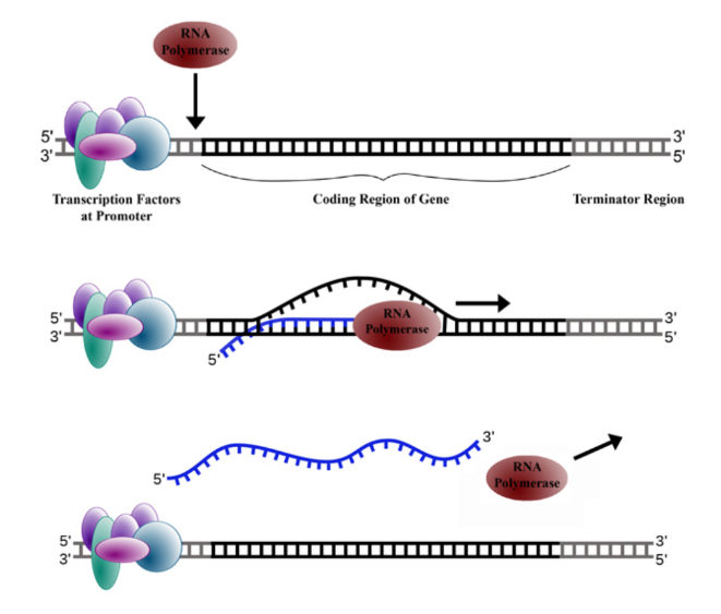
* Promoter: The non-coding sequence that is responsible for the initiation of transcription (functions as a binding site for RNA polymerase)
* Coding Sequence: The region of DNA that is transcribed by RNA polymerase
* Terminator: The sequence that is responsible for terminating transcription
* Promoter: The non-coding sequence that is responsible for the initiation of transcription (functions as a binding site for RNA polymerase)
* Coding Sequence: The region of DNA that is transcribed by RNA polymerase
* Terminator: The sequence that is responsible for terminating transcription
44
New cards
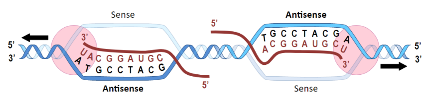
Sense vs Antisense strands
The antisense (or template) strand is transcribed (sequence is
complementary to RNA transcript)
The sense (or coding) strand is not transcribed (sequence
identical to transcript – except T / U)
complementary to RNA transcript)
The sense (or coding) strand is not transcribed (sequence
identical to transcript – except T / U)
45
New cards
Transcription of mRNA (Recall enzyme involved)
* RNA polymerase attaches to the promoter on the Anti-Sense or Template strand and separates the DNA strands.
* It then moves along the DNA in the 3' to 5' direction, pairing up RNA nucleotides with their DNA complements and adding them to the 3’ end of the growing RNA molecule.
* Once RNA polymerase has gone past the terminator, the enzyme releases the completed mRNA and detaches from the DNA.
* It then moves along the DNA in the 3' to 5' direction, pairing up RNA nucleotides with their DNA complements and adding them to the 3’ end of the growing RNA molecule.
* Once RNA polymerase has gone past the terminator, the enzyme releases the completed mRNA and detaches from the DNA.
46
New cards
mRNA Processing
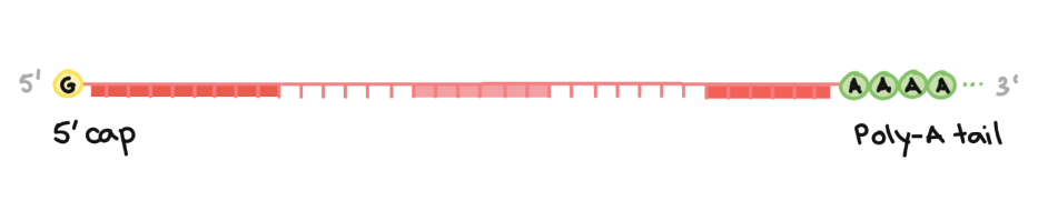
* The ends of the RNA do not code for a protein. RNA processing begins with alteration of these ends.
* A 5' cap is added to the beginning of the RNA transcript, and a 3' poly-A tail is added to the end. (mainly for protection/ to avoid damage from enzymes
* A 5' cap is added to the beginning of the RNA transcript, and a 3' poly-A tail is added to the end. (mainly for protection/ to avoid damage from enzymes
47
New cards
Introns
Portions of the coding segment that do not actually code for protein, are removed, known as introns
48
New cards
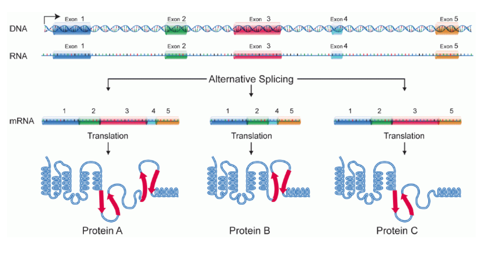
Exons
The remaining sections of the mRNA that are spliced together during mRNA processing
* The number of exons and the way they are spliced varies.
* This creates variations in the translated polypeptide chain.
* The number of exons and the way they are spliced varies.
* This creates variations in the translated polypeptide chain.
49
New cards
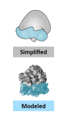
Ribosomes - Summary
* Ribosomes are the site of polypeptide synthesis (translation)
* They are made of protein (stability) and rRNA (catalytic)
* Ribosomes contain two distinct subunits: Small subunit contains an mRNA binding site Large subunit contains three tRNA binding sites (A, P, E)
* Ribosomes exist freely in the cytosol are bound to rough ER
* They can differ in size (70S = prokaryote ; 80S = eukaryote)
50
New cards
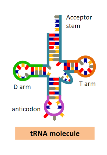
tRNA - Structure and function
Transfer RNA carries specific amino acids to the ribosome
* Transfer RNA molecules have four key regions:
* Acceptor stem (carries the amino acid)
* Anticodon (complementary to an mRNA codon)
* T arm (associates with the ribosome)
* D arm (associates with a tRNA-activating enzyme)
* Transfer RNA molecules fold into a cloverleaf structure
* Transfer RNA molecules have four key regions:
* Acceptor stem (carries the amino acid)
* Anticodon (complementary to an mRNA codon)
* T arm (associates with the ribosome)
* D arm (associates with a tRNA-activating enzyme)
* Transfer RNA molecules fold into a cloverleaf structure
51
New cards
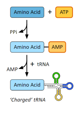
Synthesis of tRNA - Summary
* tRNA-activating enzymes join ATP to an amino acid which creates a ‘charged’ amino acid–AMP complex
* The phosphorylated amino acid is then linked to a specific tRNA molecule and the AMP is released
* The energy in the ‘charged’ amino acid is then used for peptide bond formation
* The phosphorylated amino acid is then linked to a specific tRNA molecule and the AMP is released
* The energy in the ‘charged’ amino acid is then used for peptide bond formation
52
New cards
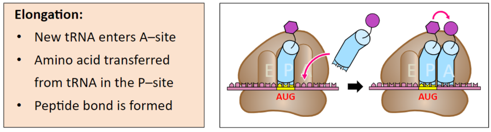
Translation - Recall steps
* Initiation: Assembly of an active ribosomal complex on an mRNA sequence
* Elongation: A new amino acid is added to a developing peptide chain based on the mRNA codon and tRNA anticodon match
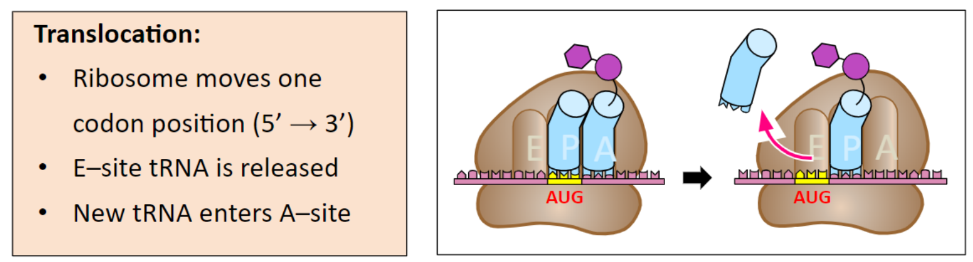
* Translocation: The ribosome moves to the next codon position
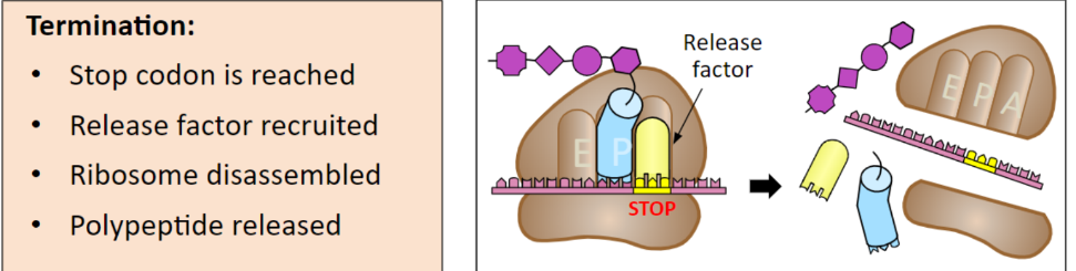
* Termination: Ribosomal complex and polypeptide dissociate from mRNA
\
Note: The processes of elongation and translocation are sequentially repeated as the ribosome moves along the transcribed mRNA sequence in a 5’ → 3’ direction
* Elongation: A new amino acid is added to a developing peptide chain based on the mRNA codon and tRNA anticodon match
* Translocation: The ribosome moves to the next codon position
* Termination: Ribosomal complex and polypeptide dissociate from mRNA
\
Note: The processes of elongation and translocation are sequentially repeated as the ribosome moves along the transcribed mRNA sequence in a 5’ → 3’ direction
53
New cards
Initiation

54
New cards
Elongation
55
New cards
Translation
56
New cards
Termination
57
New cards
The genetic code - Recall start + stop codon
* Peptides are formed from 20 different amino acids in different orders and combinations.
* The mRNA codons code for particular amino acids.
* AUG codes for methionine, the starting amino acid for all peptide chains.
* Three special base triplets -- UAA, UAG, and UGA -- do not code for amino acids, but instead act as stop codons.
* Redundancy - many amino acids have more than one codon.
* The mRNA codons code for particular amino acids.
* AUG codes for methionine, the starting amino acid for all peptide chains.
* Three special base triplets -- UAA, UAG, and UGA -- do not code for amino acids, but instead act as stop codons.
* Redundancy - many amino acids have more than one codon.

58
New cards
Proteins - Summary
* In prokaryotes, the absence of a nuclear membrane allows translation to occur immediately after transcription
* In eukaryotes, translation will occur at one of two distinct locations:
* Free ribosomes (cytosolic) synthesise proteins for use primarily within the cell
* Bound ribosomes (rough ER) synthesise proteins for secretion (or lysosomes)
* Proteins produced by the rough endoplasmic reticulum are typically transported via vesicles to the Golgi apparatus for secretion
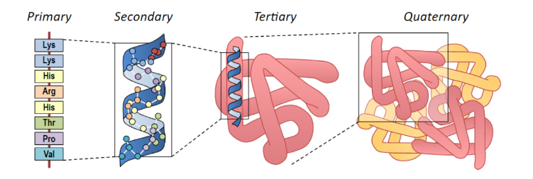
* Polypeptides fold into unique shapes which may be essential to their function and role
* Their final structure is determined by the sequence of amino acids that was coded for in the gene that was transcribed and translated
* In eukaryotes, translation will occur at one of two distinct locations:
* Free ribosomes (cytosolic) synthesise proteins for use primarily within the cell
* Bound ribosomes (rough ER) synthesise proteins for secretion (or lysosomes)
* Proteins produced by the rough endoplasmic reticulum are typically transported via vesicles to the Golgi apparatus for secretion
* Polypeptides fold into unique shapes which may be essential to their function and role
* Their final structure is determined by the sequence of amino acids that was coded for in the gene that was transcribed and translated
59
New cards
Mutation + Causes
* alterations in the DNA in chromosomes.
* Mutations may occur randomly and spontaneously.
* They may also be induced by environmental factors.
* Spontaneous mutations
* Arise from errors in replication
* Genes mutate at different rates
* Induced mutations
* Mutations can be induced by mutagens (environmental factors that cause a change in DNA)
60
New cards
List 3 main environmental causes
* Radiation
* Viruses
* chemicals
* Viruses
* chemicals
61
New cards
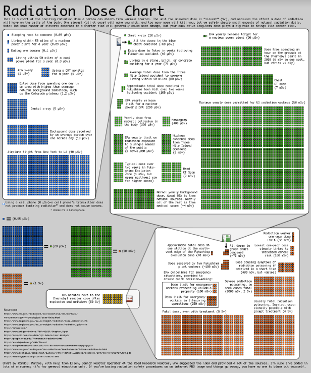
Radiation as a cause for mutations
Ionising radiation has high energy and causes reactive molecules (free radicals) that damage DNA
* Nuclear radiation causes many different cancers
* Fallout from nuclear weapons
* Nuclear workers → Children
* Ultra-violet radiation is linked to skin cancers
* Nuclear radiation causes many different cancers
* Fallout from nuclear weapons
* Nuclear workers → Children
* Ultra-violet radiation is linked to skin cancers
62
New cards
Viruses as a cause for mutations
* Viruses insert their genetic material into the DNA of the host cell
* This can disrupt the host’s genes and potentially lead to cancer
* This can disrupt the host’s genes and potentially lead to cancer
63
New cards
Chemicals as a cause for mutations
A large number of chemicals may interact directly with DNA. However, many are not necessarily mutagenic by themselves, but through metabolic processes in cells they produce mutagenic compounds.
Ex inc:
* Reactive Oxygen Species (ROS) such as hydrogen peroxide
* Benzene
Ex inc:
* Reactive Oxygen Species (ROS) such as hydrogen peroxide
* Benzene
64
New cards
Cancer
* Cancer = cells with uncontrolled cell growth and division
* Normally genes control the rate of cell division
* Damaging these genes (E.g. proto-oncogenes and tumour suppressor genes) causes uncontrolled cell division
* Any type of cell can develop cancer, but it is more common in cells with rapid rates of cell division
* Normally genes control the rate of cell division
* Damaging these genes (E.g. proto-oncogenes and tumour suppressor genes) causes uncontrolled cell division
* Any type of cell can develop cancer, but it is more common in cells with rapid rates of cell division
65
New cards
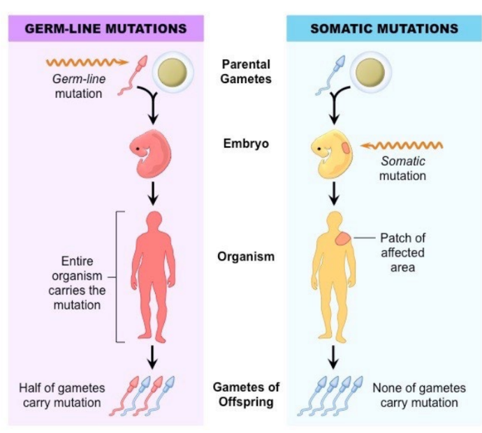
Gametic vs Somatic
* The location of a mutation determines whether or not it will be inherited.
* Most mutations occur in **somatic cells** and are not inherited.
* **Gametic (germ line)** mutations occur in the cells of the gonads (which produce sperm and eggs) and may be inherited.
* Most mutations occur in **somatic cells** and are not inherited.
* **Gametic (germ line)** mutations occur in the cells of the gonads (which produce sperm and eggs) and may be inherited.
66
New cards
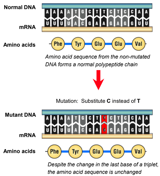
‘Same-sense’ mutation
a change in the third base of a codon still codes for the same amino acid.
* Are neutral mutations → have little/ no effect on the organism
* Are neutral mutations → have little/ no effect on the organism
67
New cards
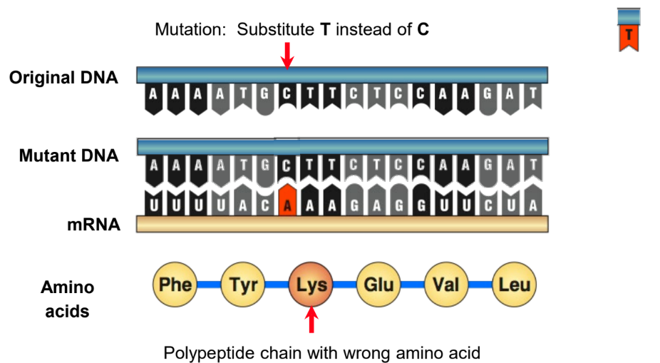
Missense mutation
* A single base is substituted by another (point mutation).
* Usually results in coding for a new amino acid in the polypeptide chain.
* Usually results in coding for a new amino acid in the polypeptide chain.
68
New cards
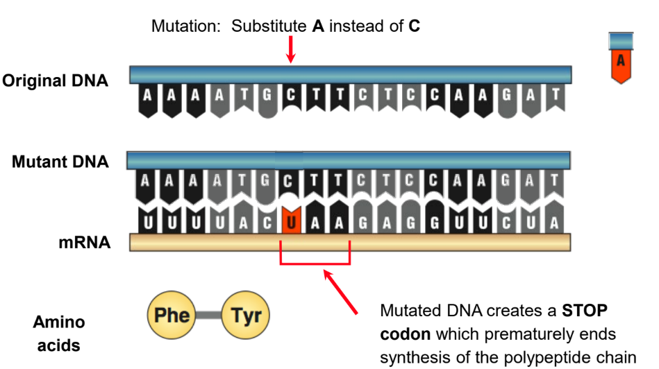
Nonsense Substitution
* A single base is substituted by another.
* This results in a new triplet that does not code for an amino acid.
* This may be an instruction to terminate the synthesis of the polypeptide chain.
* This results in a new triplet that does not code for an amino acid.
* This may be an instruction to terminate the synthesis of the polypeptide chain.
69
New cards
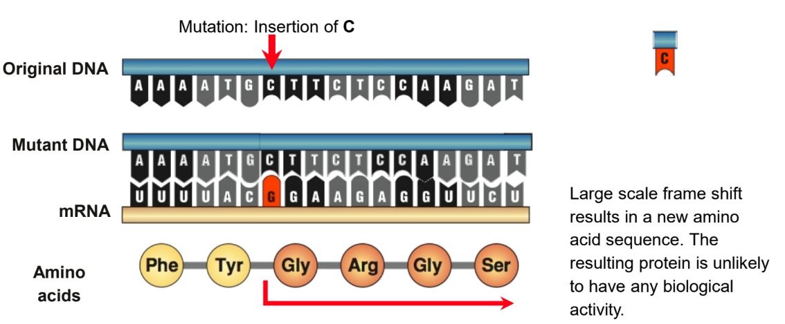
Reading Frame Shift
* A single base is inserted or deleted, upsetting the reading sequence for all those after it.
* A reading frame shift results in new amino acids in the polypeptide chain from the point of insertion onwards.
* The resulting protein will be significantly different most likely non-functional.
* A reading frame shift results in new amino acids in the polypeptide chain from the point of insertion onwards.
* The resulting protein will be significantly different most likely non-functional.
70
New cards
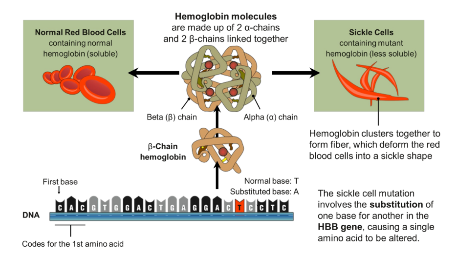
Sickle cell disease
* Incidence: Most common in people of African ancestry.
* West Africans: 1% (10-45% are carriers)
* West Indians: 0.5%
* Gene type: Autosomal recessive mutation (HBB) on chromosome 11 which results in the substitution of a single nucleotide in the HBB gene coding for the beta chain of hemoglobin.
* mutation responsible for causing sickle cell disease is a point substitution mutation (substitution of a valine for a glutamic acid in the beta-chain).
\
Symptoms include the following:
* Pain, ranging from mild to severe, in the chest, joints, back, or abdomen
* Swollen hands and feet
* Jaundice
* Repeated infections, particularly pneumonia and meningitis
* Kidney failure
* Gallstones (at an early age)
* Strokes (at an early age)
* Anaemia
71
New cards
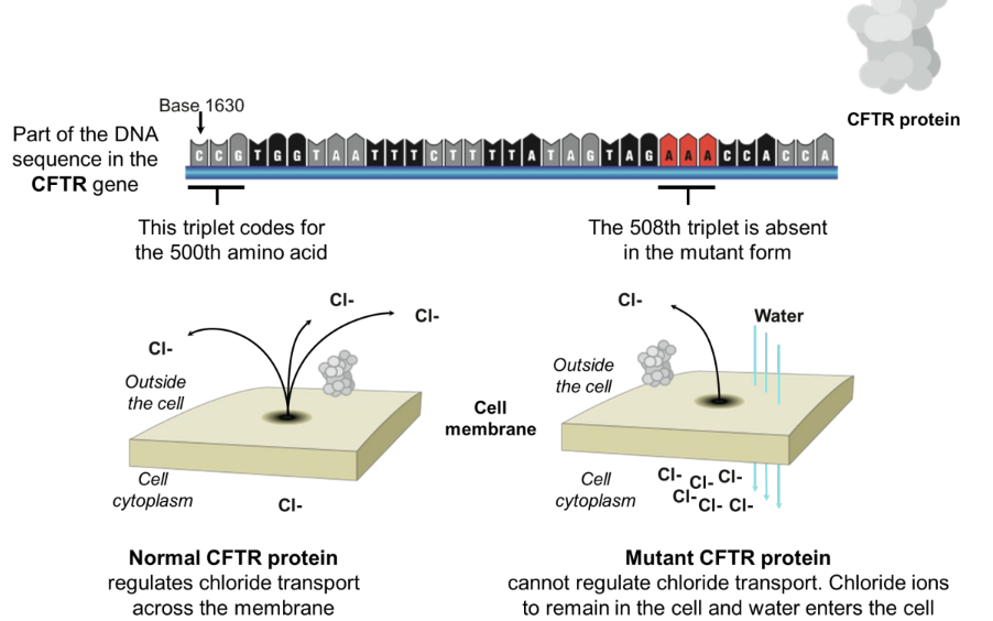
Cystic Fibrosis
* Synonyms: Mucoviscidosis, CF
* Incidence: Varies with populations:
* Asians: 1 in 10 000
* Caucasians: 1 in 20-28 are carriers
* Mutation type: Autosomal recessive. Over 500 different recessive mutations of the CFTR gene have been identified:
* Deletions, missense, nonsense, terminator codon
* The mutation causing 70% of cystic fibrosis cases is a gene mutation (delta F508) involving a triplet deletion.
\
Symptoms:
* Infertility (both)
* Disruption of the following glands:
* the pancreas
* intestinal glands
* biliary tree (biliary cirrhosis)
* bronchial glands (chronic lung infections)
* sweat glands (high salt content of which becomes depleted in hot environments)
72
New cards
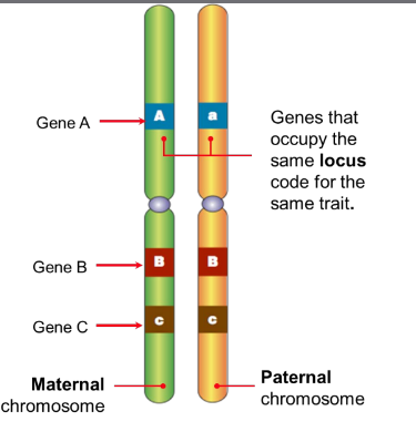
Allele
Genes occupying the same position (locus) on homologous chromosomes
Alleles are versions of the same gene that code for a variant of the same protein.
Alleles are versions of the same gene that code for a variant of the same protein.
73
New cards
Inheritance patterns
* Autosomal codominance pattern
* Autosomal dominance pattern
* Autosomal recessive pattern
* Autosomal dominance pattern
* Autosomal recessive pattern
74
New cards
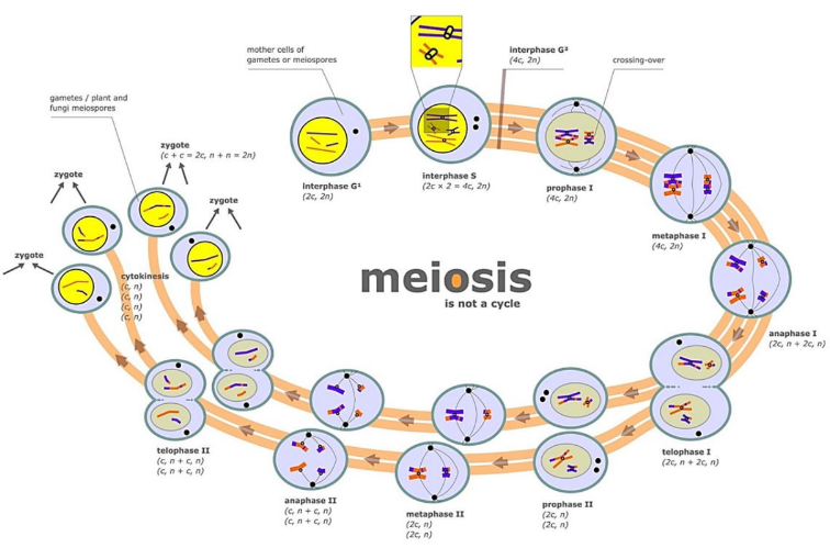
Meiosis
is the cell division process of making haploid sex cells
(gametes)
(gametes)
75
New cards
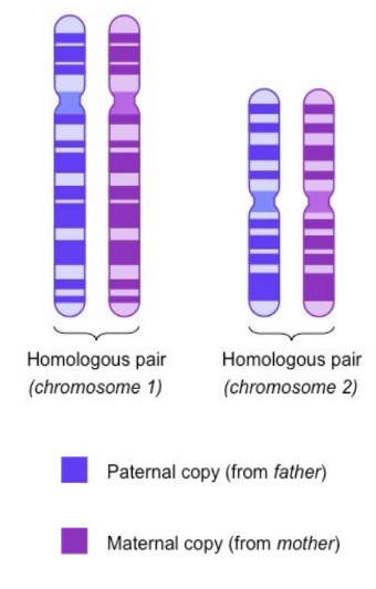
Homologous chromosomes
* Homologous chromosomes are chromosomes that share:
* The same structural features (e.g. same size, same banding patterns, same centromere positions)
* The same genes at the same loci positions (while the genes are the same, alleles may be different)
* Homologous chromosomes must be separated in gametes (via meiosis) prior to reproduction, in order to prevent chromosome numbers continually doubling with each generation
IE (homo - same, logus - location)
76
New cards
Interphase (Meiosis)
* DNA is replicated during the S phase of interphase
* Replicated chromosomes will consist of genetically identical sister chromatids that remain attached to each other at the centromere
* Replicated chromosomes will consist of genetically identical sister chromatids that remain attached to each other at the centromere
77
New cards
Prophase I
* During Prophase I, homologous chromosomes become connected via synapsis
* This allows connected chromosomes to be arranged for a reduction division
* The connected homologous chromosomes are known as bivalents (or tetrads)
* This allows connected chromosomes to be arranged for a reduction division
* The connected homologous chromosomes are known as bivalents (or tetrads)

78
New cards
Bivalents/ Tetrads
Paired chromosomes during prophase 1 meiosis 1
79
New cards
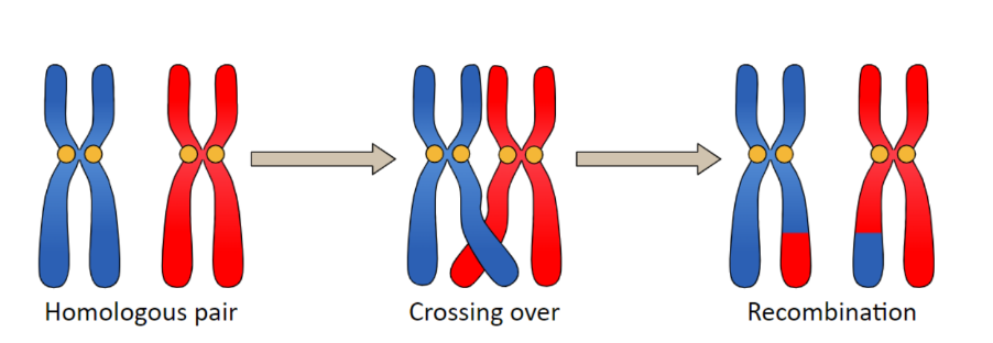
Synapsis ; List significance too
the process where non-sister chromatids recombine with their homologous partner and exchange sections of DNA
* Allows exchange of genetic information → leads to genetic diversity which is important in species resilience
* Allows exchange of genetic information → leads to genetic diversity which is important in species resilience
80
New cards
Chiasma; chiasmata
Physical connections between paired chromosomes
81
New cards
Metaphase I
* Homologous chromosomes align at the metaphase plate
* During Metaphase I, homologous chromosomes line up in a random orientation, referred to as independent assortment
* There is an equal chance of the resulting gamete containing either of the pair
* During Metaphase I, homologous chromosomes line up in a random orientation, referred to as independent assortment
* There is an equal chance of the resulting gamete containing either of the pair
82
New cards
Anaphase I
* The chromosomes with two sister chromatids are separated, and they begin to migrate to the opposite poles.
* This separation is achieved because of the contraction of the spindle fibres attached to each chromosome’s centromere.
* This separation is achieved because of the contraction of the spindle fibres attached to each chromosome’s centromere.
83
New cards
Telophase I
* Two daughter cells are formed with each daughter containing only one chromosome of the homologous pair
* However, there are still 2 copies of that single chromosome
* Cytokinesis occurs concurrently, sometimes referred to as interkinesis
* However, there are still 2 copies of that single chromosome
* Cytokinesis occurs concurrently, sometimes referred to as interkinesis
84
New cards
Meiosis II
* Seperates cells formed from meiosis one so that each cell only has one copy of the chromosome
* No interphase
* No interphase
85
New cards
Prophase II
* DNA does not replicate
* New spindles form in both daughter cells
* New spindles form in both daughter cells
86
New cards
Metaphase II
Chromosomes (sister chromatids) align at the metaphase plate.
87
New cards
Anaphase II
Centromeres divide and sister chromatids migrate separately to each pole
88
New cards
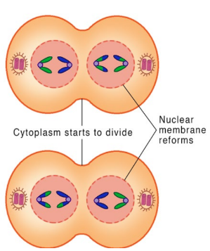
Telophase II
Chromosomes decondense, nuclear membrane reforms, cells divide
89
New cards
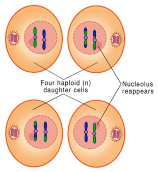
Cytokinesis
* Four haploid (single set; think half = hap) daughter cells are obtained
* These cells are likely to all be genetically distinct due to the crossing over that occurs during Prophase I (recombination of sister chromatids)
* These cells are likely to all be genetically distinct due to the crossing over that occurs during Prophase I (recombination of sister chromatids)
90
New cards
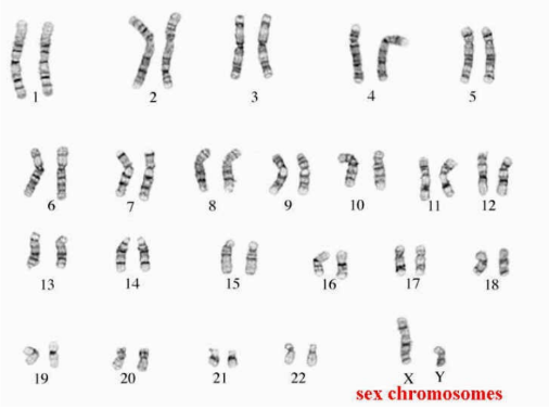
Karyotypes
a picture of all of the chromosomes found in an individual
* can be used to diagnose certain genetic problems, either before birth (using CVS or amniocentesis) or after birth
* can be used to diagnose certain genetic problems, either before birth (using CVS or amniocentesis) or after birth
91
New cards
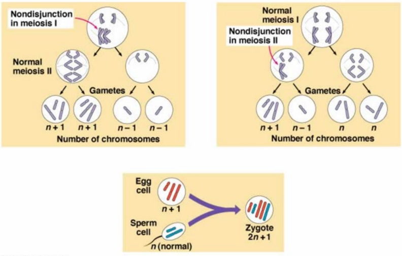
Non-disjunction
chromosomes failing to separate
correctly
correctly
92
New cards
Aneuploidy
* **Monosomy** refers to lack of one chromosome of the normal complement.
* **Trisomy** refers to the presence of three copies, instead of the normal two, of a particular chromosome.
* **Trisomy** refers to the presence of three copies, instead of the normal two, of a particular chromosome.
93
New cards
Other DNA abnormalities (Meiosis) - Name 3
**Structural abnormalities** Sometimes, chromosomes break, leading to changes in chromosome structure:
* Parts of the chromosome are deleted
* Parts of the chromosome are duplicated
* Parts of the chromosome are attached to other chromosomes, turned upside down etc.
**Duplication**: if a fragment joins the homologous chromosome, then that region is repeated
**Translocation:** a fragment of a chromosome is moved ("trans-located") from one chromosome to another - joins a non-homologous chromosome.
94
New cards
**Homozygous vs Heterozygous**
Homozygous
* When there is the same allele for a particular gene on both chromosomes of a homologous pair.
Heterozygous
* When there is an alternative allele for a particular gene on one chromosome of a homologous pair.
* When there is the same allele for a particular gene on both chromosomes of a homologous pair.
Heterozygous
* When there is an alternative allele for a particular gene on one chromosome of a homologous pair.
95
New cards
Autosome vs sex chromosomes
An autosome is one of the numbered chromosomes, as opposed to the sex chromosomes. Humans have 22 pairs of autosomes and one pair of sex chromosomes (XX or XY)
96
New cards
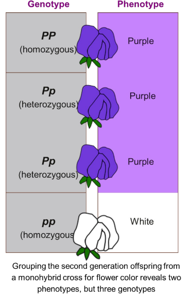
Genotype vs phenotype
The **genotype** of an organism refers to its genetic make-up.
The **phenotype** of an organism refers to its observable features or traits. (think phenotype=physical!)
* Dominant alleles are labelled with a capital letter and recessive ones with a lower case letter.
* A heterozygote will be a carrier for a recessive allele.
The **phenotype** of an organism refers to its observable features or traits. (think phenotype=physical!)
* Dominant alleles are labelled with a capital letter and recessive ones with a lower case letter.
* A heterozygote will be a carrier for a recessive allele.
97
New cards
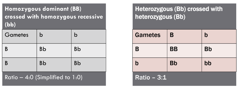
Dominance & Recessiveness
* A trait is **dominant** when it appears in the phenotype of a heterozygote. It will mask the expression of a recessive trait.
* A **recessive** trait only appears in the phenotype of homozygotes. They do not appear in the phenotype of heterozygotes.
* A **recessive** trait only appears in the phenotype of homozygotes. They do not appear in the phenotype of heterozygotes.
98
New cards
Punnet squares
99
New cards
Independent assortment
* During first stage of meiosis chromosomes line up, but the way they pair is random leading to different possible combinations
* Leads to genetic information
* Leads to genetic information
100
New cards
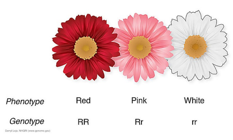
Incomplete dominance
Hybrids/heterozygotes have an appearance that is intermediate or blended between the phenotypes of the parents.