A level Bio - Mrs Glover
1/222
There's no tags or description
Looks like no tags are added yet.
Name | Mastery | Learn | Test | Matching | Spaced | Call with Kai |
|---|
No analytics yet
Send a link to your students to track their progress
223 Terms
What is cell theory?
a unifying concept in biology that states:
all living organisms are made up of one or more cell
cells are the basic functional unit of living organisms
new cells are produced from pre-existing cells
Units for biology

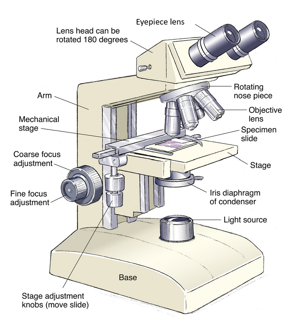
Diagram of a microscope?
What is resolution?
the minimum distance apart that two objects can be in order for them to appear as separate items
Resolving power depends on the wavelength or form of radiation used
greater resolution means greater clarity and a clearer image
How to calculate magnification?
magnification = image size/ actual size
What is cell fractionation?
The process of breaking up cells and separating out the different organelles they contain.
3 stages: homogenisation, filtration, ultracentrifugation.
Stages of cell fractionation
Tissue is cut up + placed in a solution which is - cold (to reduce enzyme activity), buffered (so pH does not fluctuate), and same water potential (to prevent osmotic gain/loss of water)
Homogenisation: blender-like machine that grinds up cells to break plasma membrane of cells + release organelles into a solution called the homogenate.
Filtration: Homogenate is filtered through a gauze to separate smaller organelles from cell debris, leaving a filtrate of organelles
Ultracentrifugation: Filtrate is spun in a centrifuge, initially at slow speed. Heaviest organelles nuclei forced to bottom of tube where they form a pellet. Fluid at top of tube (supernatant) is removed. Mixture spun again at higher speed and repeated to produce solutions of pure organelles.
Light microscopes?
Light microscopes use a pair of convex glass lenses that can resolve images that are 0.2um apart. This is the wavelength of light and therefore restricts the resolution that a light microscope resolve to.
Electron microscopes?
Electron microscopes can be used to look at objects that are closer than 0.2um apart.
2 main types:
Transmission electron microscope (TEM)
Scanning electron microscope (SEM)
Light vs electron microscopes?
electron - cannot view living specimens as system must be a vacuum
electron - complex staining process required
electron - specimens have to be very thin
SEM has lower resolution than TEM, but both have higher resolution than light
TEM is 2D, SEM is 3D
light - images in colour, black and white in electron
How do we measure the actual size of an object in a light microscope?
eyepiece graticule is used: a tiny printed scale placed in the eyepiece lens of a light microscope
has its own unit: eye piece unit (EPU)
must be calibrated to work out size of object being measured
stage micrometre is placed on stage and used to measure the graticule scale depending on magnification used
Ultrastructure of eukaryotic animal cells?
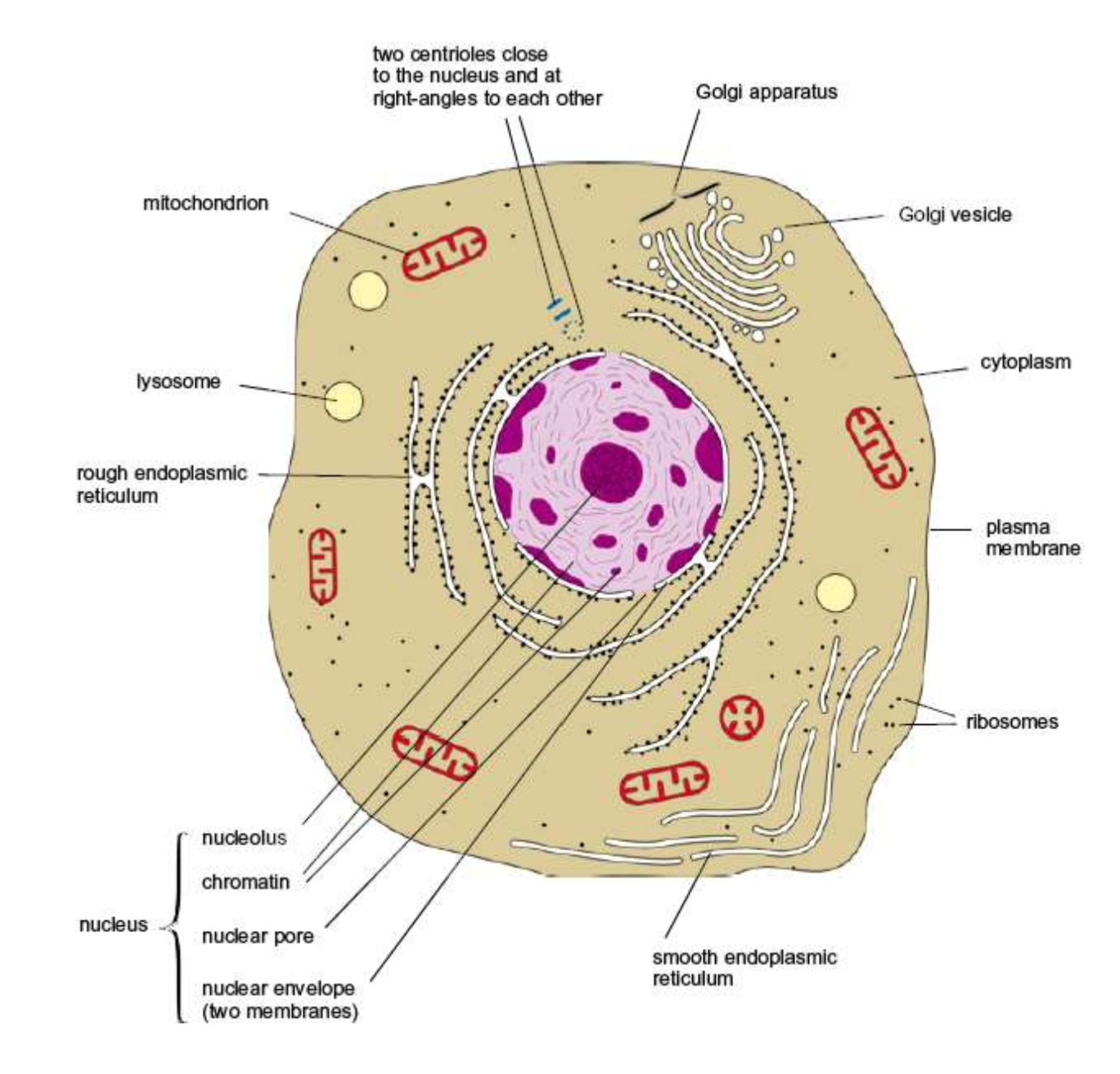
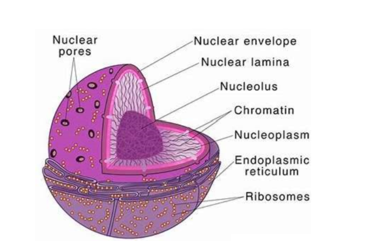
Structure and function of nucleus?
Nucleus is a double-membrane organelle, contains the genetic material and manufactures rRNA and ribosomes
Nuclear envelope is continuous with endoplasmic reticulum and often has ribosomes on surface
Contains nuclear pores that allow some molecules to move in and out of the nucleus
It also contains chromatin and a nucleolus which is the site of ribosome production
A granular jelly like material called nucleoplasm makes up the bulk of the nucleus
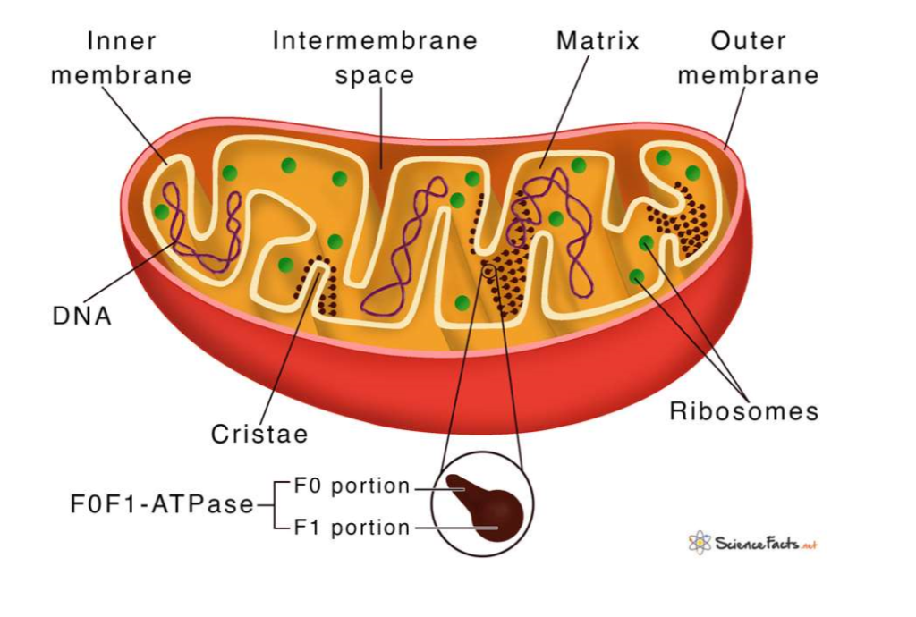
Structure and function of mitochondria?
Mitochondria are double-membrane organelles, responsible for generating energy through respiration
Inner membrane is folded into cristae, increasing the surface area for reactions
Matrix, the internal fluid, contains enzymes
Circular DNA and ribosomes allow them to synthesize some proteins independently
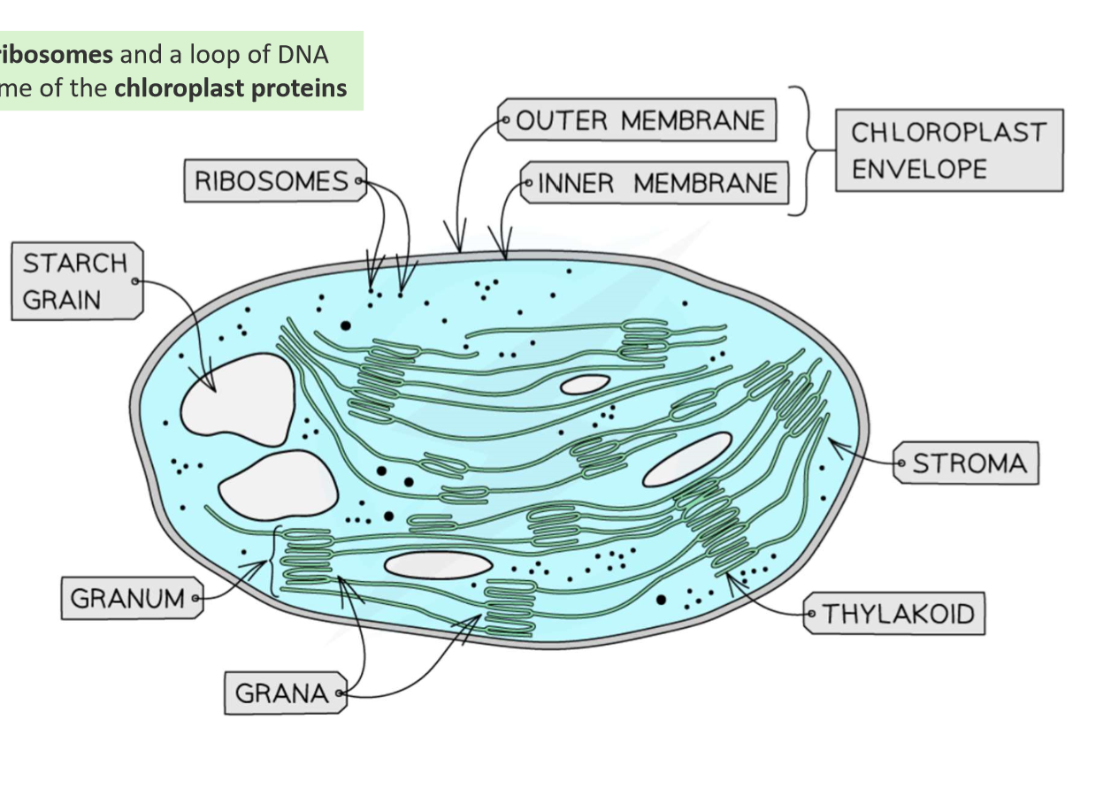
Structure and function of chloroplasts?
Chloroplasts are double-membrane organelles found in plant cells, responsible for photosynthesis.
Thylakoids, stacked into grana, house chlorophyll for capturing light energy.
Stroma, the fluid surrounding the grana, contains enzymes for the light-independent reactions
Circular DNA and ribosomes, allowing them to synthesize some of their own proteins
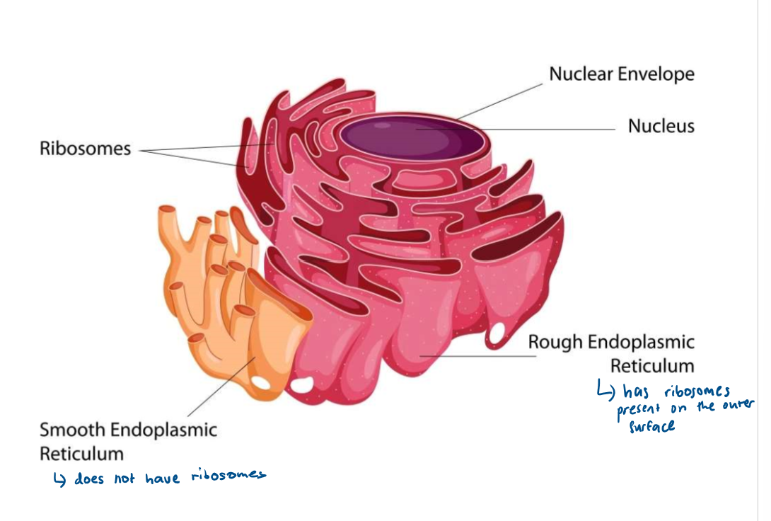
Structure and function of rough endoplasmic reticulum?
series of flattened sacs enclosed by a membrane with ribosomes on the surface. RER folds and processes proteins made on the ribosomes.
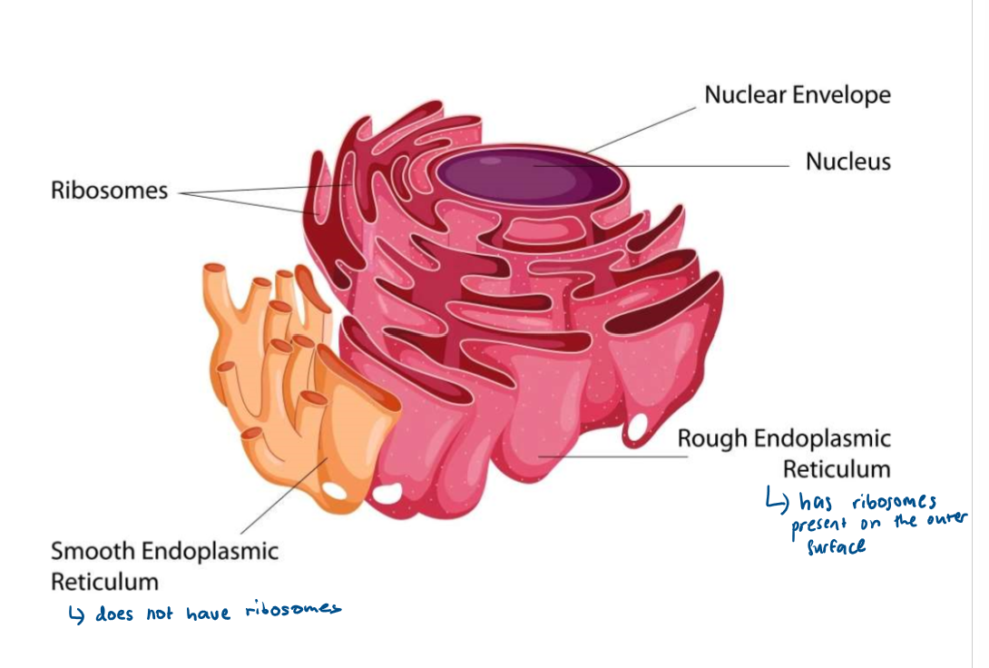
Structure and function of smooth endoplasmic reticulum?
system of membrane bound sacs that produces and processes lipids
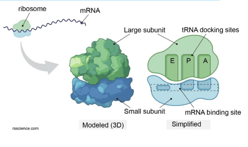
Structure and function of ribosomes?
small, non-membrane-bound organelles and are the site of protein synthesis
2 types: 70S (smaller and found in prokaryotic cells and organelles) and 80S (bigger and found in eukaryotic cells)
Contain rRNA and proteins
Structure and function of Golgi apparatus?
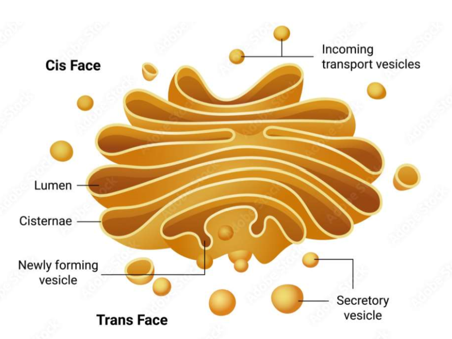
series of fluid filled sacs called cisternae with vesicles surrounding the edges
Receives proteins and lipids from the rough endoplasmic reticulum, modifies them, then sorts them and transports them in vesicles that are regularly pinched off from the ends of the cisternae
Vesicles may move to the surface where they fuse with the membrane and secrete substances outside the cell
Structure and function of lysosomes?
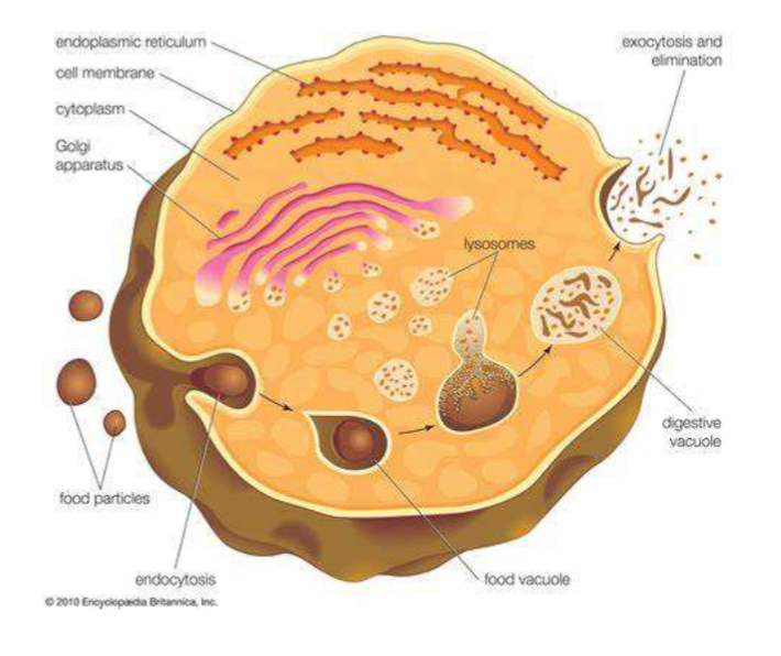
vesicles containing digestive enzymes called lysozymes
Lysozymes hydrolyse cell walls of certain bacteria, are excreted outside the cell to destroy material around them (exocytosis), and digest worn out/dead cells and organelles
Structure and function of cell wall?
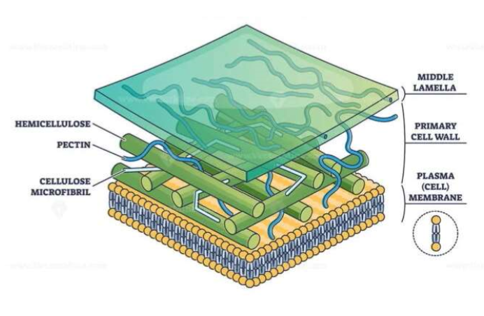
provides structural support, protects the cell from pathogens and prevents the cell bursting from water pressure due to osmosis
It is composed of microfibrils of cellulose embedded in a matrix
The middle lamella cements adjacent cells together
Structure and function of vacuole?
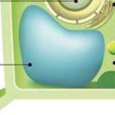
fluid-filled sac bound by a membrane called the tonoplast
Stores water, nutrients, and waste products, and helps maintain cell turgor, which is important for structural support
What is an artefact?
things you can see under the microscope that aren’t actually part of the specimen such as dust, air bubbles or fingerprints
occur during preparation of a sample
What is a mesosome?
a complex infolding of the inner membrane in prokaryotic cells
still debated by scientists whether its an organelle or an artefact
Stages of binary fission?
circular DNA replicates and both copies attach to the cell membrane
plasmids also replicate
cell membrane begins to grow between two DNA molecules and begins to pinch inward, dividing cytoplasm in two
a new cell wall forms between the two molecules of DNA dividing into 2 identical daughter cells, each with a single copy of the circular DNA and a variable number of the copies of the plasmids
How do viruses replicate?
attach to their host cell using attachment proteins
inject their nucleic acid into the host cell
host cell starts to produce viral components, nucleic acid, enzymes and proteins
these are assembled into new viruses which are released, damaging the host cell
Why do viruses only infect one cell type?
they have specific attachment proteins with specific tertiary structures
different host cells have different receptors so viruses are only able to bind to host cells with complementary receptor proteins
Ultrastructure of prokaryotic cells?
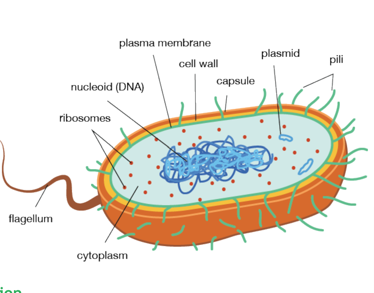
Organelles in prokaryotic organisms?
flagellum, nucleoid, capsule, cell wall, cytoplasm, cell membrane, plasmid, ribosomes
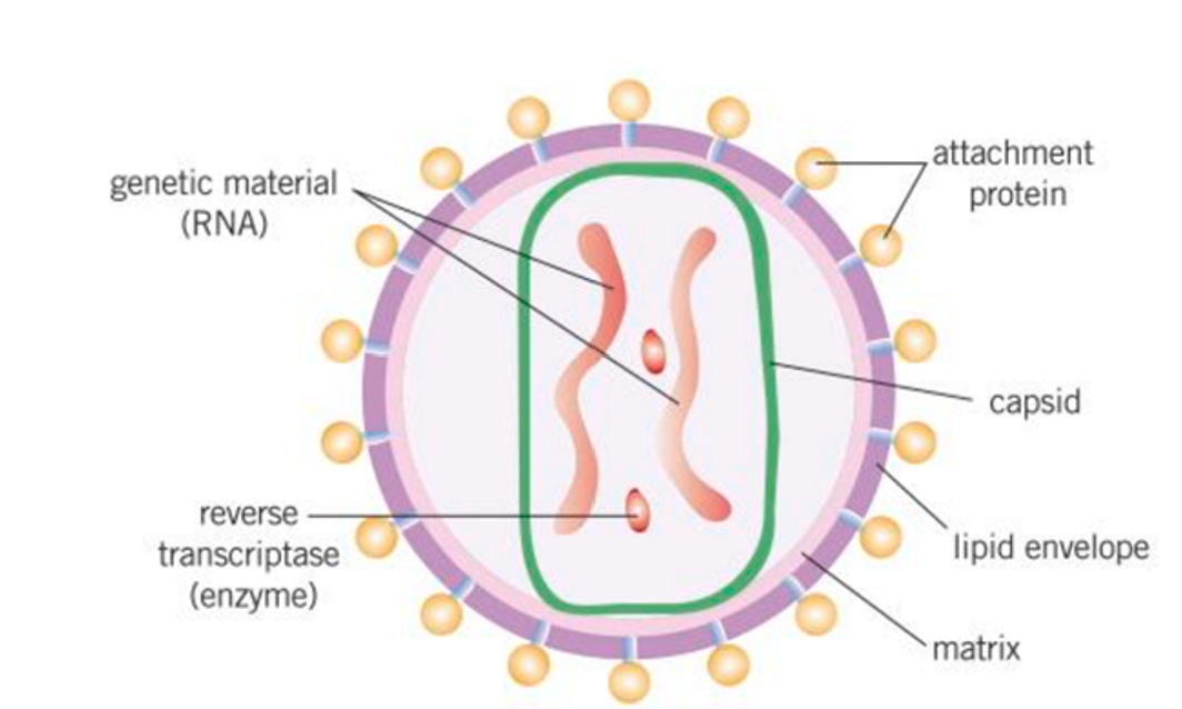
Structure of viruses?
contain nucleic acids (DNA or RNA) but genetic material can only multiply inside living host cells
nucleic acid enclosed within capsid
some viruses are surrounded by lipid envelope
lipid envelope has attachment proteins to allow virus to identify and attach to host cell
Mitosis vs meiosis?
mitosis - produces 2 daughter cells that are diploid, used to produce body cells for growth, repair and replacement
meiosis - produces 4 daughter cells that are haploid, used to produce gametes
What is a chromosome?
compact X shaped form of chromatin formed during cell division
chromatids (identical arms of chromosome) are joined together by centromere
What is chromatin?
DNA associated with proteins when it is not wound up tightly as a chromosome
What is the purpose of mitosis?
growth, repair and replacement, reproduction
What are the stages of mitosis?
interphase, prophase, metaphase, anaphase, telophase, cytokinesis
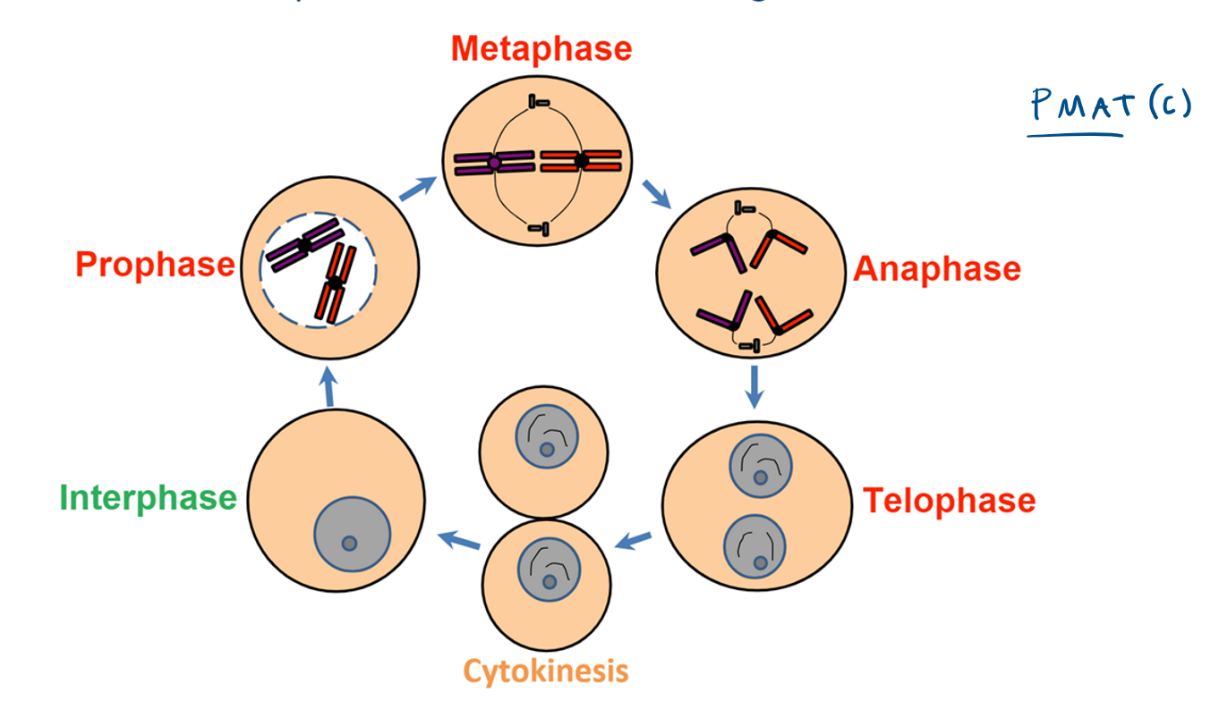
What happens in interphase?
period of time during which the cell is not dividing
chromosomes condense, organelles are replicated, protein synthesis
DNA is replicated and checked for errors to avoid mutations
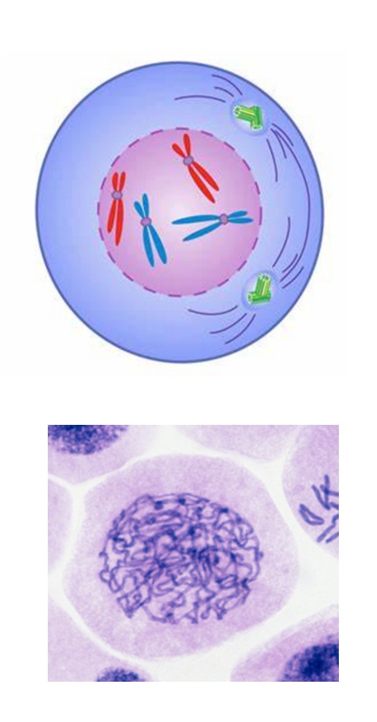
What happens during prophase?
chromosomes condense
nuclear envelope breaks down
spindle fibres start to form
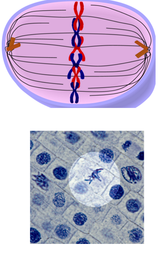
What happens during metaphase?
chromosomes align down equator of cell
attach to spindle fibres by centromeres
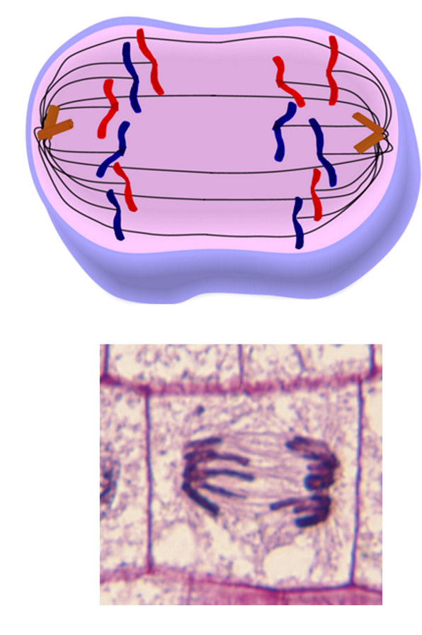
What happens during anaphase?
chromatids separate
pulled to opposite poles by spindle fibres
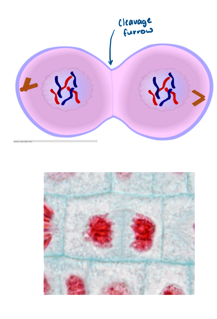
What happens during telophase?
chromosomes reach poles of cell
nuclear envelope reforms
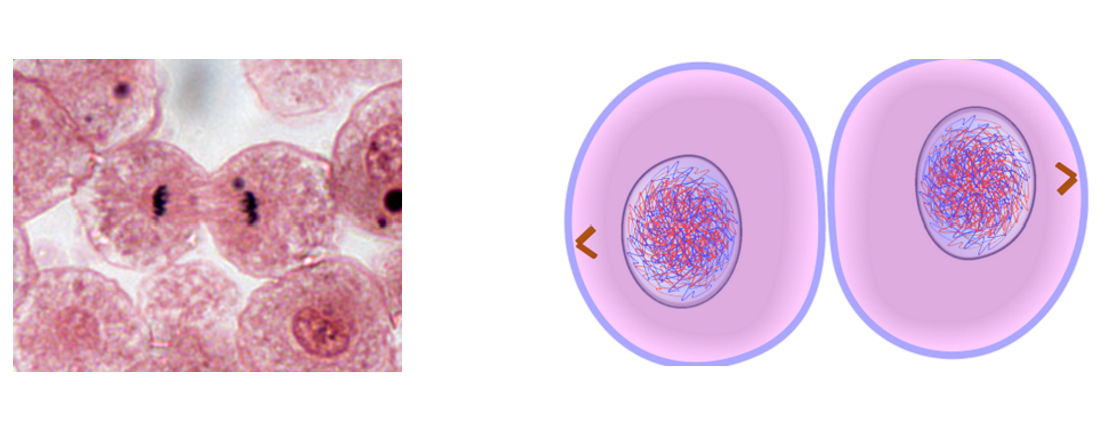
What happens in cytokinesis?
cytoplasm constricts and divides to form two identical daughter cells
chromosomes uncondense
What is the cell cycle?
the regulated sequence of events that occurs between one cell division and the next
the length of a cell cycle varies amongst organisms
three phases: interphase, nuclear division (PMAT) and cytokinesis
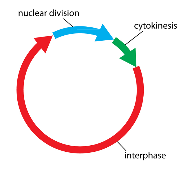
What are the phases of the cell cycle and what happens in each stage?
mitosis: nuclear division (PMAT)
cytokinesis: cell division
interphase: organelles are replicated, DNA is replicated, protein synthesis, DNA is checked for errors to avoid mutations
What is cancer?
a group of diseases caused by damage to the genes that regulate mitosis and the cell cycle, leading to uncontrolled growth and division of cells
What is a tumour and when do they become cancerous?
a group of abnormal cells
tumours become cancerous if they change from benign to malignant
Benign vs malignant tumours?
Benign Tumours:
Slow growth, normal-looking nucleus.
Cells: Well-differentiated, produce adhesion molecules (stay in original tissue).
Encapsulated (surrounded by dense tissue).
Localised effects – rarely life-threatening but may disrupt organ function.
Treatment: Surgery, rarely reoccur.
Malignant Tumours:
Rapid growth, darker/larger nucleus (abnormal DNA).
Cells: De-differentiated, lack adhesion molecules → spread and form secondary tumours in surrounding tissues
Systemic effects (e.g., weight loss, fatigue).
Treatment: Surgery + chemotherapy/radiotherapy.
High reoccurrence risk.
What treatments are used for cancer?
chemotherapy prevents DNA from replicating and inhibiting the metaphase stage by interfering with spindle formation
they also disrupt cell cycles of normal cells so cells such as hair cells are also damaged
What is mitotic index and how do you calculate it?
the ratio between the number of a population's cells undergoing mitosis to its total number of cells
mitotic index = number of cells with visible chromosomes / total number of cells
RP2 - Preparation of stained squashes of cells from plant root tips; use of an optical microscope to identify the stages of mitosis, and calculation of a mitotic index
used to observe mitosis because the root tip contains meristematic tissue, where cells divide rapidly
Soften the tissue with heated HCl to break down cell walls
Stain the root tip to make the chromosomes visible under a microscope.
Squash the root tip gently to create a single layer of cells for clear observation of mitotic stages.
Use microscope to count cells with chromosomes visible and total number of cells.
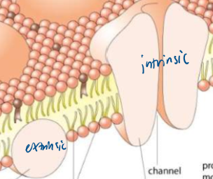
What is the cell membrane made up of and diagram?
phospholipids, proteins (extrinsic and integral), glycolipids, cholesterol, glycoproteins
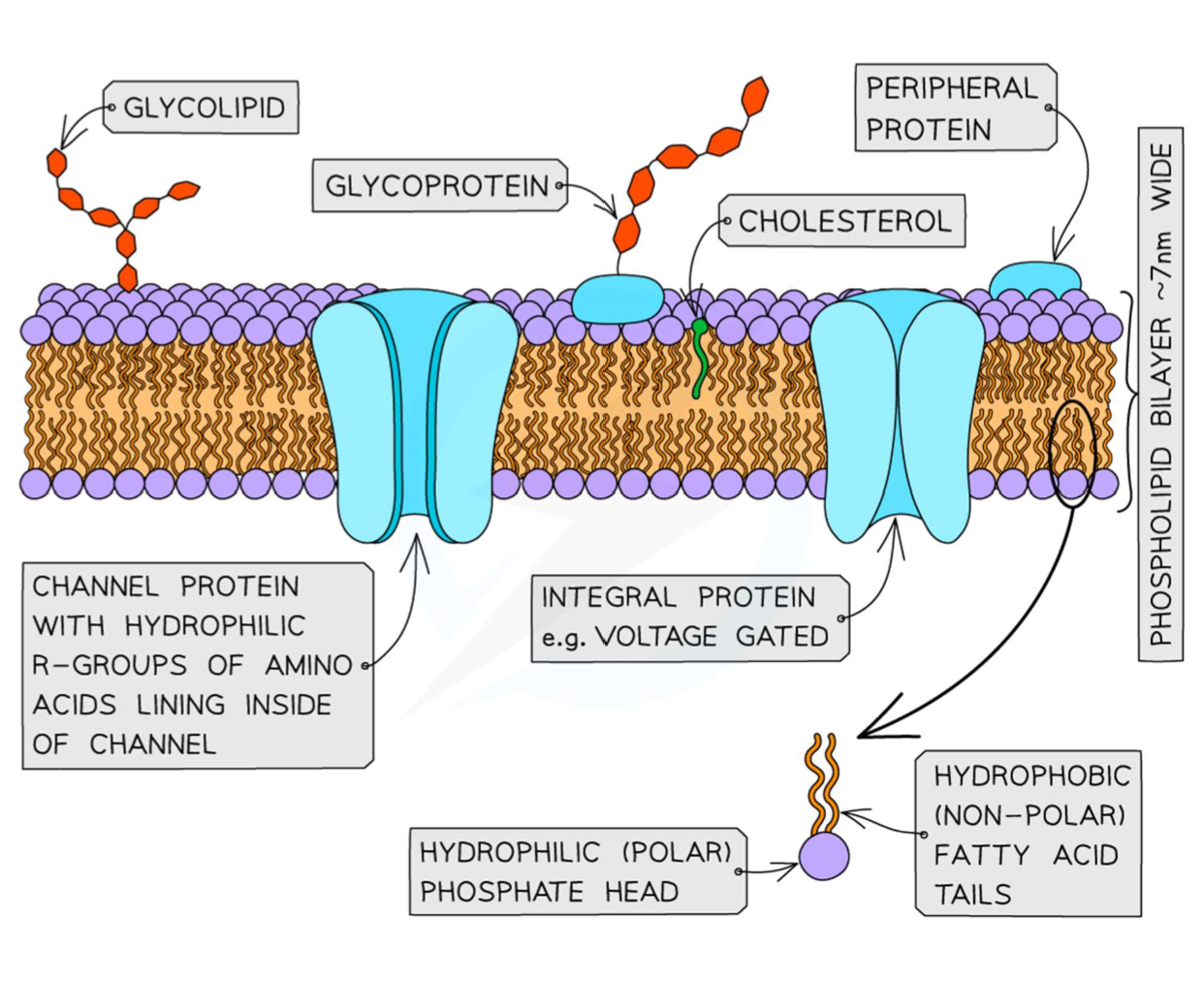
What is the role of the phospholipid bilayer in a cell membrane?
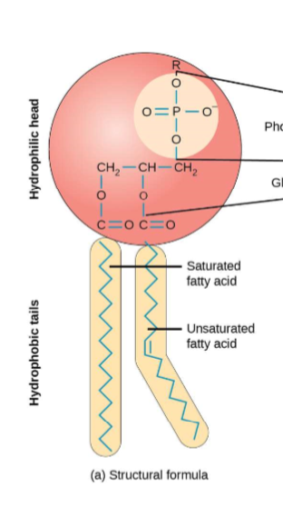
hydrophilic phosphate heads orientate towards water, hydrophobic fatty acid tails away from water
allow simple diffusion of small non-polar and lipid-soluble molecules
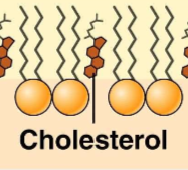
What is the role of cholesterol in cell membranes?
hydrophobic molecule that restricts movement of phospholipids
regulates fluidity especially at high temperatures
prevents leakage of water and dissolved ions (decreases permeability)
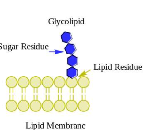
What are glycolipids and glycoproteins their role in cell membranes?
glycolipids - carbohydrate bonded to a lipid
glycoproteins - carbohydrates bonded to external proteins
act as cell surface receptors
allow cells to stick together and form tissues
glycoproteins can also act as neurotransmitters
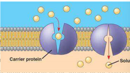
What is the role of carrier proteins in a cell membrane?
integral protein used in facilitative diffusion and active transport
binds with the ions or molecules, then change shape to move the molecules across the membrane
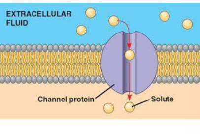
What is the role of channel proteins in a cell membrane?
integral protein used in facilitative diffusion
form water-filled tubes to allow water-soluble ions to diffuse across the membrane
selective due to diameter and charged groups
some are open all the time and some are gated and open in the presence of a specific ion
What is the fluid mosaic model?
the arrangement of molecules in the cell membrane
fluid - the structure is constantly changing in shape due to the moving phospholipids
mosaic - the proteins are embedded into the bilayer and vary in shape size and pattern
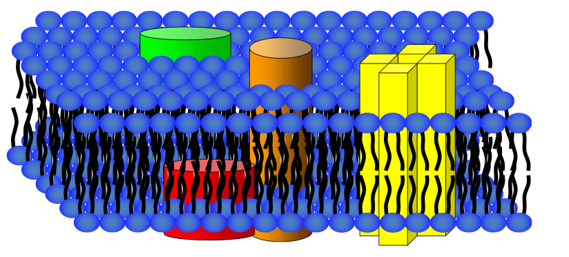
What are extrinsic and intrinsic proteins?
intrinsic proteins - span the entire width of the phospholipid bilayer, e.g. carrier and channel proteins
extrinsic proteins - occur in the surface of the bilayer and do not extend across it, give mechanical support to the membrane
RP4 - investigation into the effect of a named variable (temp/ethanol) on the permeability of cell membranes?
Prepare beetroot discs of equal size and rinse them to remove excess pigment.
Place the discs in solutions with varying temperatures/ ethanol concentrations
Leave the samples for a set time and then remove the beetroot discs.
Measure the absorbance of the solution with a colorimeter.
Conclusion:
Higher temperatures increase membrane permeability, causing more pigment to be released, as phospholipids will have more kinetic energy and therefore move and create gaps in the membrane allowing pigment to leak out.
Higher ethanol concentrations increase membrane permeability, as ethanol causes lipids to dissolve (phospholipids), allowing pigment to leak through.
What is a colorimeter and how is it used?
A colorimeter is an instrument used to measure the percentage absorbance or transmission of light through a solution, which helps determine the concentration of a solute/ pigment in the solution.
use distilled water to calibrate to zero
place solution in a cuvette
higher % absorbance = higher concentration
What is the definition of diffusion?
the net movement of molecules or ions from an area of higher concentration to an area of lower concentration until equilibrium is reached
What are the two types of diffusion?
simple diffusion - the process by which small, non-polar molecules (such as oxygen and carbon dioxide) are transported across a membrane
facilitated diffusion - the process by which charged ions and polar molecules are transported across a membrane by channel and carrier proteins
What factors affect the rate of simple diffusion?
temperature - higher temperature gives molecules more kinetic energy so increases diffusion rate
concentration gradient - steeper concentration gradient increases diffusion rate
surface area - larger surface area increases diffusion rate
diffusion distance - shorter distance increases diffusion rate
What is the definition of osmosis?
the net movement of water molecules from an area of higher water potential to an area of lower water potential through a partially permeable membrane
What is water potential (Ψ)?
the pressure created by water molecules
measured in kPa
the more solute added the lower the water potential
pure water has a water potential of zero (so water potential of a solution will always be negative)
Osmosis exam question key mark scheme:
solution on left is hyper/iso/hypotonic and has higher/lower concentration of solute molecules and a higher/lower water potential
both solute and water molecules are in random motion due to kinetic energy
water molecules diffuse via osmosis from higher water potential to lower water potential down the concentration gradient
dynamic equilibrium is reached and there is no more net movement of water
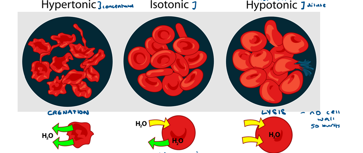
What are hypertonic, isotonic and hypotonic solutions?
hypertonic - higher concentration of solute and a lower water potential
isotonic - same water potential inside and outside of cell
hypotonic - higher water potential and a lower concentration of solute
What happens to animal cells in hypertonic, isotonic and hypotonic solutions?
hypertonic - crenation, water moves out of the cell
isotonic - dynamic equilibrium, water moves into and out of the cell at the same rate
hypotonic - lysis, water moves into the cell, cell may burst as no cell wall
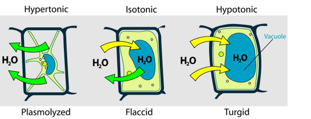
What happens to plant cells in hypertonic, isotonic and hypotonic solutions?
hypertonic - plasmolysed, water moves out of cell, protoplast completely pulled away from cell wall
isotonic - flaccid, incipient plasmolysis, dynamic equilibrium, water moves into and out of cell at the same rate, protoplast beginning to pull away from cell wall
hypotonic - turgid, water moves into the cell, protoplast pushed against cell wall
RP3 - water potential of plant tissue
Create a sucrose dilution series (e.g. 0.0, 0.2, 0.4, 0.6, 0.8, 1.0 mol dm⁻³)
Cut identical cylinders of potato using a cork borer to control size/surface area.
Blot dry to remove excess water and weigh initial mass
Put one cylinder in each sucrose solution for 20 mins.
Control variables: Temperature, time, volume of solution.
Blot dry again and record final mass.
Calculate % Mass Change = Final mass−Initial mass / Initial mass×100
this enables comparison as plant tissues had different initial masses
Plot Calibration Curve with concentration on x axis and percentage change in mass on y axis
Find Water Potential use a ψ table in a textbook to convert x-intercept concentration to kPa.
What is the definition of active transport?
the movement of molecules or ions into or out of a cell from an area of lower concentration to an area of higher concentration, using ATP and carrier proteins.
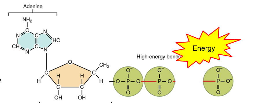
How is energy released from ATP?
ATP (adenosine triphosphate) is broken down into:
ADP (adenosine diphosphate) and an inorganic phosphate (Pi) group when water is added in a hydrolysis reaction
catalysed by the enzyme ATP hydrolase
ATP + H2O → ADP + Pi + energy
bonds between the phosphate groups in ATP are unstable and store a lot of energy
breaking the final bond releases lots of energy
this reaction is reversible (phosphorylation catalysed by ATP synthase)
What is the process of active transport?
Specific molecules/ ions bind to receptor on carrier proteins in the cell membrane
ATP is hydrolysed into ADP and inorganic phosphate (Pi), releasing energy
energy allows carrier protein to change shape, transporting molecule/ion across the membrane to the other side
molecule or ion is released on the other side and carrier protein reverts to its original shape
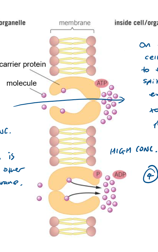
What adaptations do cells have to increase rate of absorption of molecules (4)?
folded membrane and microvilli for large surface area
large number of channel and carrier proteins for active transport and facilitative diffusion
many mitochondria for aerobic respiration
membrane-bound digestive enzymes to maintain concentration gradient
How does movement across membranes occur by co-transport?
two different substances bind and move simultaneously via a co-transporter protein
one substance moves against concentration gradient
the other moves down concentration gradient
What is a sodium-potassium pump?
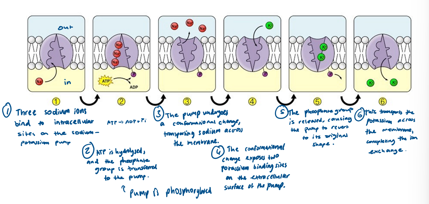
integral protein that exchanges 3 sodium ions with 2 potassium ions
a key example of active transport where multiple ions are transported
Stages for a sodium-potassium pump?
Three sodium ions bind to specific sites on the pump inside the epithelial cell.
ATP binds to the pump and is hydrolysed to ADP + Pi.
The phosphate group binds to the pump and causes the pump to change shape, moving the sodium ions out of the cell.
Two potassium ions bind to specific sites on the pump on the extracellular side.
The phosphate group is released, causing the pump to return to its original shape.
The potassium ions are transported back into the cell.
What is an antigen?
foreign molecule (protein/ glycoprotein/ glycolipid) that stimulates an immune response leading to production of antibodies
How are cells identified by the immune system?
Each type of cell has specific molecules on its surface that identify it
Proteins have a specific tertiary structure (or glycoproteins / glycolipids)
What type of cells and molecules can the immune system identify?
Pathogens (e.g. viruses, fungi, bacteria)
Cells from other organisms (e.g. organ transplants)
Abnormal body cells (e.g. tumour cells or virus-infected cells)
Toxins released by some bacteria
What is a non-specific immune response?
a response that is immediate and the same for all pathogens
physical barriers and phagocytosis
Physical barriers (non specific response)
skin, stomach acid, cilia in respiratory system, mutualistic bacteria in gut, enzymes in intestines
What types of white blood cell are there and their functions?
phagocytes - ingest and destroy pathogens by phagocytosis
T lymphocytes - recognise antigens of antigen presenting cells
B lymphocytes (B for Body so humoral)- recognise free antigens (in blood or tissues)
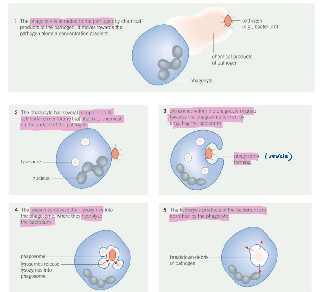
What are the stages of phagocytosis (non-specific immune response)?
Phagocyte recognises foreign antigens on pathogen
Engulfs pathogen by surrounding it with its cell membrane
Phagosome forms and fuses with lysosome (phagolysosome)
Lysozymes hydrolyse and digest pathogen
Phagocytosis leads to antigen presentation
Stimulates the specific immune response
What is a specific immune response?
a response that is slower and specific to each pathogen
cell mediated response and humoral response
What is an antigen presenting cell?
a cell that displays foreign antigens on its cell surface membrane
T lymphocytes types and function
cell mediated immunity
have cell surface receptors called T cell receptors which have a similar structure to antibodies and are specific to each antigen
4 types: T helper cells, T killer cells, T memory cells, T regulator cells
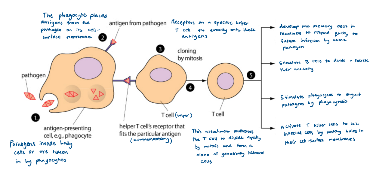
What are the stages of the cell mediated response?
pathogen invades
Phagocyte engulfs pathogen and becomes an APC
Helper T cell with a complementary receptor binds to the antigen
Helper T cell is activated to divide by mitosis to form clones:
Some differentiate into cytotoxic T cells which release perforin
Some stimulate phagocytosis
Some differentiate into memory T cells
Some activate B cells
B lymphocytes
mature in the bone marrow
humoral immunity
2 types: B plasma cells and B memory cells
What are the stages of the humoral response?
specific B cell with complementary receptor takes up antigens by endocytosis to become an APC
helper T cells with complementary receptors bind to these processed antigens and activate the B cell
B cells divide rapidly by mitosis to form clones:
some differentiate into plasma B cells which secrete antibodies
some differentiate into memory B cells which circulate in blood for secondary immune response
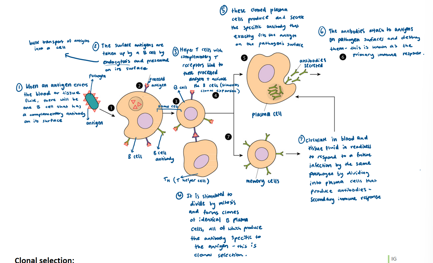
What is clonal selection?
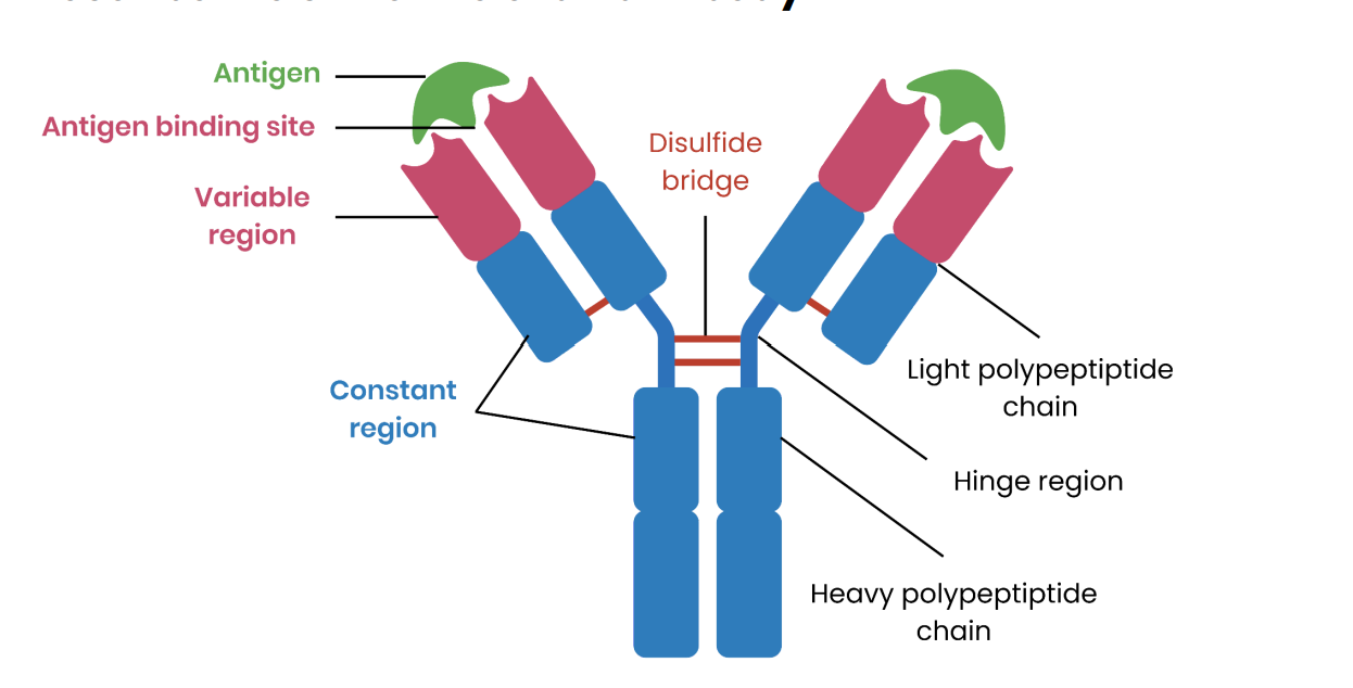
What are antibodies?
proteins synthesised by B cells in response to specific antigens
Antibody structure?
antigen binding site - 2 of them so can bind to more than one pathogen for agglutination
hinge region - allows flexibility
variable region - different for each antibody as tertiary structure complimentary to antigen
constant region - same in all antibodies, site to bind to immune system cells
How do antibodies prepare antigens for destruction?
agglutination - clumping of pathogens together so they cannot enter host cells and can be more easily located by phagocytes
opsonisation - antibodies coat pathogens to make them easier for phagocytes to recognize and engulf
anti-toxin - antibodies bind to toxins produced by pathogens, neutralizing them and preventing them from causing harm
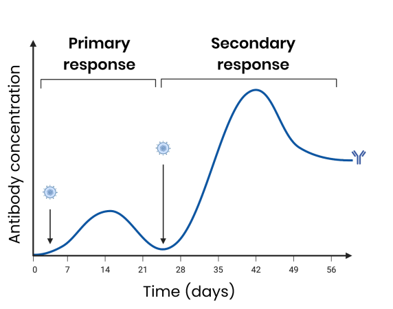
What are the differences between the primary and secondary immune response?
primary immune response - first exposure to antigen
antibodies are produced slower and at a lower concentration
B plasma cells are stimulated to produce specific antibodies
memory cells produced
secondary immune response -second exposure to antigen
antibodies produced faster and at a higher concentration
B memory cells rapidly undergo mitosis to produce many B plasma cells
B plasma cells produce specific antibodies
What is a vaccine?
injection of antigens from attenuated pathogens/ antigen fragments / modified toxins / genetically engineered DNA which stimulates formation of memory cells
How do vaccines provide protection to individuals against disease?
Antigens from dead or attenuated pathogens are injected
Specific B lymphocyte with a complementary receptor binds to the antigen
B cell takes up antigen by endocytosis and presents it on its surface
Helper T cell with a complementary receptor binds to the antigen
B cell divides by mitosis to form clones
Some differentiate into plasma cells which secrete antibodies
Some differentiate into memory B cells
On secondary exposure, memory B cells divide rapidly
Antibodies are produced faster and in higher concentrations
Why might vaccines fail to eliminate a disease?
individuals may develop the disease immediately after vaccination and then infect others
mutations of pathogens leading to antigen variability and short lived immunity
may be too many varieties of a pathogen to develop a vaccine
vaccination may fail to induce immunity in those with defective immune systems
What makes a vaccination programme successful?
economically viable
few side effects
method to produce, store and transport vaccines
method to administer vaccines efficiently
vaccinate majority of population and achieve herd immunity
What is immunity?
the ability of an organism to resist infection
What is herd immunity?
when a significantly large proportion of the population is vaccinated
spread of pathogen is reduced as
large proportion of population are immune so do not become ill from infection
unvaccinated people are less likely to come into contact with someone who is infected