times on slide 38 not important, just know general process
Cardiac Muscle
Striated/Have intercalated discs
Centrally located nuclei
Y shape allows muscle fibers to interweave producing efficient contraction mechanism
Myofibrils are less dense/organized
Alternate with abundant mitochondria
t tubules are larger/more numerous
reduced sarcoplasmic reticulum
diads instead of triads
makes up myocardium of heart
capable of generating endogenous action potentials
What is rest membrane potential at?
-55
Pacemaker potential process
1) slow depolarization/pacemaker potential
2) rapid depolarization
3) action potential
4) rapid repolarization
Phase 1: Pacemaker potential
opening of voltage gated Na+ channels (funny channels) and voltage gated transient Ca2+ channels
closure of voltage gated K+ channels
Phase 2: Rising Phase/Depolarization
Opening of long-lasting voltage gated Ca2+ channels
large influx of Ca2+
Phase 3: Falling Phase/Repolarization
opening of voltage gated K+ channels
closing of long lasting voltage gated Ca2+ channels
Potassium Efflux
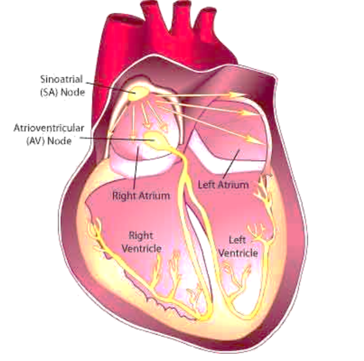
Sinoatrial/SA Node
located in right atrial wall just inferior/above to superior vena cava
generates 75 action potentials
pacemaker (has sinus rhythm)
no contractile elements, but connected directly to atrial fibers which makes it spread faster
Internodal Pathway
SA to AV node
Atria/Ventricles separate by non conductive fibrous tissue
AV is only electrical connection
Interartial pathway
right → left atrium
thru gap junctions
really fast
Atrioventricular/AV node
located in upper part of interarterial septum
uses internodal pathway to spread depolarization
Bundle of His
AV bundle
only electrical connection btwn atria/ventricles
left and right branch
Purkinje fibers
modified muscle fibers with few myofibrils
controls ventricles and papillary muscles
tighten chordae tenineae
open tricuspid/mitral valve
Control of heart rhythm
1) pacemaker (SA node) generates wave of signals to contract
2) signals delayed getting to AV node
3) signals pass to heart apex
4) signals spread thru-out ventricles
P-wave
depolarization of SA node → atria
QRS wave
ventricular depolarization/covers atrial repolarization
T-waves
ventricular repolarization
Cardiac Muscle Refractory Period
long
period which heart cant make AP
cannot go into tetany/summation
Regulation of Stroke Volume
Regulated by: Preload, contractility, and afterload
Explain the process happening in this image
fluids flow from the SA node into the right and left atrium
it flows faster into the left atrium and slower into the right
the fluids then make contact with the AV node, travels quickly into the left and right ventricles, and spreads out