WSU BIOLOGY 315 Lab Exam 3
1/460
There's no tags or description
Looks like no tags are added yet.
Name | Mastery | Learn | Test | Matching | Spaced | Call with Kai |
|---|
No analytics yet
Send a link to your students to track their progress
461 Terms
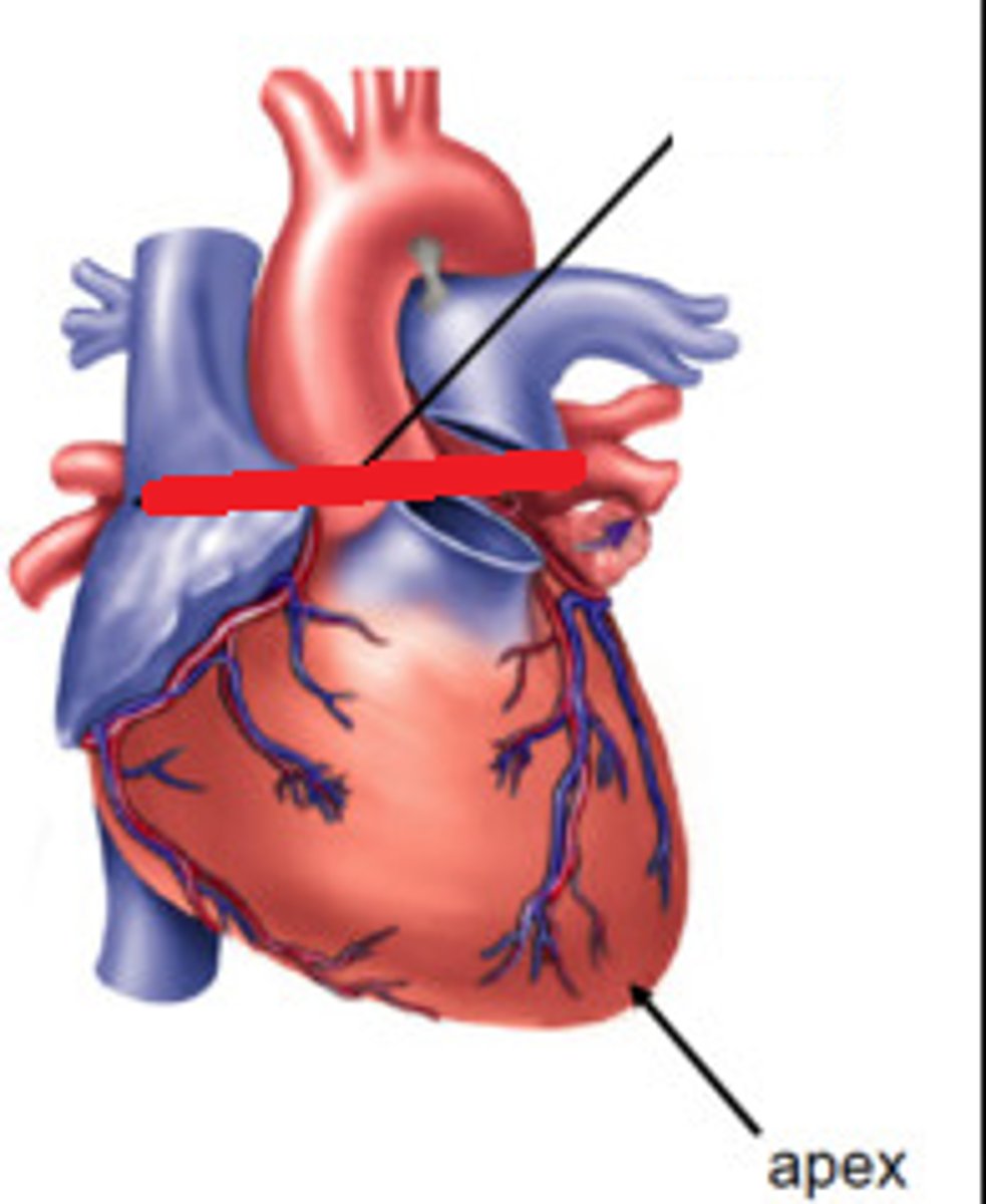
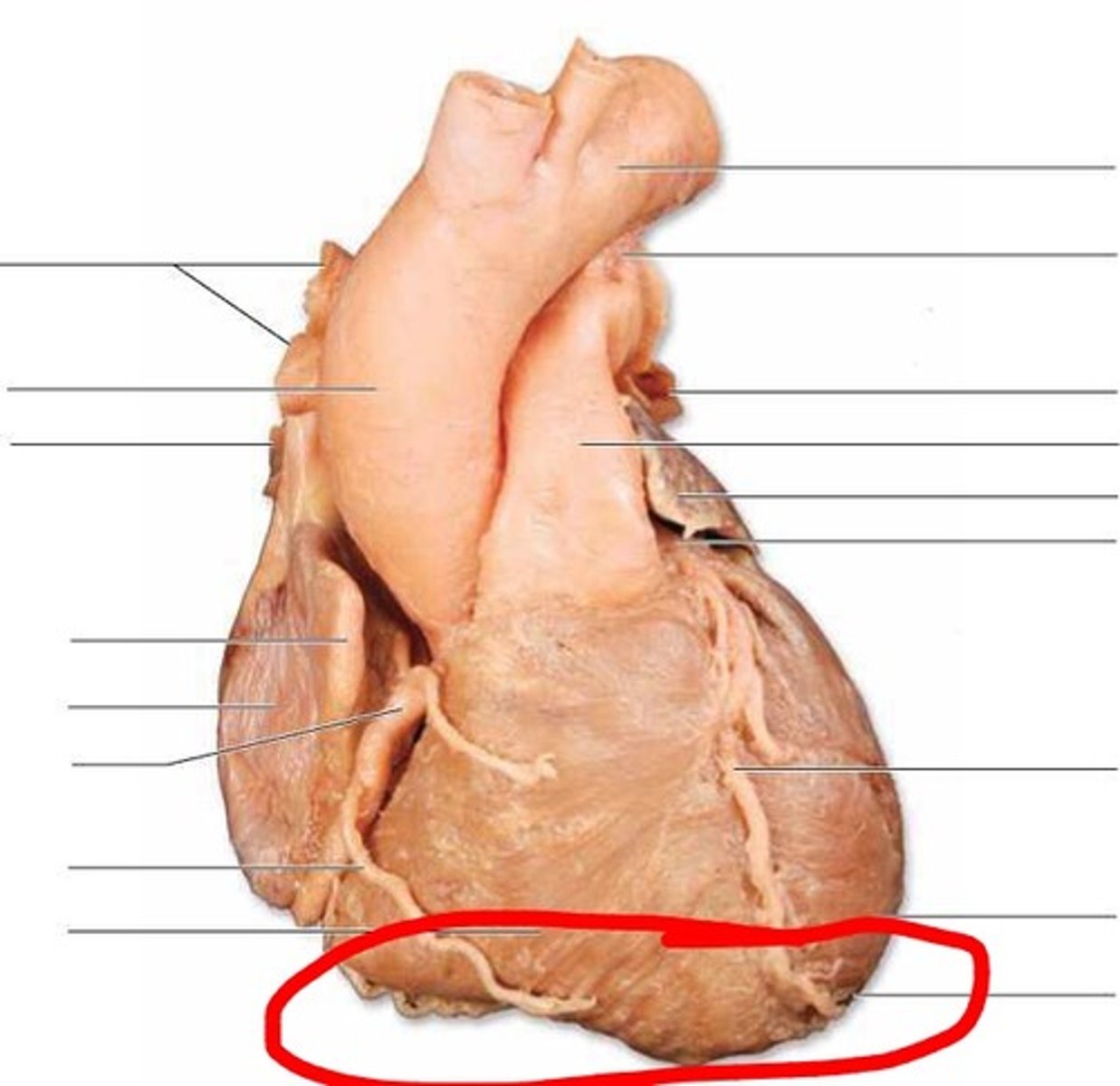
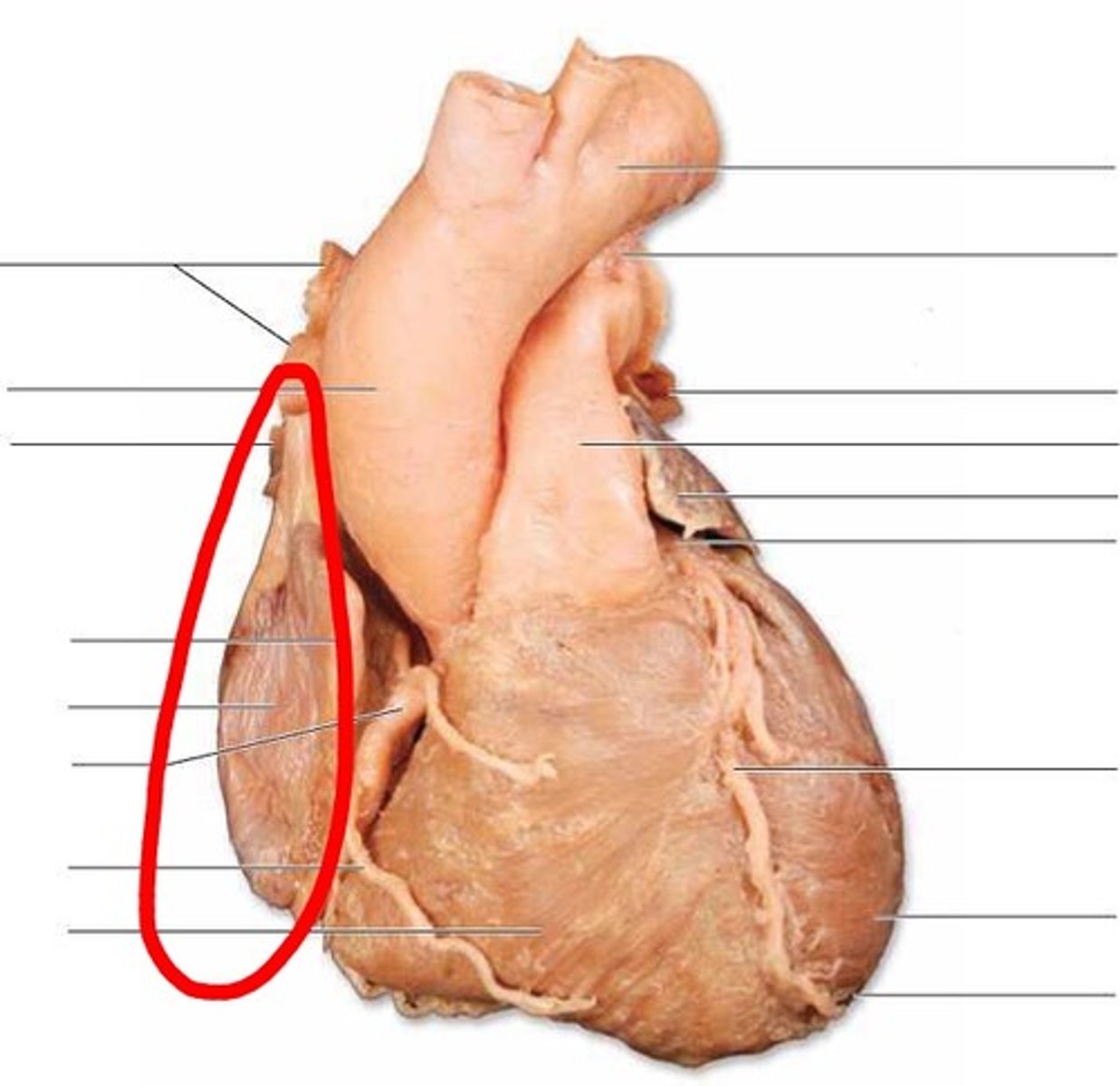
Apex of Heart
ft - bottom left side
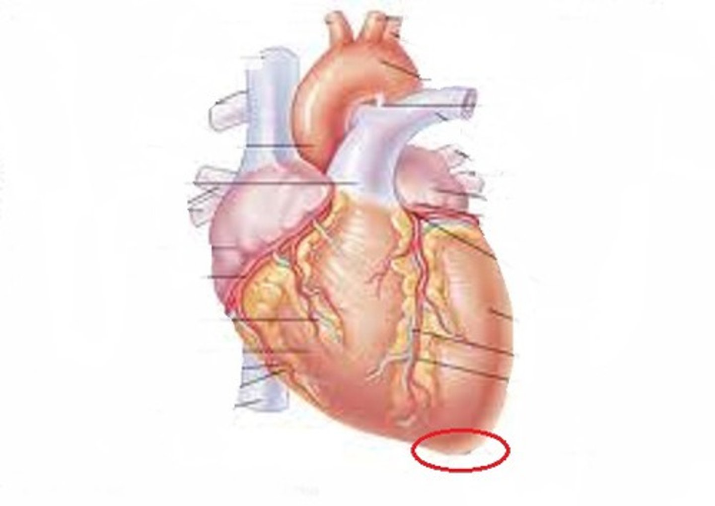
Base of Heart
TA will choke heart
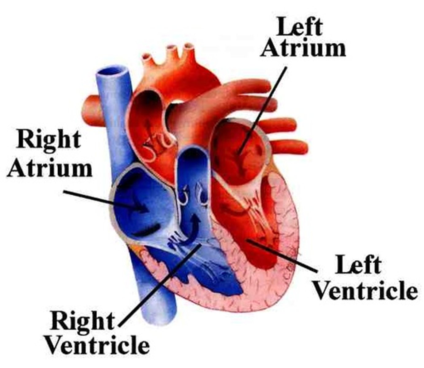
Right Atrium of the Heart
chamber TA will tap it
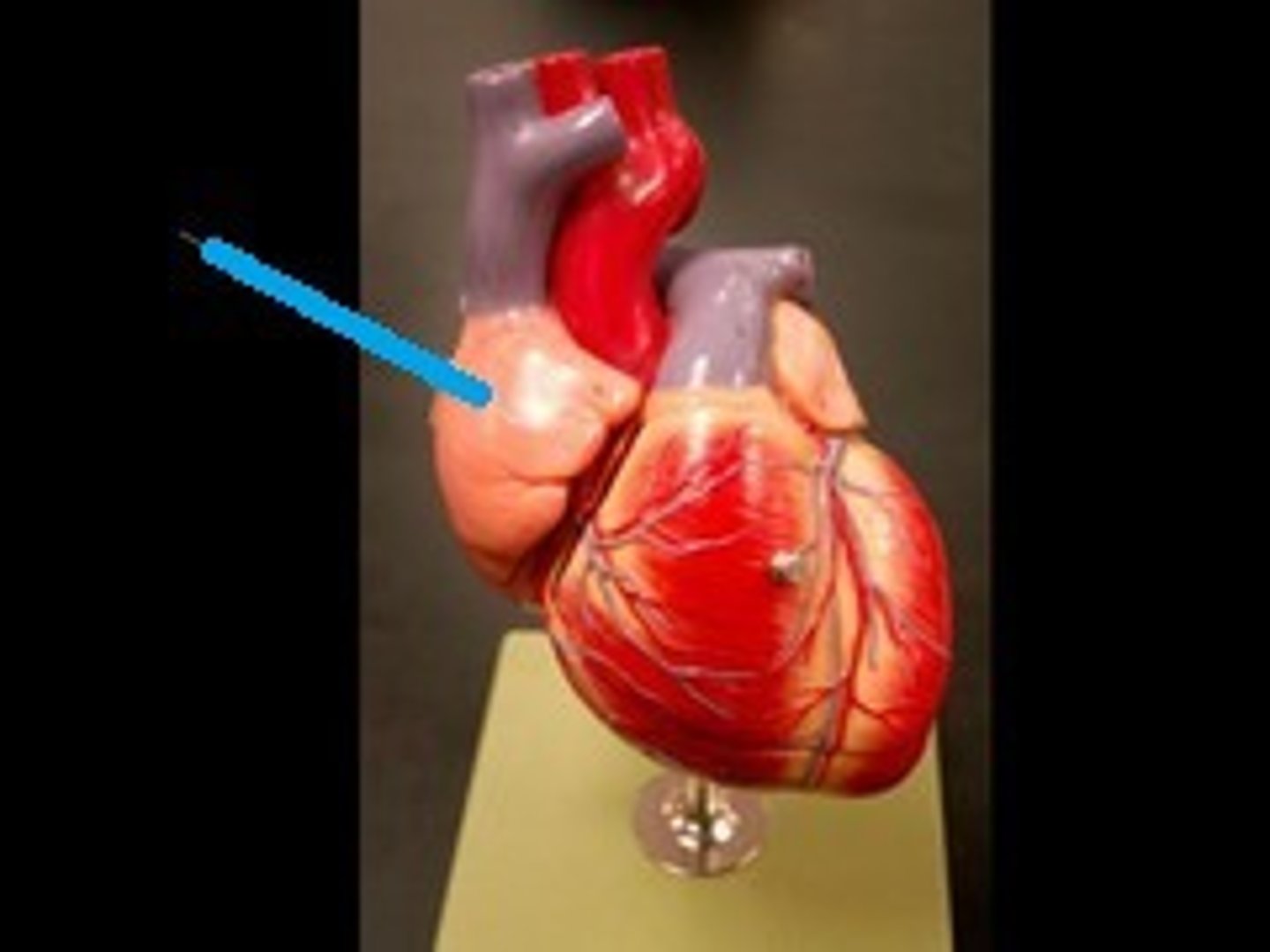
Right Auricle of Heart
feature, black nub posterior side
Above atrium
Left Atrium of the Heart
chamber

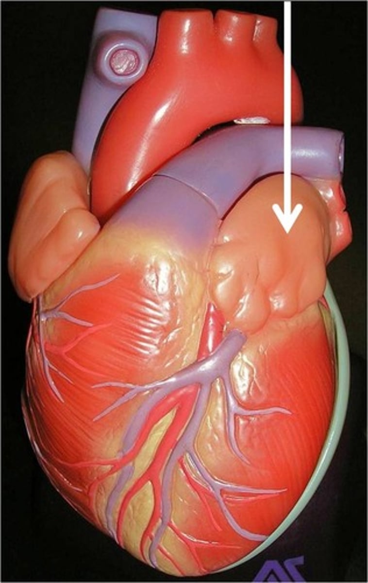
Left Auricle
ft will lift elephant shaped on apex side

Right Ventricle of the Heart
chamber
Left Ventricle of Heart
chamber
Anterior Surface of the Heart
hand in front- region
Diaphragmatic Surface of Heart
hand below heart
region
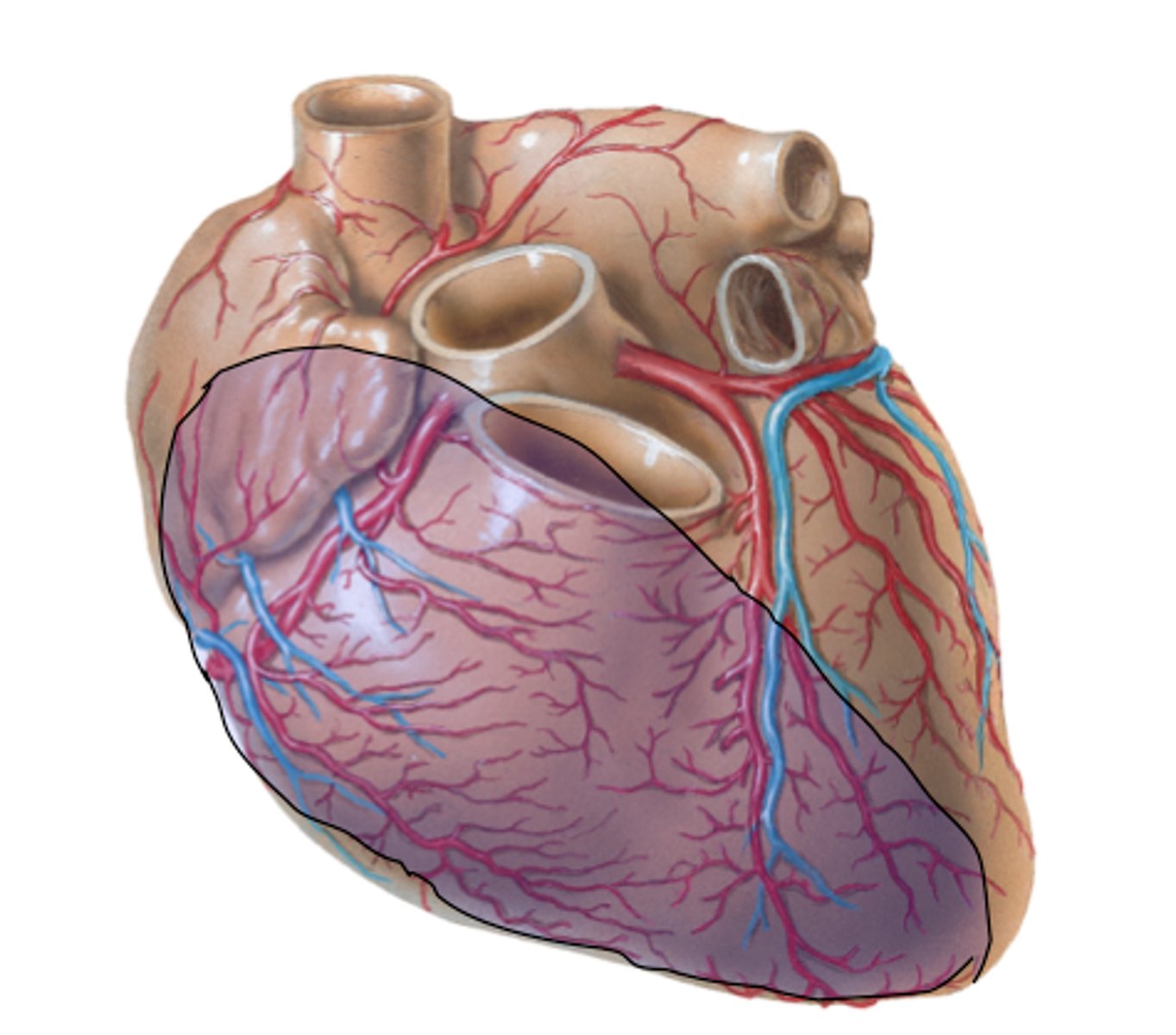
Right Pulmonary Surface of Heart
region
Left Pulmonary Surfaces
region
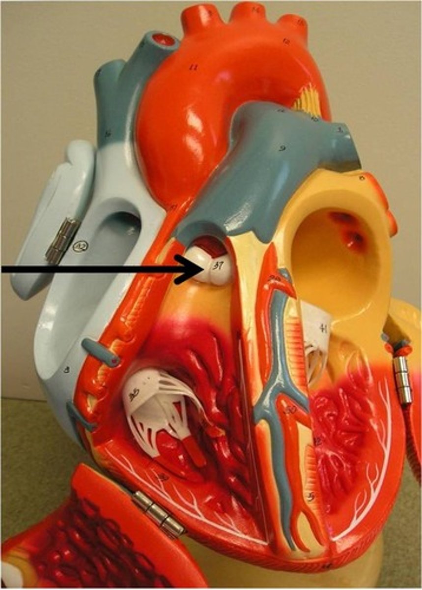
Right Border of Heart
Opposite apex
boundary
Left Border of Heart
Apex side
boundary
Inferior Border of Heart
blue area
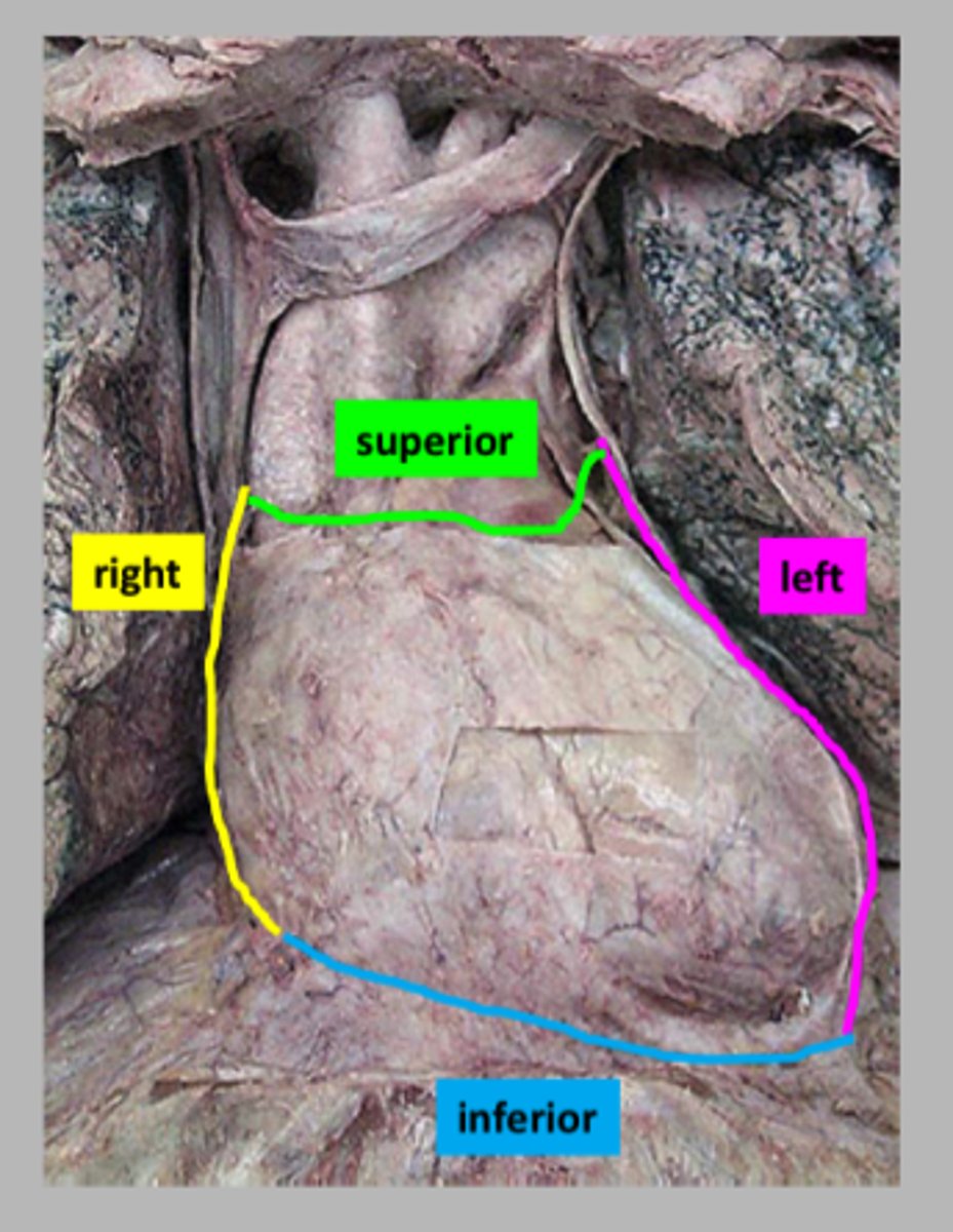
Superior Border of Heart
green
boundary

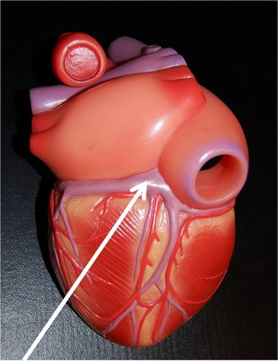
Coronary Sulcus
depression
under aorta+trunk
below all tubes, probe laying down above base
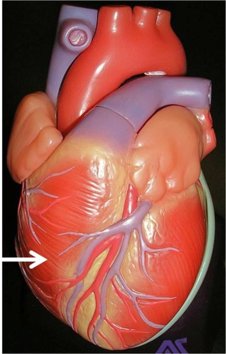
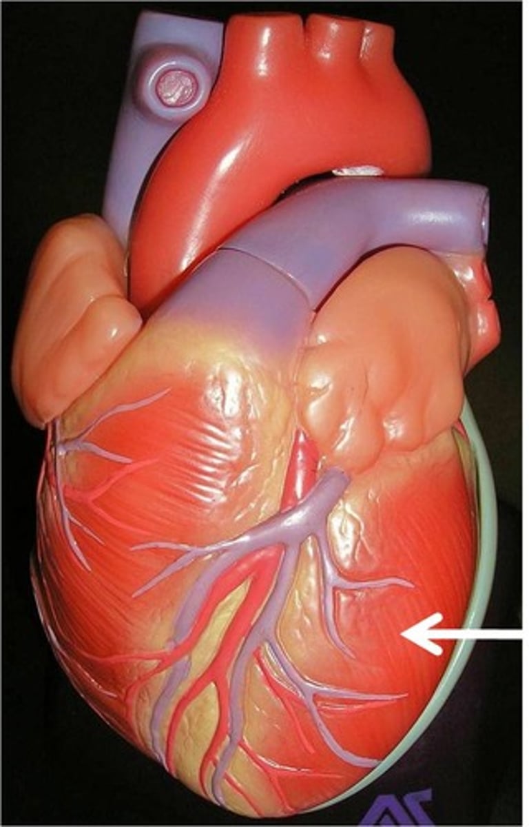
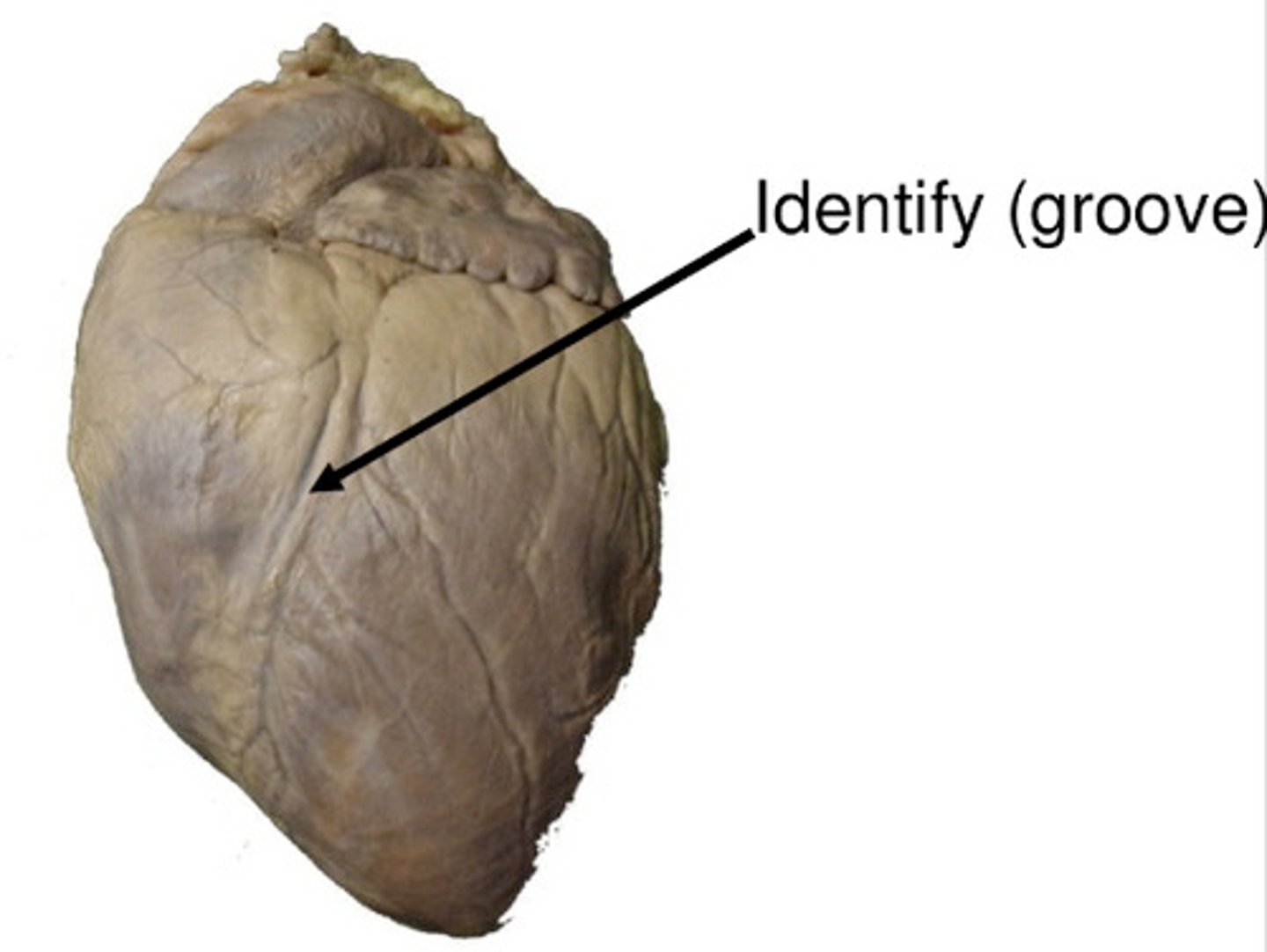
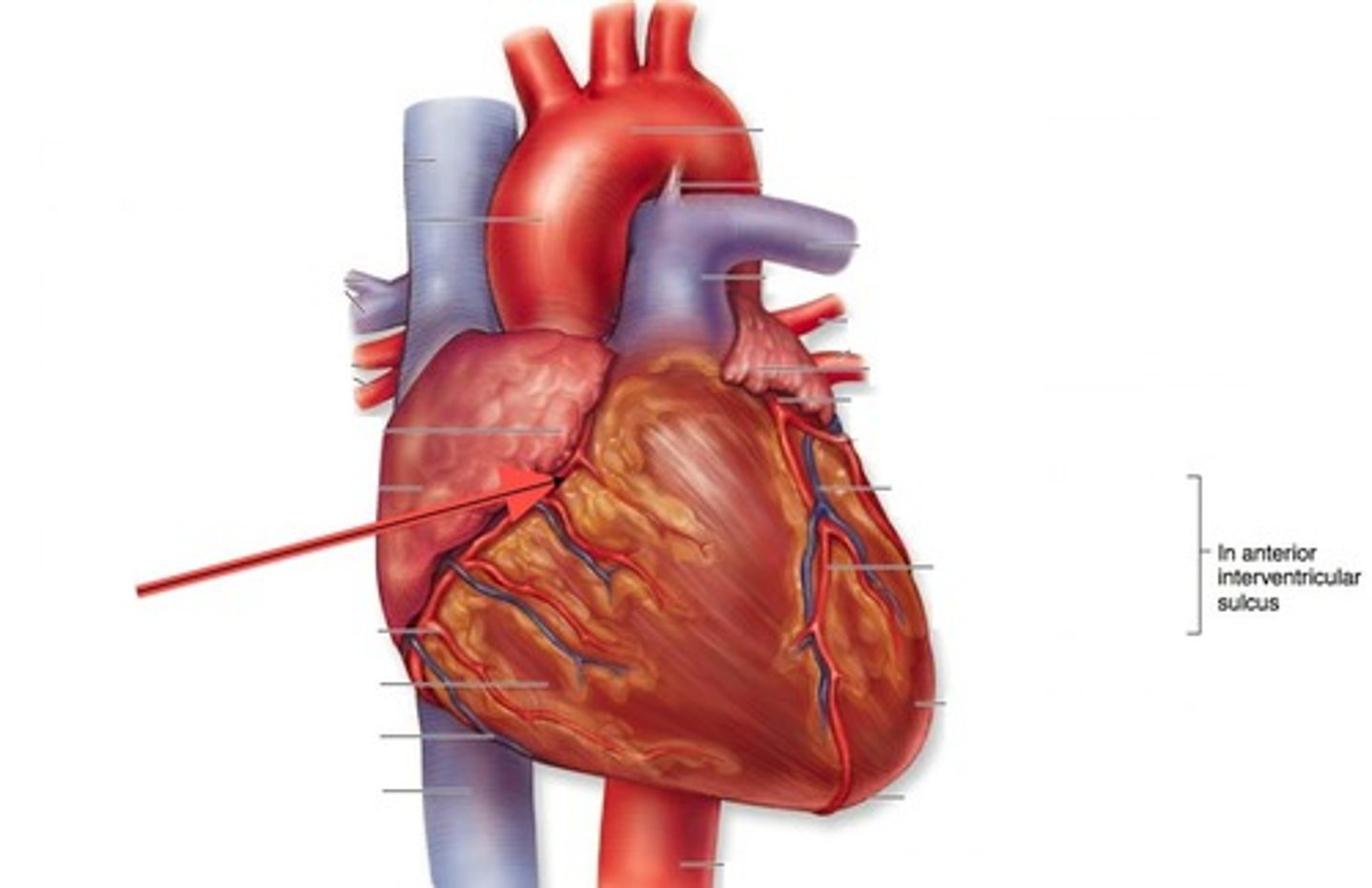
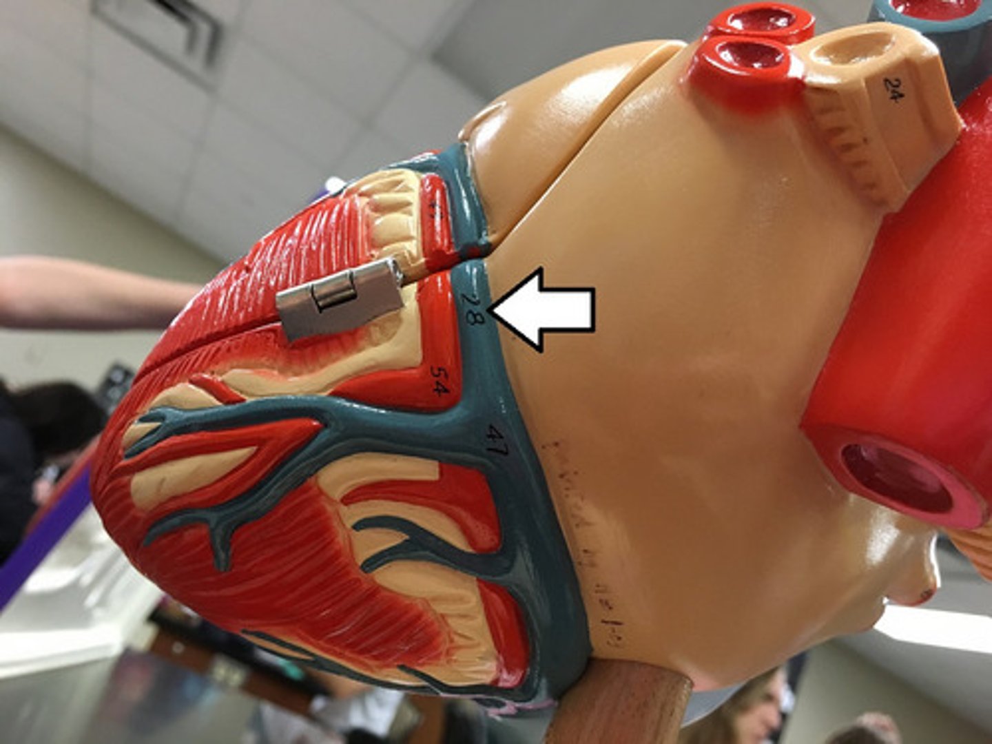
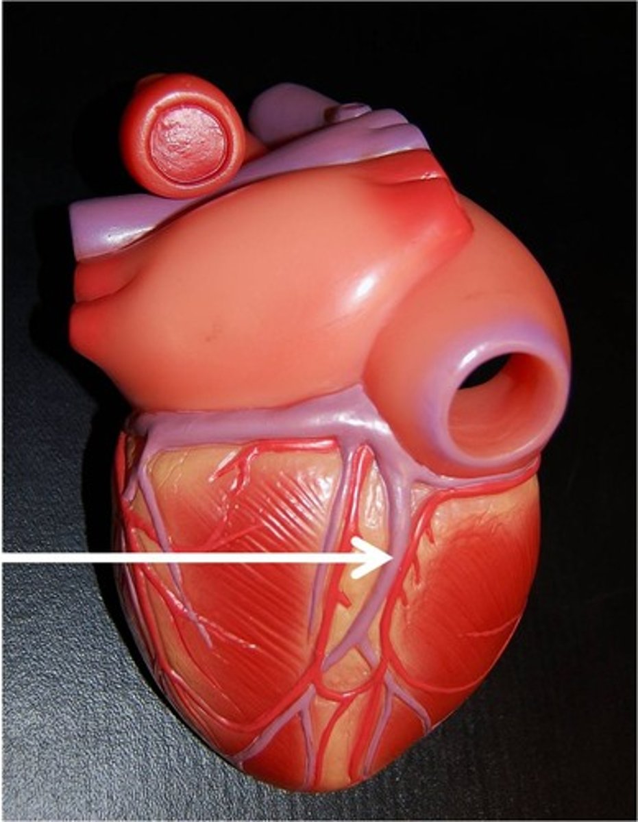
Anterior Interventricular Sulcus
depression on anterior heart
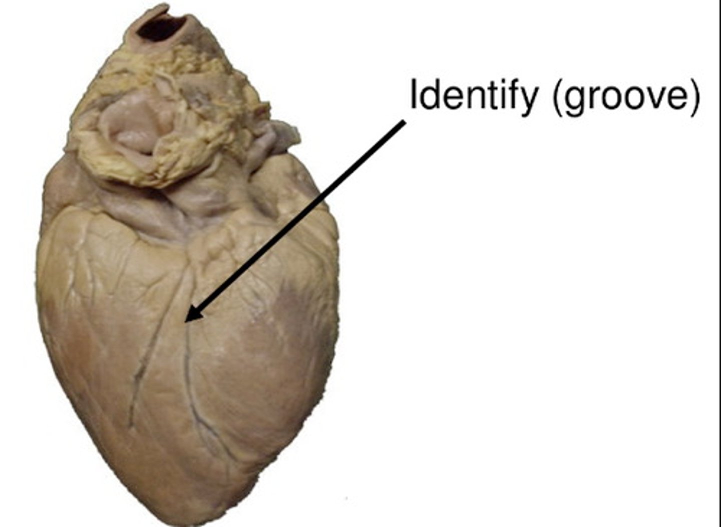
Posterior Interventricular Sulcus
depression on posterior side
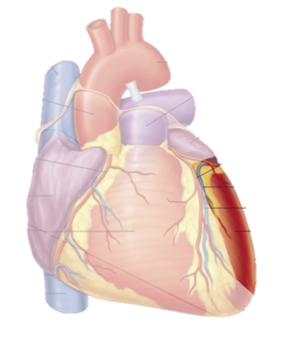
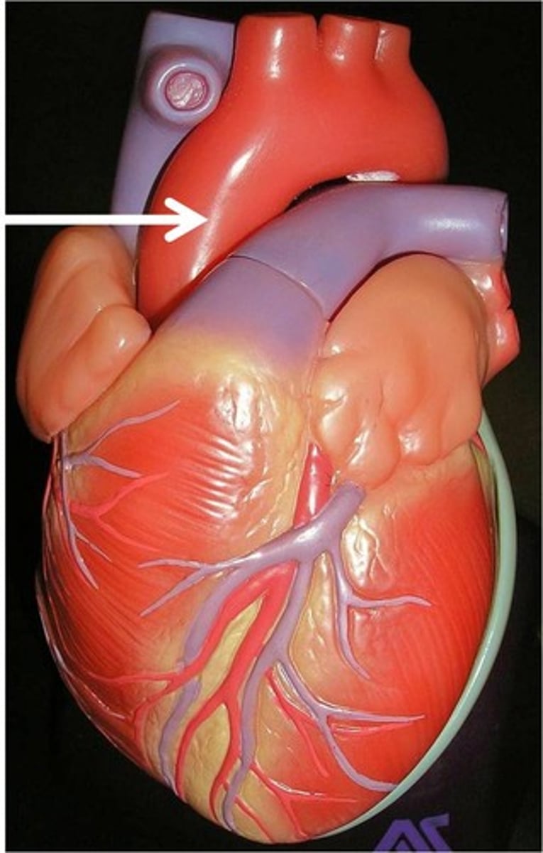
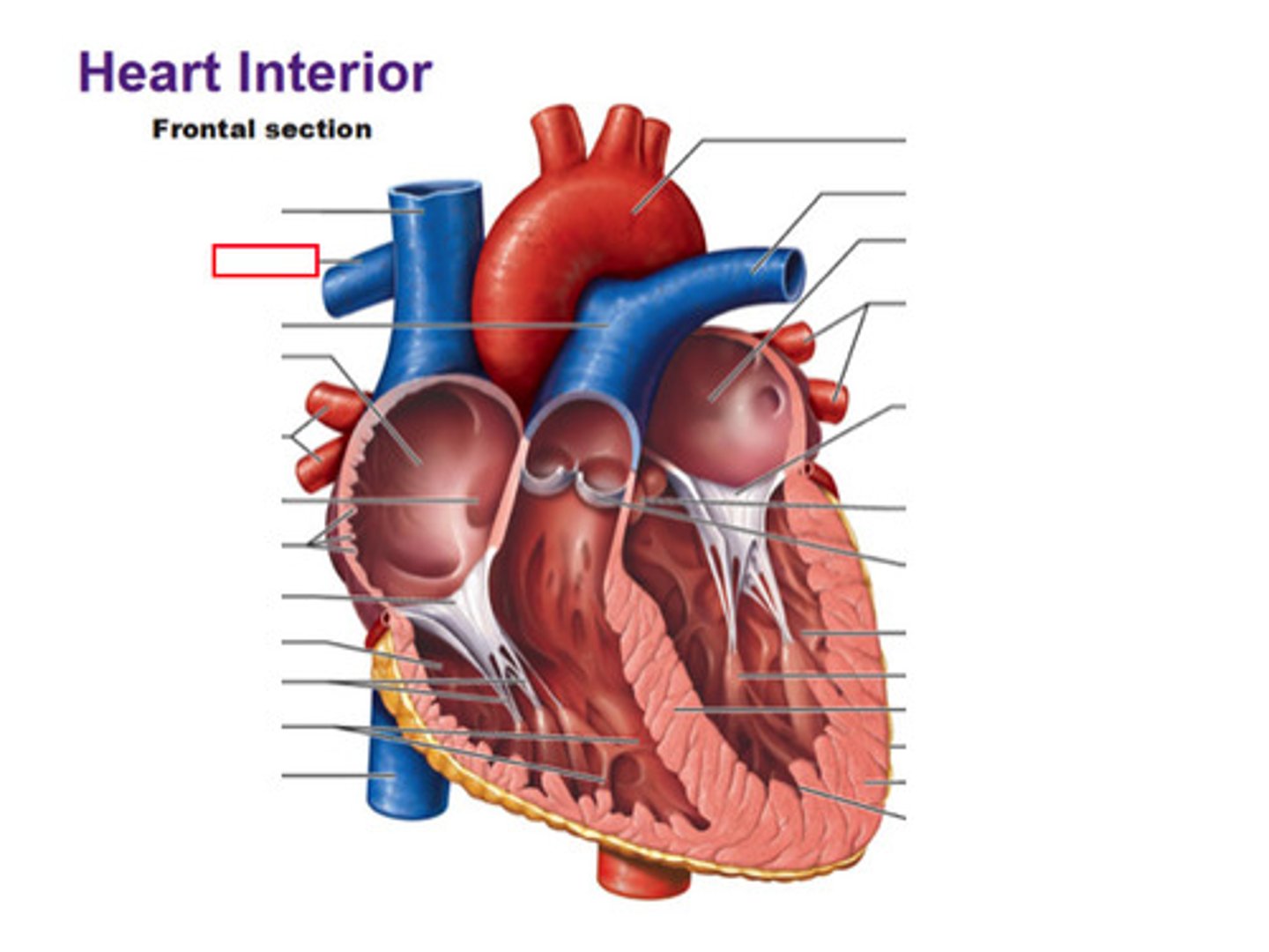
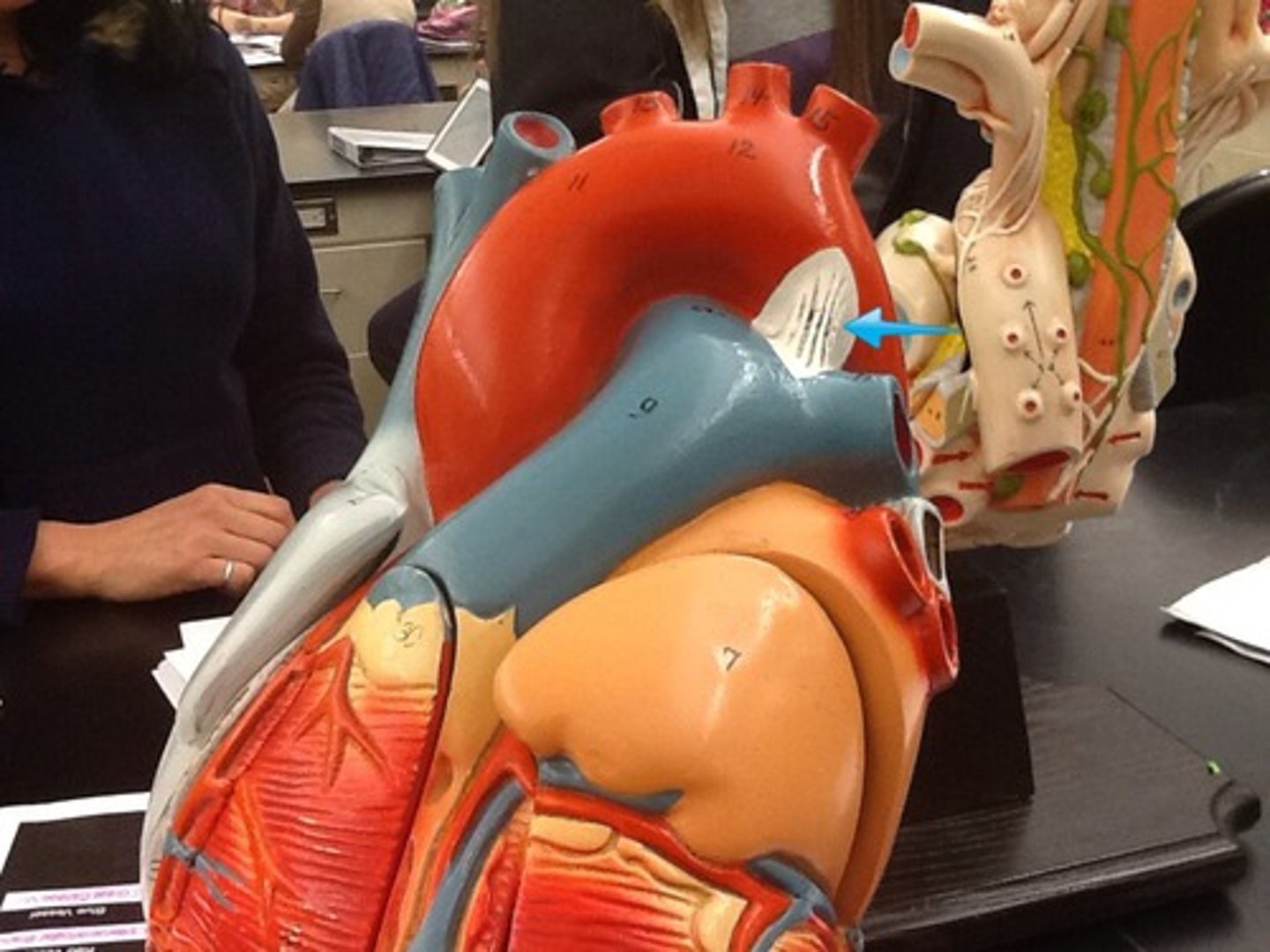
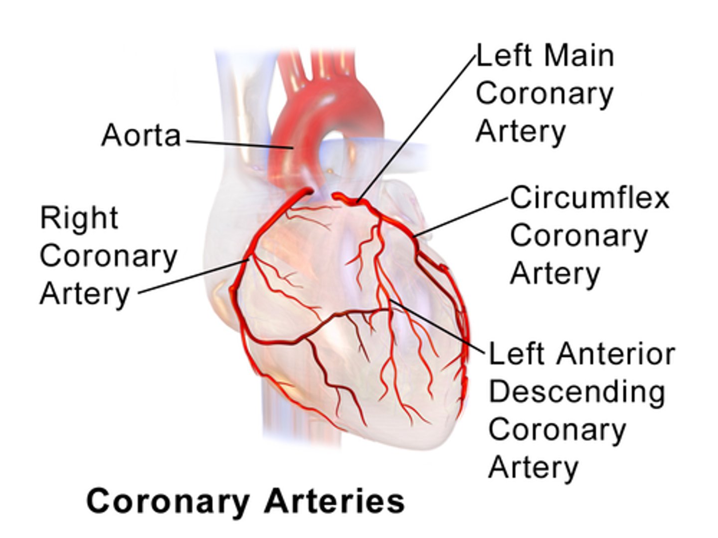
Aorta
Largest whole structure- with tubes
leaves left ventricle
Ascending Aorta
First portion
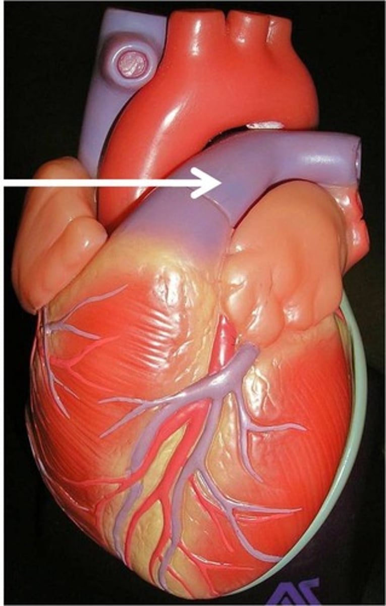
Arch of Aorta
Follows ascending aorta
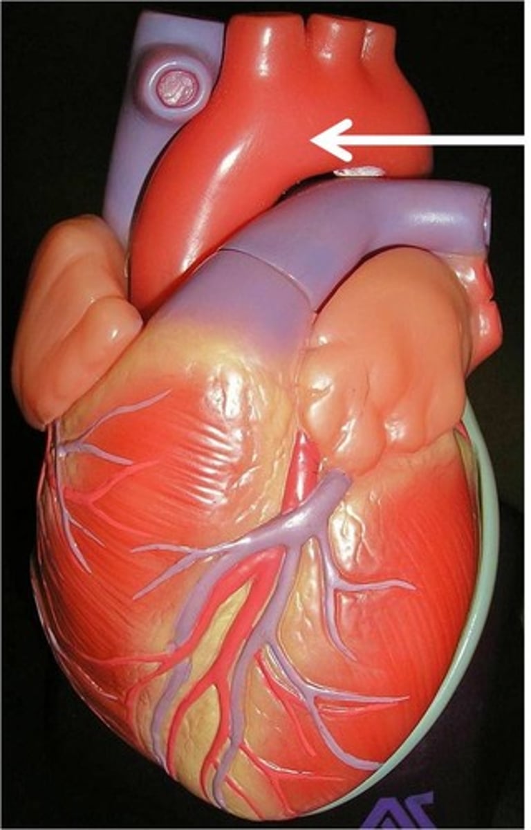
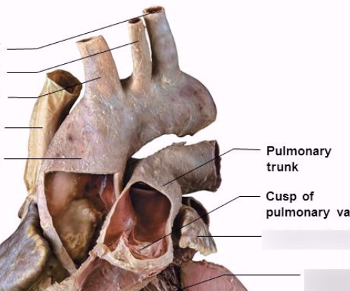
Pulmonary Trunk
collective structure
below aorta
Left Pulmonary Artery
On the left side of heart (below)
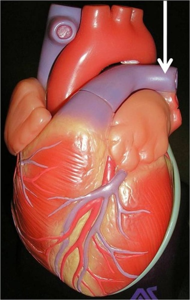
Right Pulmonary Artery
right side of heart (above)
backside of heart
Ligamentum Arteriosum
black clot
Webbing on small heart
Ligamentum Arteriosum Board Question
Vestige of the fetal ductus arteriosus,
which shunted blood in the pulmonary trunk
away from the lungs in the fetus
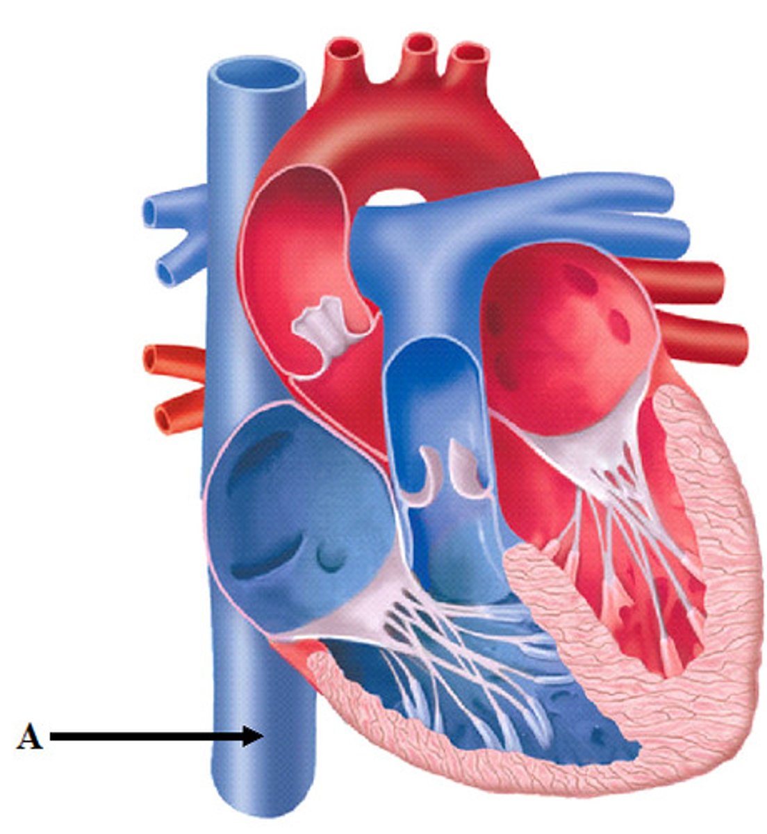
Superior Vena Cava
top part of probe
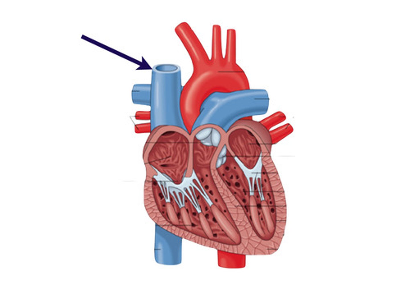
Inferior Vena Cava
bottom part of probe
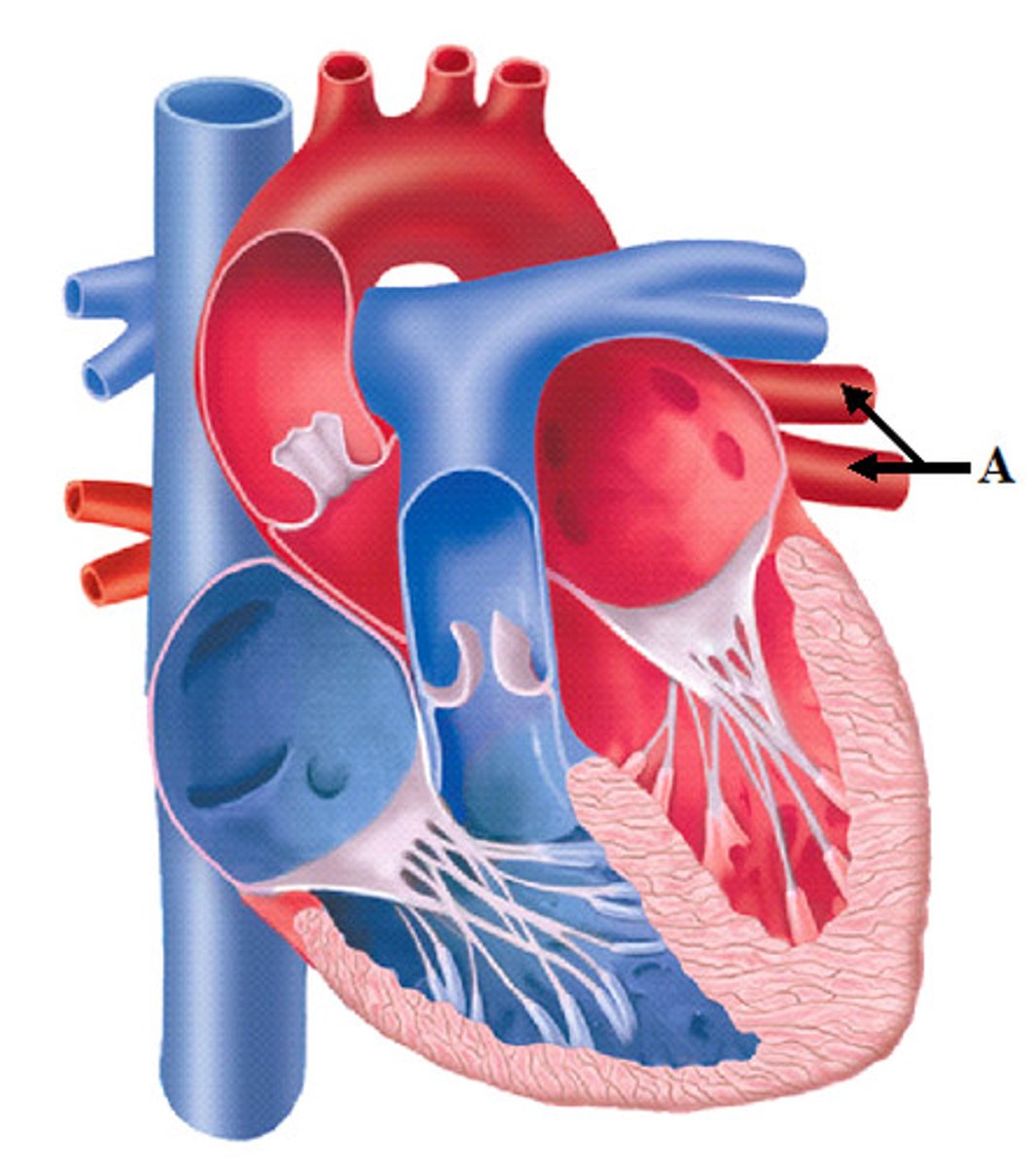
Pulmonary Veins
collective structure
all 4 probes in

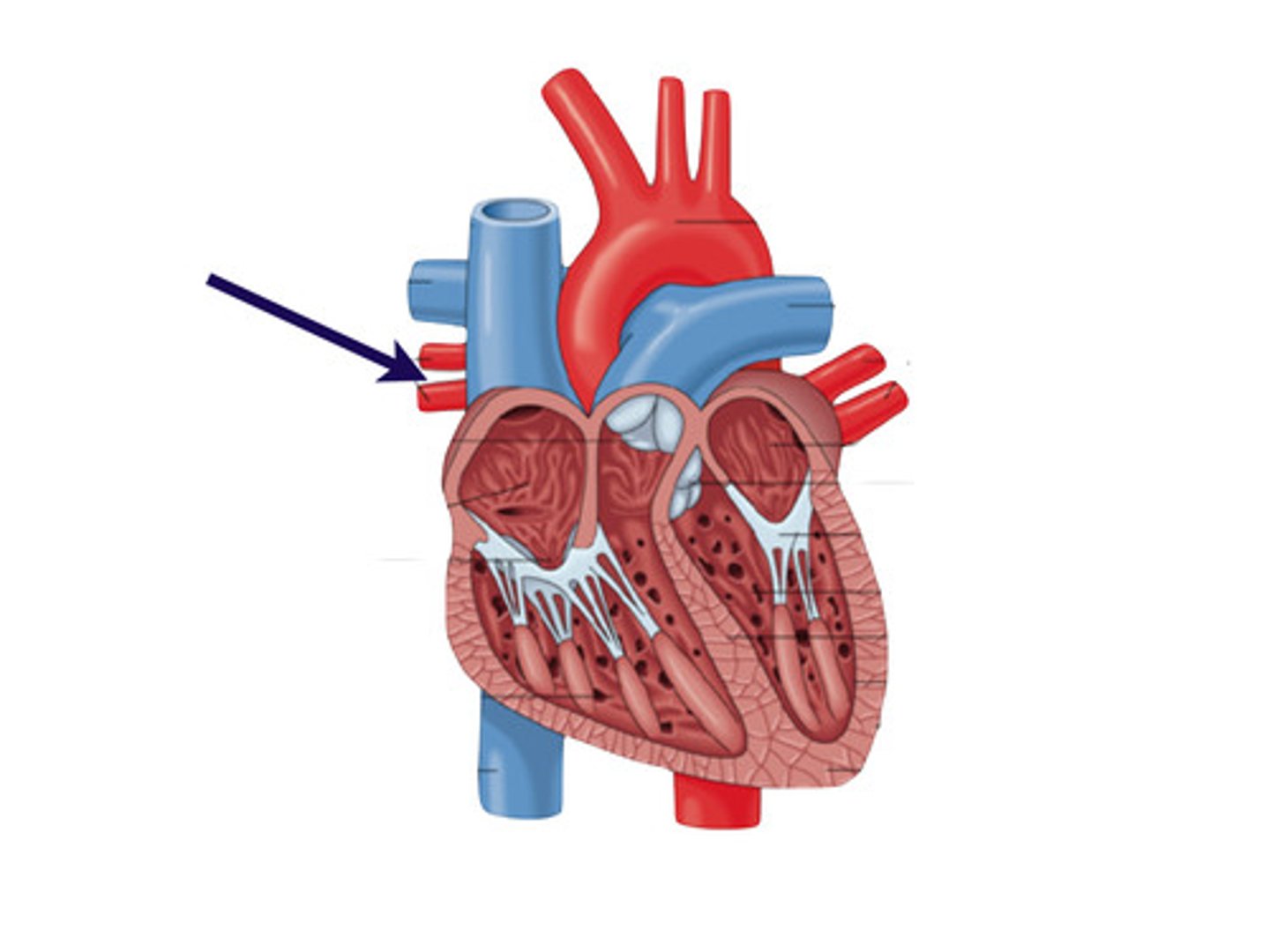
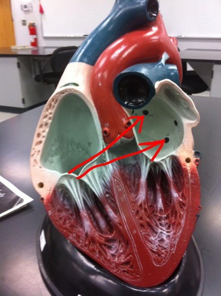
Left Superior Pulmonary Veins
Upper tube, apex-side of probe
Left Inferior Pulmonary Veins
Lower tube, apex-side of probe
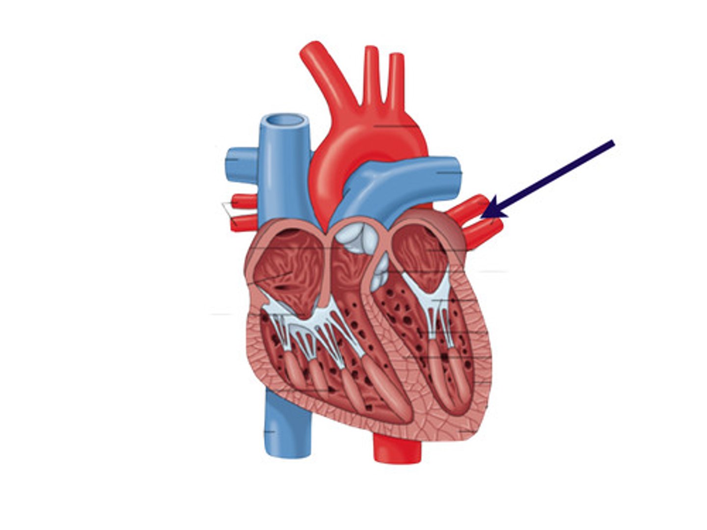
Right Superior Pulmonary Veins
Upper tube, probe side opposite the apex

Right Inferior Pulmonary Veins
Lower tube, probe side opposite the apex
Coronary Arteries
located in coronary sulcus
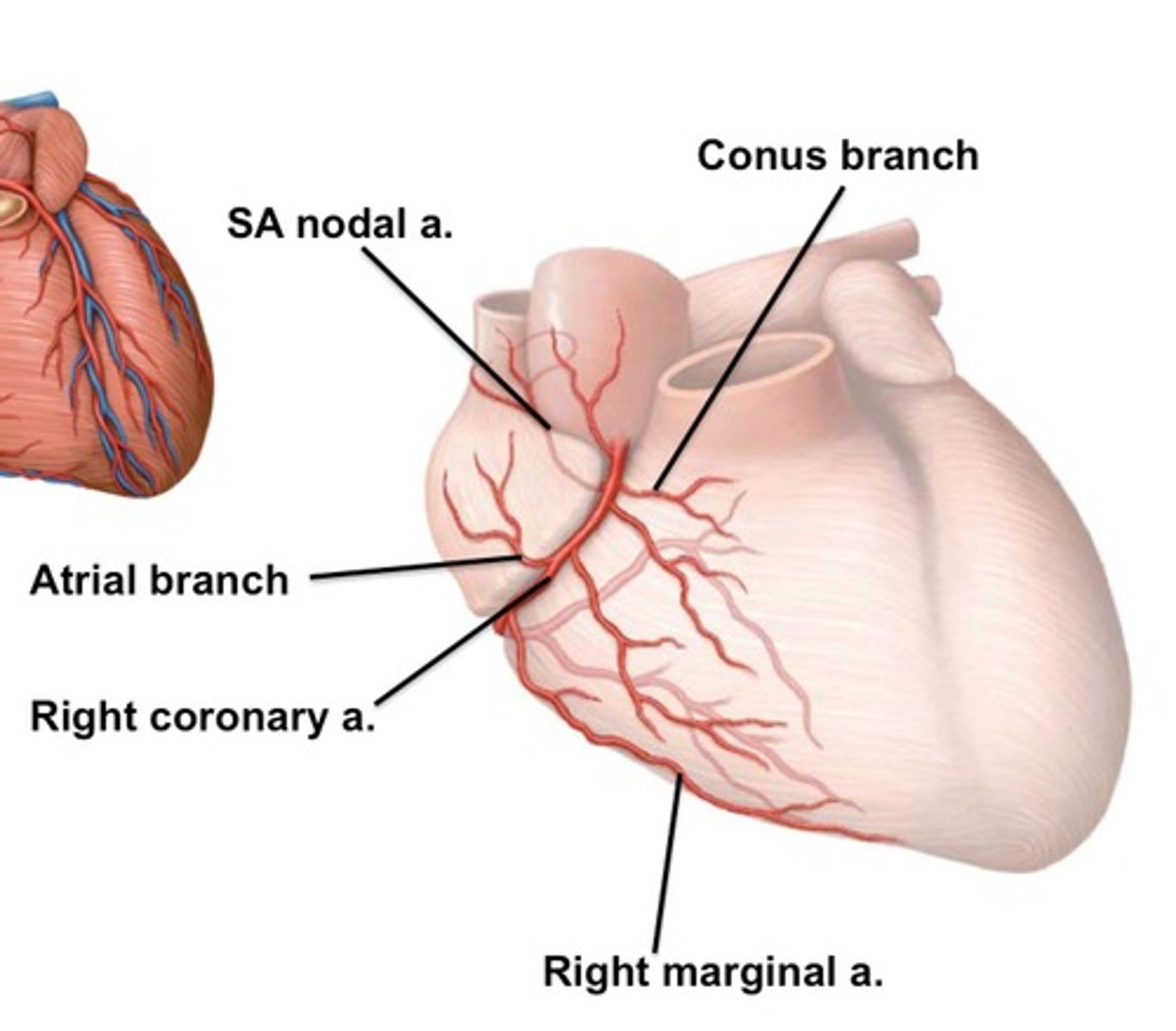
Right Coronary Artery
Under right auricle
Right Marginal Artery
Below right coronary artery
y shaped
Posterior Interventricular Artery
st
inside posterior interventricular sulcus
Left Coronary Artery
Under left auricle OR
peel aorta + trunk→ sitting in coronary sulcus on top
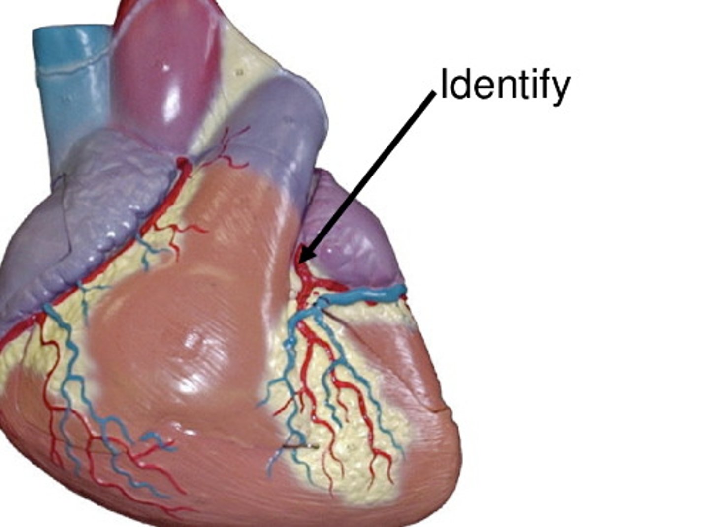
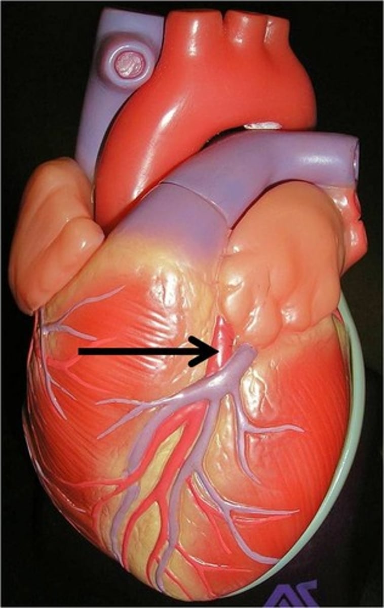
Anterior Interventricular Artery
st
in anterior interventricular sulcus
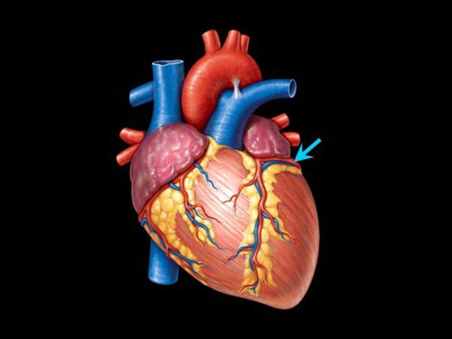
Circumflex Artery
Squiggly, on left ventricle
Right atrium
part of left ventricle
The Right Coronary Artery Supplies:
Left atrium
part of the right ventricle
The Left Coronary Artery Supplies:
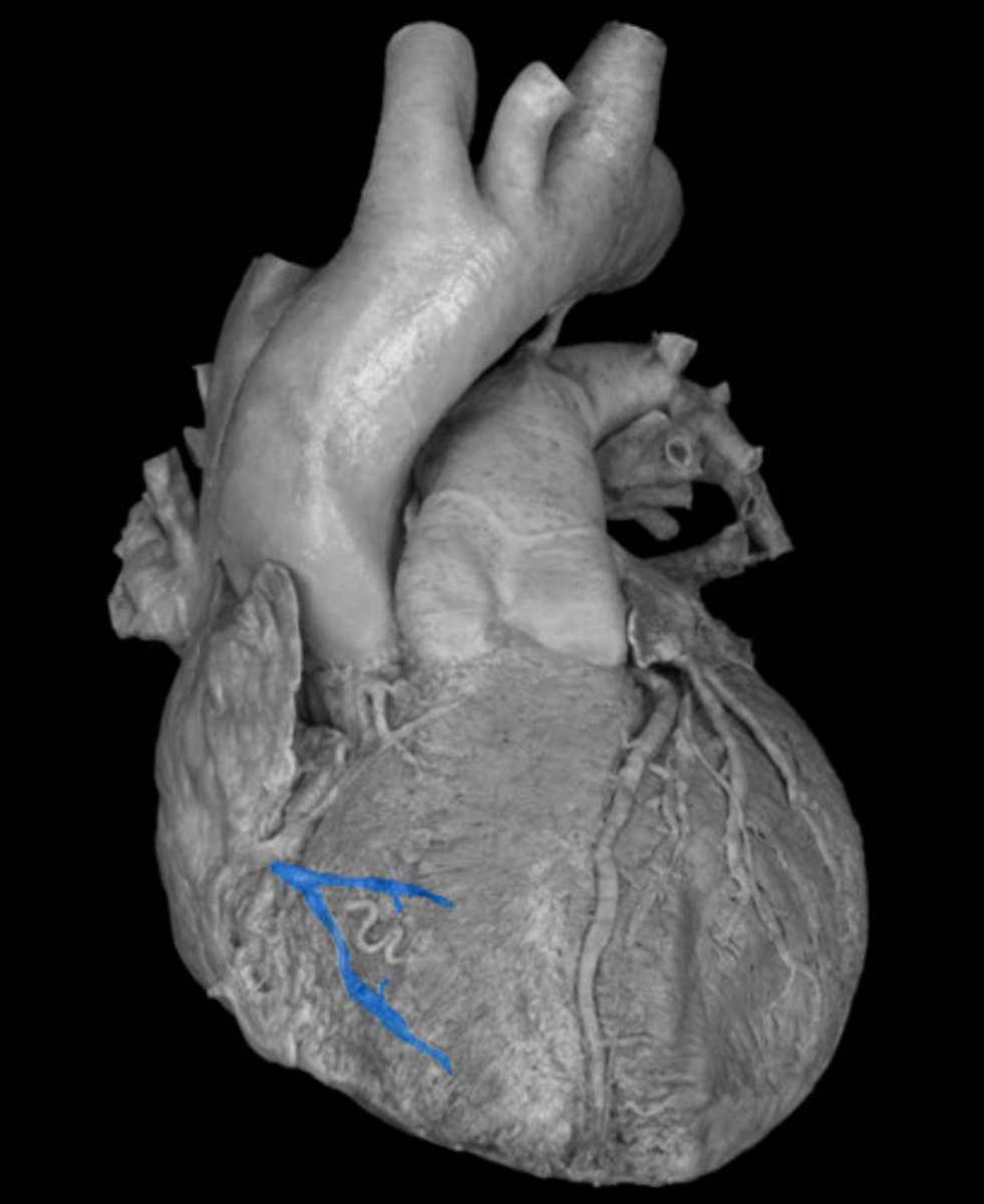
Cardiac Veins
Posterior T ST
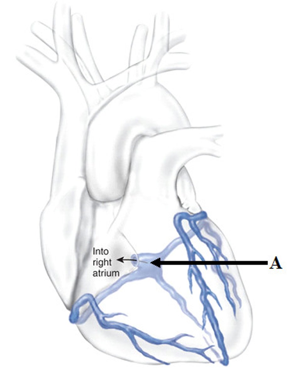
Coronary Sinus
Space
top middle of the T
Probe inside
Great Cardiac Vein
goes left of coronary sinus
ST
will be lifted
Middle Cardiac Vein
st
going downwards
base of t
Small Cardiac Vein
top right side of T
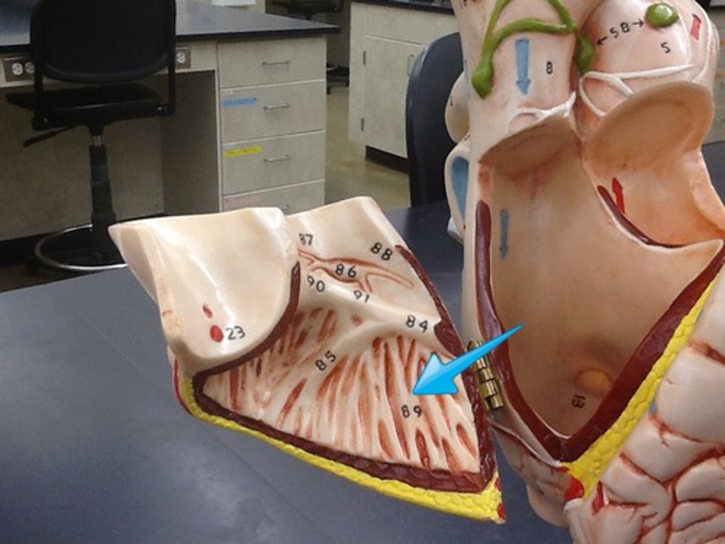
Fibrous Pericardium
covering
Outside of the flap
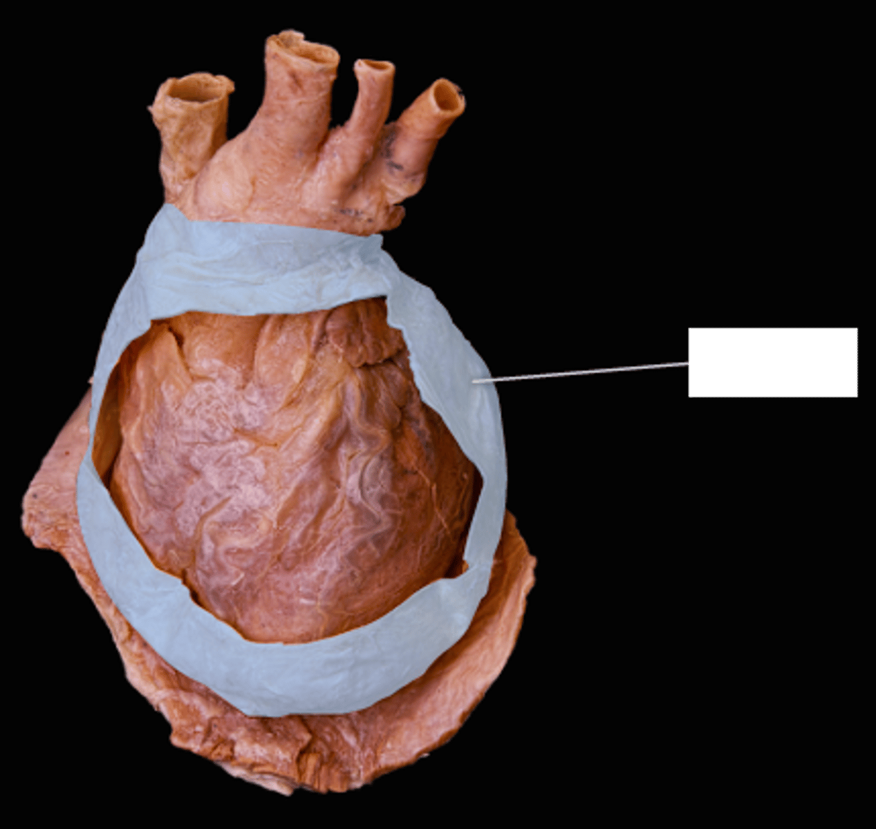
Serous Pericardium
collective covering
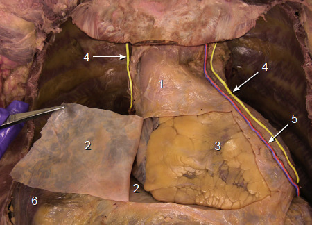
Parietal Layer of Serous Pericardium
feature
Shiny part of the flap
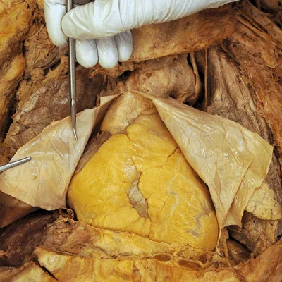
Visceral Layer of the Serous Pericardium
ft
On the heart/ touching it
pg 3
Pericardial Cavity
space
Between the flap and the heart
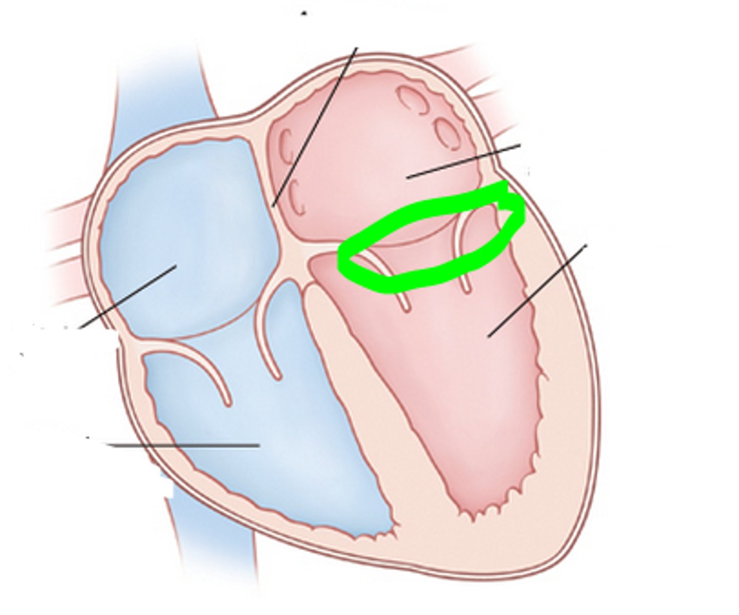
Transverse Pericardial Sinus BQ
Connects the left and right sides of the pericardial cavity
Olbique Pericardial Sinus BQ
a blind cul de sac posterior to the heart
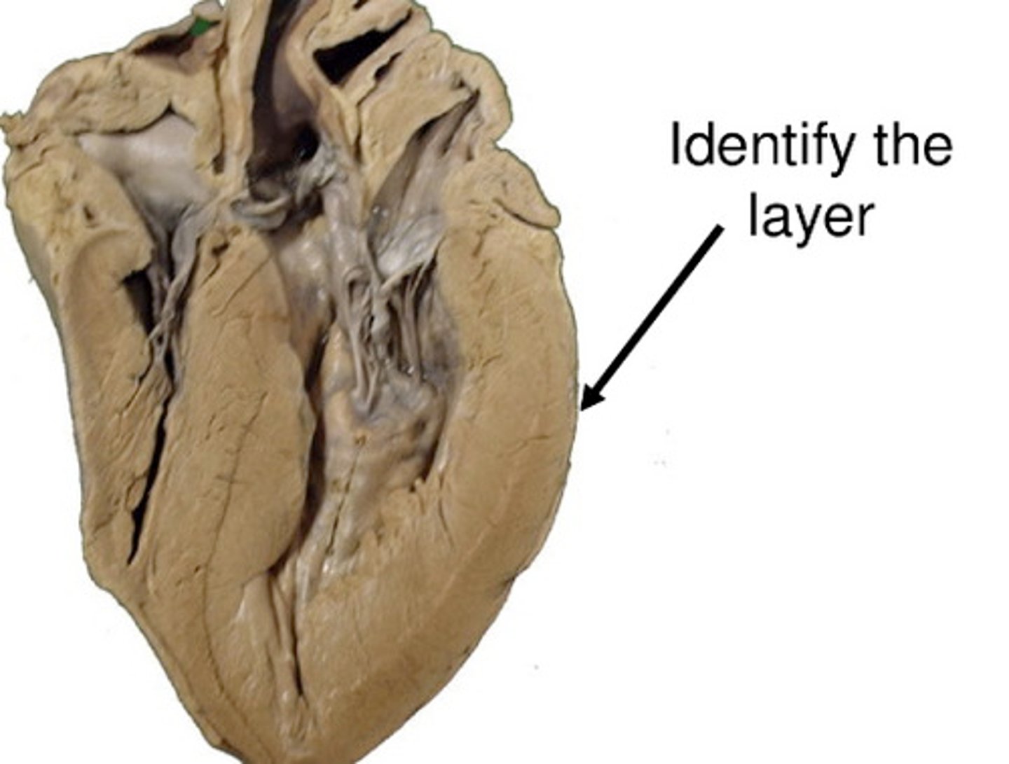
Endocardium
Inner layer of heart
Myocardium
middle layer of heart
Epicardium
Most outer layer of heart
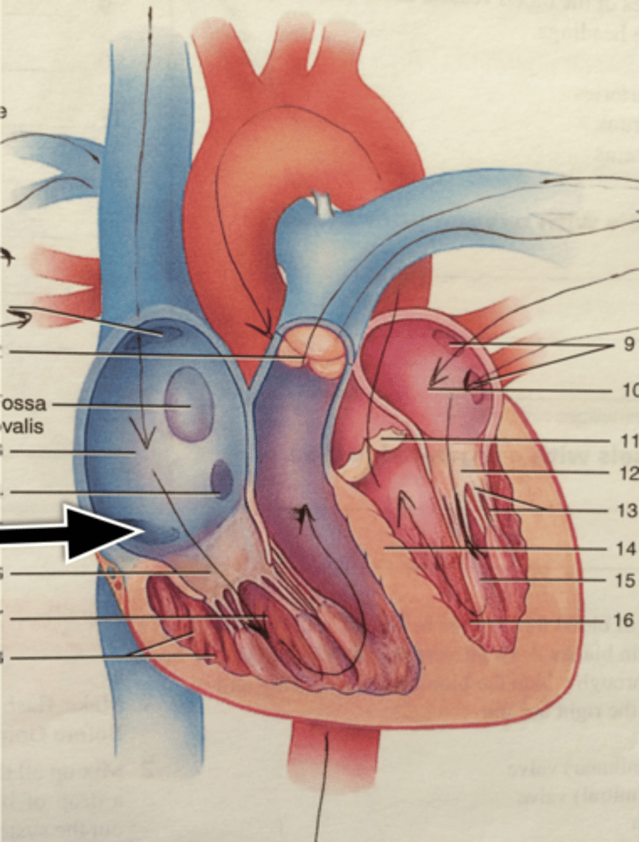
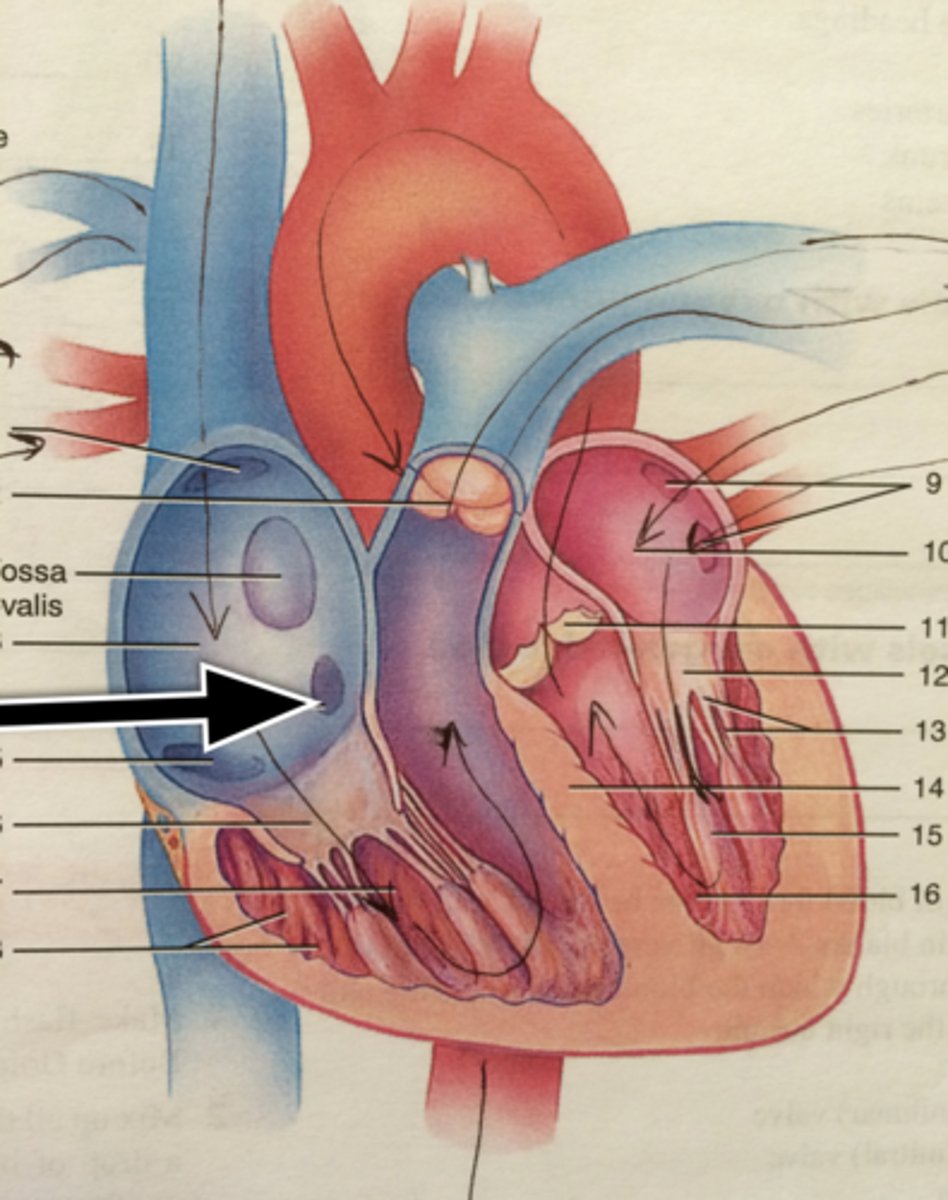
Interatrial Septum
structure
Will be pinched
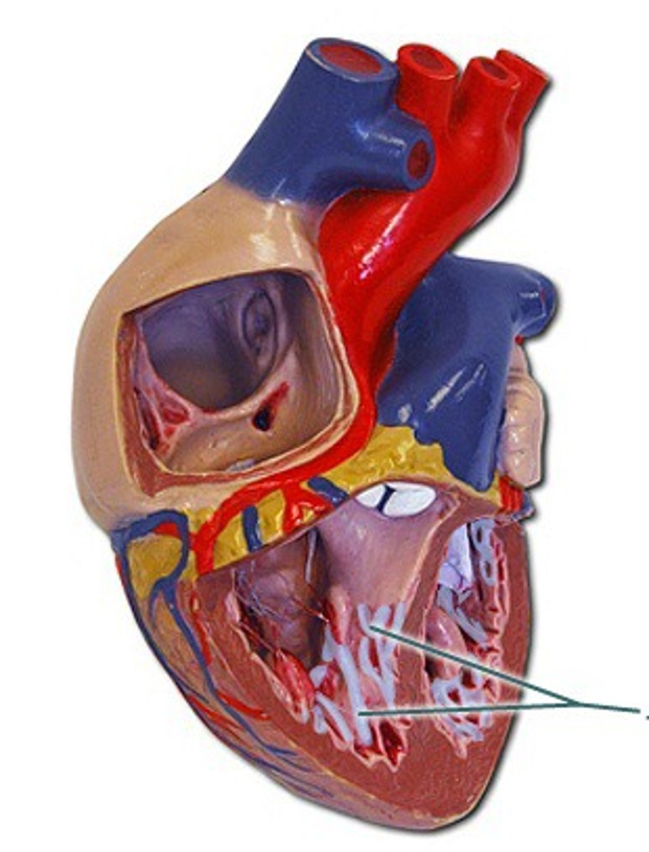
Fossa Ovalis
depression bean
tan smooth
Fossa Ovalis Board Question
oval depression- in the interatrial septum,
indicating the location of the fetal oval-
where blood was shunted from the right atrium to the left atrium in the fetus
Right Atrium
A chamber
Will be tapped
top left

Pectinate Muscle
Dark, branching ST
Sinus Venarum
smooth surface next to pectinate muscle
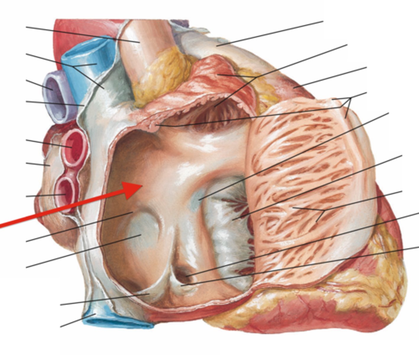
Opening of the Superior Vena Cava
opening
Top of probe
Opening of the Inferior Vena Cava
opening
Bottom of probe
Opening of the Coronary Sinus
space
next to fossa ovalis
Left Atrium
top chamber
same side apex

Left Auricle
Looks like an elephant ear
Openings of the Pulmonary Veins
collective space
all 4 probes
backpack straps
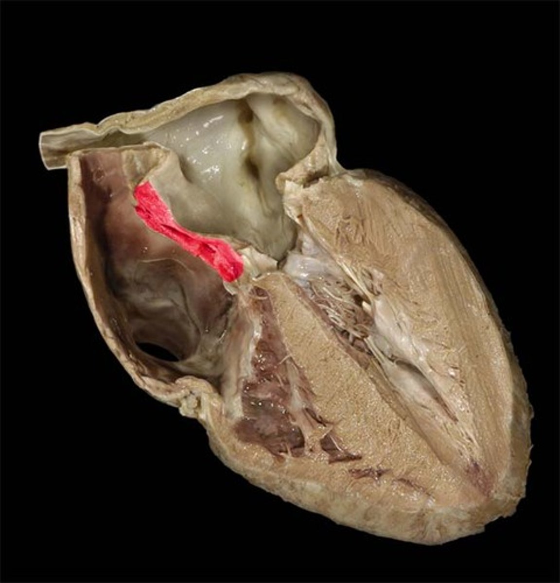
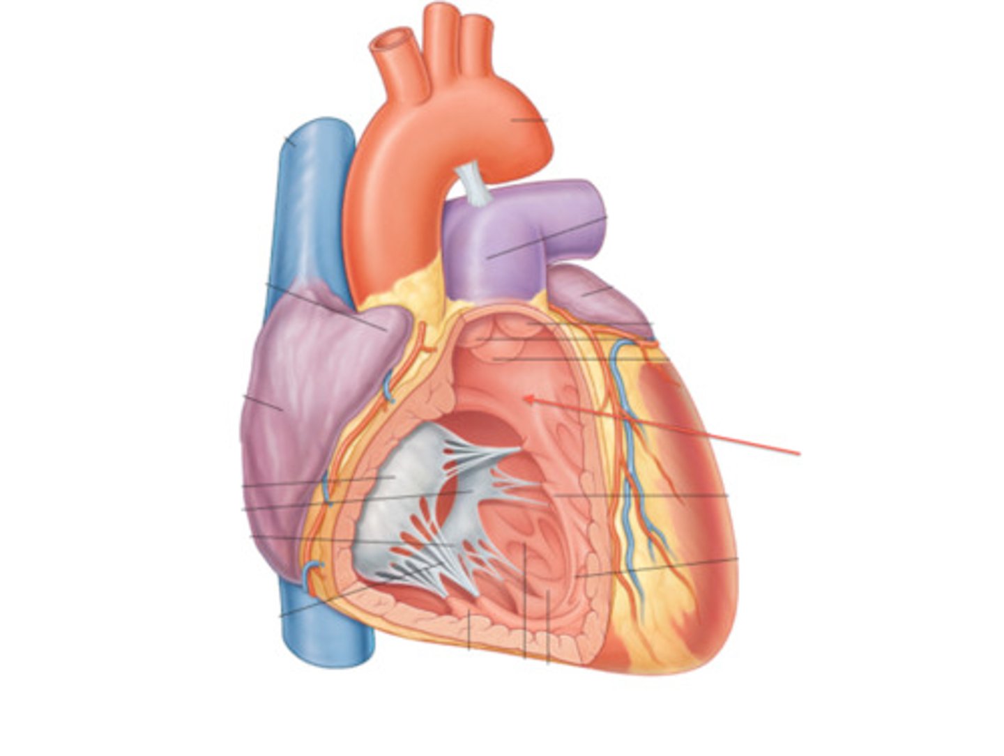
Interventricular Septum
collective structure
divides ventricles
Muscular Part of the Interventricular Septum
Thicker, inferior
portion
Membranous Part of the Interventricular Septum
Thinner, superior portion
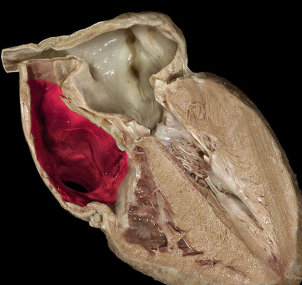
Right Ventricle
chamber below right atrium

Right Atrioventricular Orifice
space pull flap to reveal
probe inside
doorway
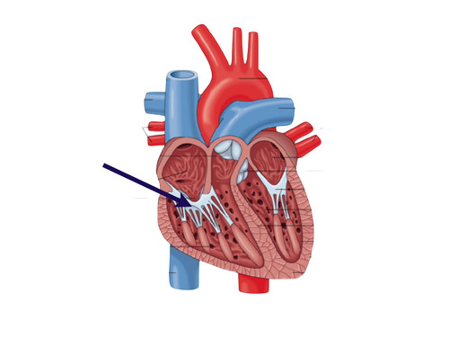
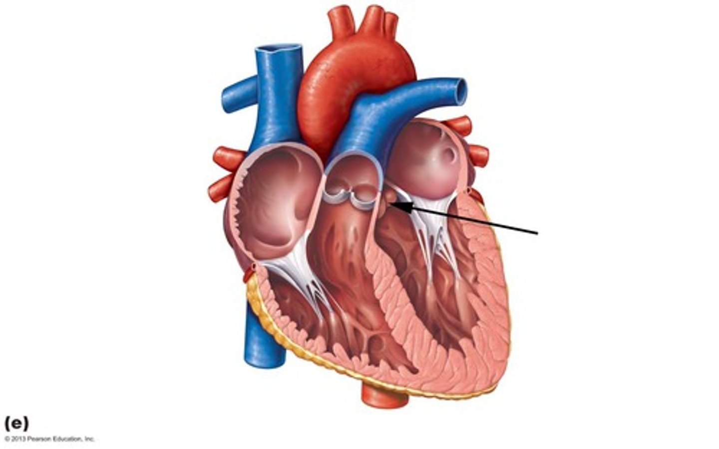
Tricuspid Valve
STRUCTURE
Probe through the right atrioventricular orifice
the door
Papillary Muscles
The carrot itself, thicker
st
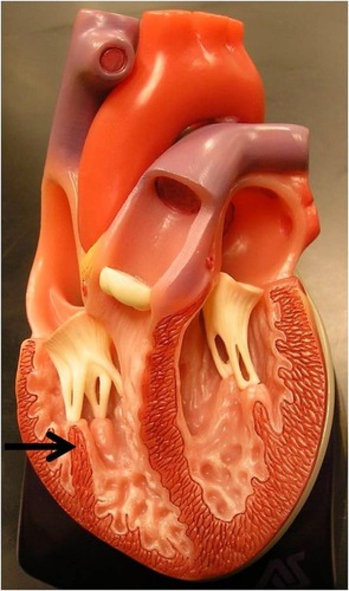
Tendinous Cords
Cords on papillary muscle
Greens of carrot
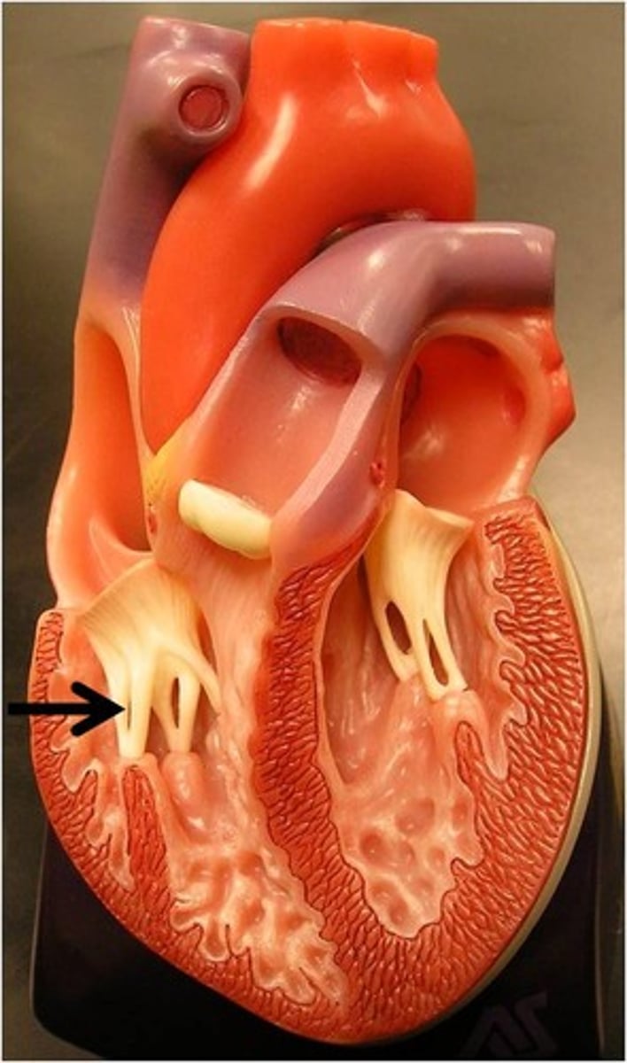
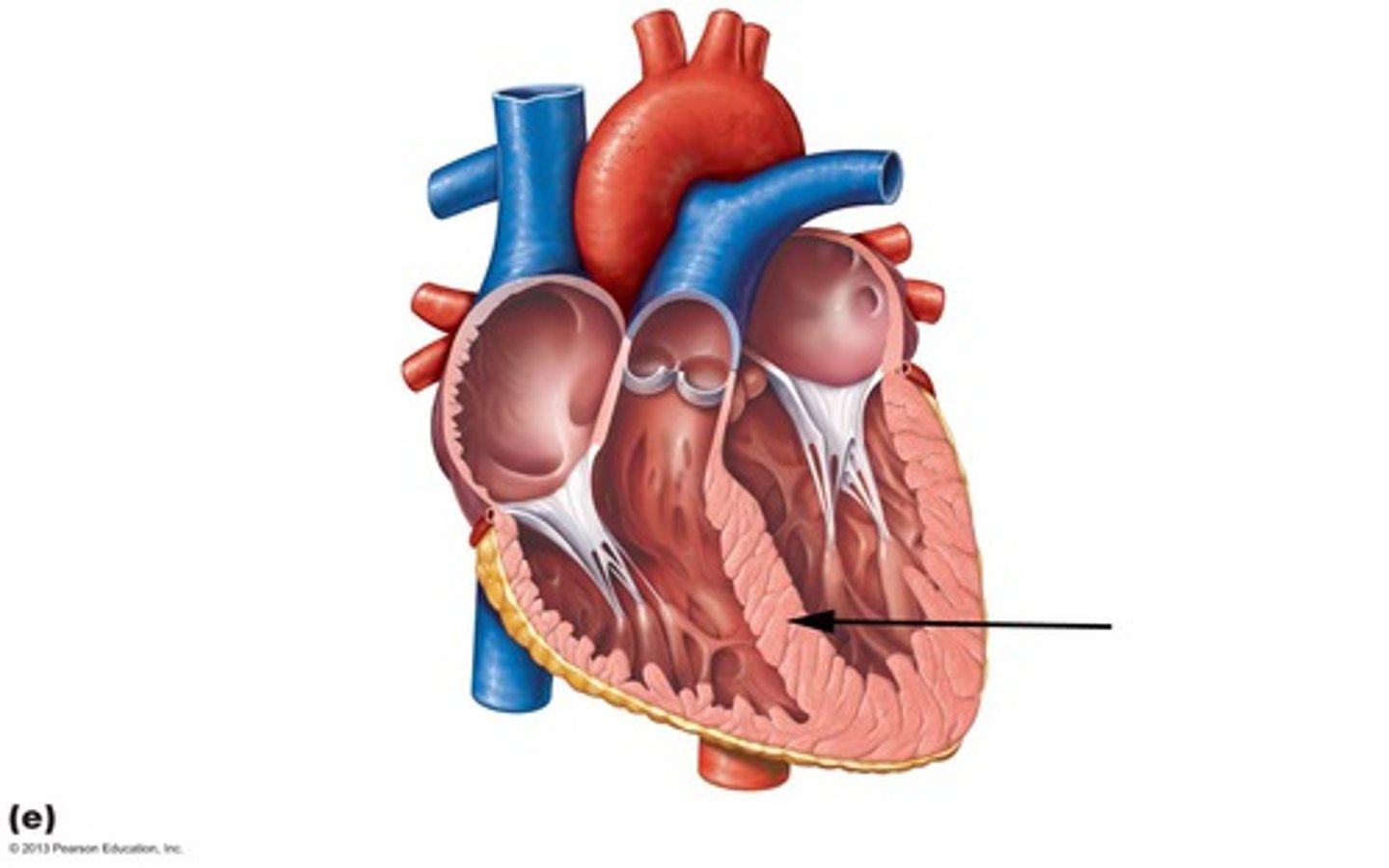
Trabeculae Carneae
Roots of the carrot/ the dirt
WALL of the right ventricle
Conus Arteriosus
Cone shaped narrowing
Opening of the Pulmonary Trunk
space
inside large hole on the non apex side of the heart

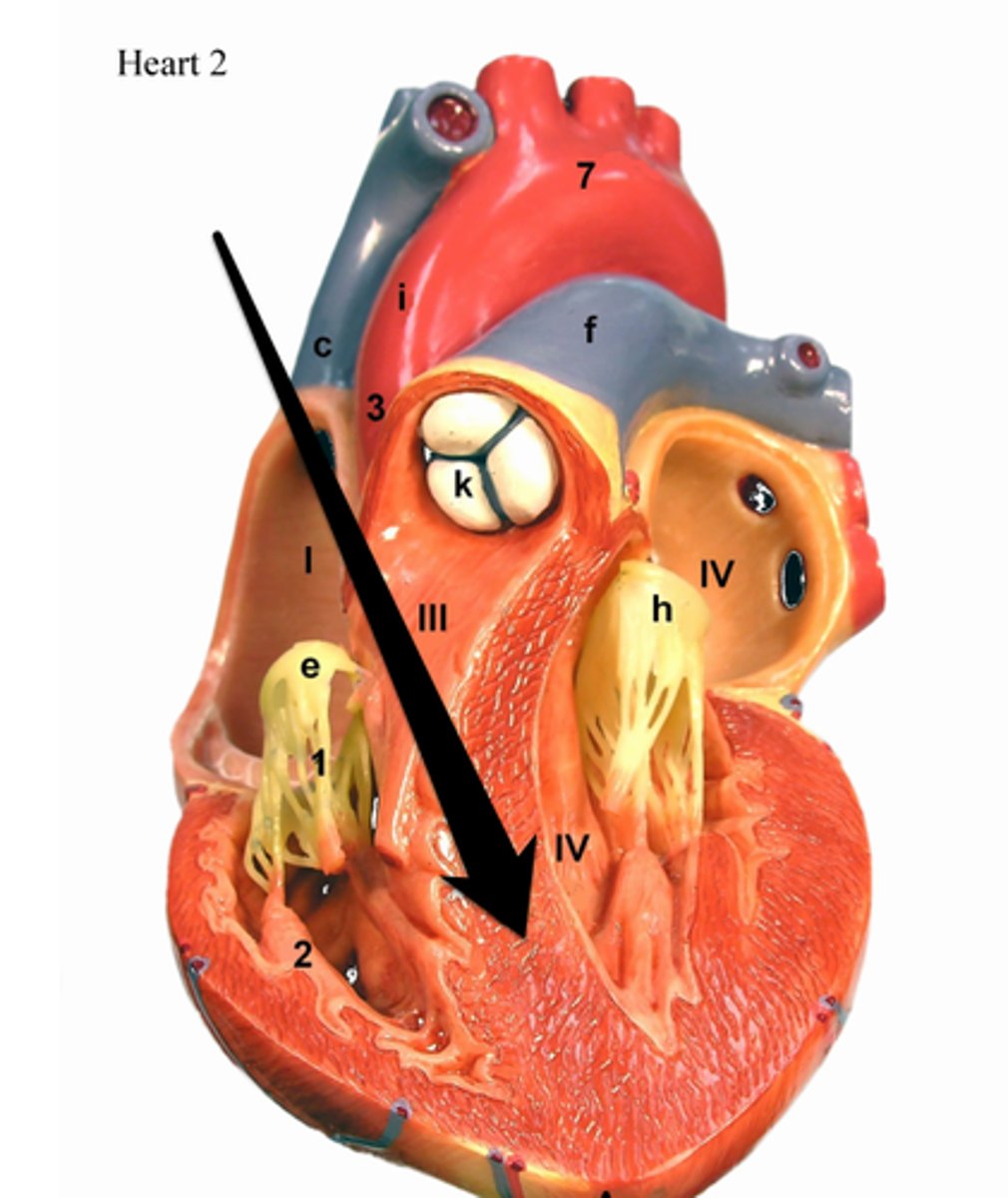
Pulmonary Valve
collective structure
opening of the space opened up to reveal
3 cusps
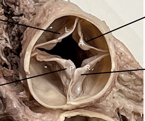
Semilunar Cusps of Pulmonary Valve
Components of the valve
Left Ventricle
chamber next to apex
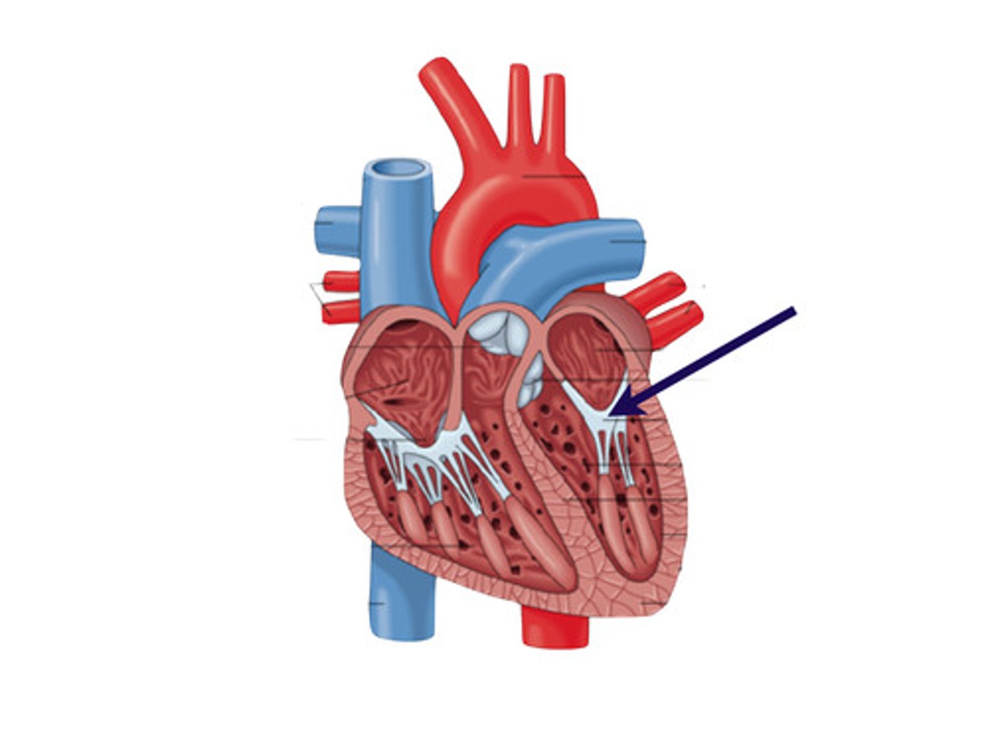
Left Atrioventricular Orifice
Space
reveals hole into left ventricle
will be seen sideways
Mitral Valve
st in left atrioventricular orifice
Aortic Vestibule
Narrowing
shiny
heart will be opened up
Opening of the Ascending Aorta
Whole space
Includes aortic vestibule
heart opened up
Aortic Valve
collective structure
heart opened up like book
Semilunar Cusps of the Aortic Valve
Components of the aortic valve

right atrium,
right ventricle,
PULMONARY artery,
LUNGS,
left atrium,
left ventricle,
AORTA,
REST OF BODY
flow of blood
where is blood oxygenated + deoxygenated
deoxygenated- vena cava, right atrium, tricuspid valve, right ventricle, pulmonary valve, pulmonary artery, lungs
oxygenated- left atrium, mitral valve, left ventricle, aortic valve, aorta, rest of body
gonadal arteries
in females these are the ovarian arteries
in males they are the testicular arteries
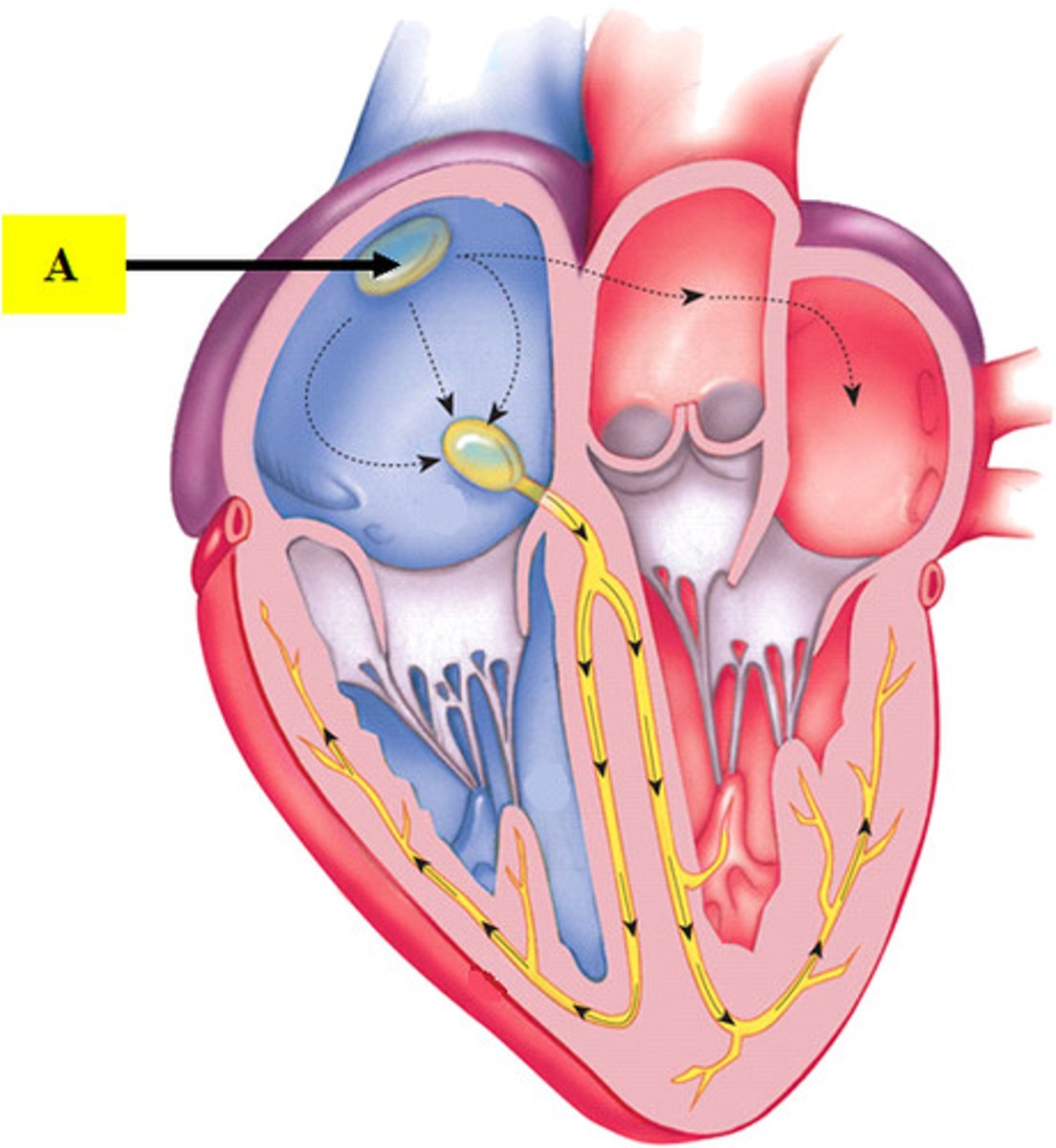
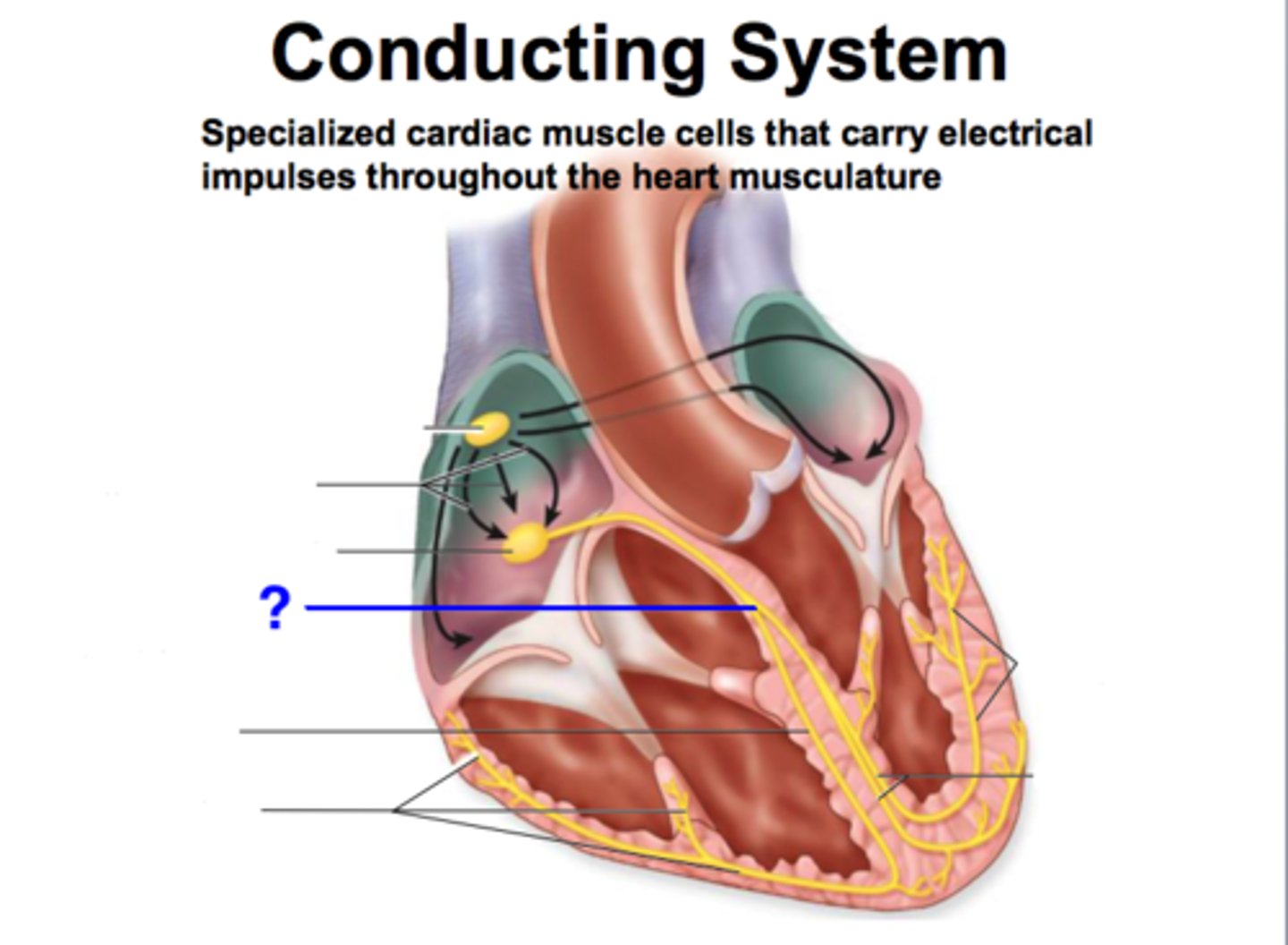
Circulation of Blood Through the Heart
Veins -> superior/inferior vena cava -> right atrium -> right ventricle (via tricuspid valve) -> pulmonary trunk (via pulmonary valve) -> left/right pulmonary arteries -> lungs -> pulmonary veins -> left atrium -> left ventricle (via mitral valve) -> aorta (via aortic valve) -> arteries -> capillaries
When Blood is Deoxygenated in Circulation Steps
Steps from veins to pulmonary arteries
Difference Between Arteries and Veins
Arteries: round, away from heart, lighter in cadaver
Veins: flat, toward heart, darker in cadaver