ANAPHY: LESSON 6 - NERVOUS SYSTEM
1/276
Earn XP
Description and Tags
AMO INI AN USA HA MAKURI NA LESSON
Name | Mastery | Learn | Test | Matching | Spaced | Call with Kai |
|---|
No analytics yet
Send a link to your students to track their progress
277 Terms
Receiving sensory input
Integrating information
Controlling muscles and glands
Controlling muscles and glands
Establishing and maintaining mental activity
major functions of the nervous system
Receiving sensory input
Sensory receptors monitor numerous external and internal stimuli. We are aware of sensations from some stimuli and other stimuli that are processed at a subconscious level.
Integrating information
The brain and spinal cord process sensory input and initiate responses, which can be immediate, stored as memory, or ignored.
Controlling muscles and glands
The nervous system controls skeletal muscles, cardiac muscle, smooth muscle, and glands, allowing it to control movements and bodily functions.
Maintaining homeostasis
The nervous system helps maintain a constant internal environment by detecting, interpreting, and responding to changes in internal and external conditions.
Establishing and maintaining mental activity
The brain is responsible for mental activities such as consciousness, memory, and thinking.
central nervous system
peripheral nervous system
nervous system can be divided into two major divisions:
Central Nervous System (CNS)
consists of the brain and spinal cord, which are major organs for processing sensory input and initiating responses.
Peripheral Nervous System (PNS)
consists of all the nervous tissue outside the CNS, including nerves and ganglia
Peripheral Nervous System (PNS)
functions to link the CNS with various parts of the body.
Sensory Division
Motor Division
Peripheral Nervous System (PNS) can be subdivided into two parts
Sensory division
or afferent division
Sensory division
conducts action potentials from sensory receptors to the CNS.
sensory neurons
neurons that transmit action potentials from the periphery to the CNS.
Motor division
or efferent division
Motor division
conducts action potentials from the CNS to effector organs, such as muscles and glands.
motor neurons
neurons that transmit action potentials from the CNS toward the periphery
Somatic nervous system
Autonomic nervous system
motor division can be further subdivided based on the type of effector being innervated:
Somatic nervous system
transmits action potentials from the CNS to skeletal muscles.
Autonomic nervous system (ANS)
transmits action potentials from the CNS to cardiac muscle, smooth muscle, and glands.
sympathetic divisions
parasympathetic divisions
autonomic nervous system is divided into
Sympathetic division
the division of the ANS that prepares the body for stressful situations and activates the "fight or flight" response.
Parasympathetic division
The division of the ANS that promotes relaxation and conserves energy.
Enteric nervous system (ENS)
is a unique subdivision of the peripheral nervous system. It has both sensory and motor neurons contained wholly within the digestive tract.
Enteric nervous system (ENS)
It can function without input from the CNS or other parts of the PNS, although it is normally integrated with the CNS by sensory neurons and ANS motor neurons.
Neurons
or nerve cells that receive stimuli, conduct action potentials, and transmit signals to other neurons or effector organs.
Cell body
dendrites
Axon
Parts of neurons
cell body
contains a single nucleus. It is the source of information for gene expression.
Dendrites
are short, often highly branching cytoplasmic extensions that are tapered from their bases at the neuron cell body to their tips.
Dendrites
usually receive information from other neurons or from sensory receptors and transmit the information toward the neuron cell body.
axon
a single long cell process extending from the neuron cell body.
axon hillock
The area where the axon leaves the neuron cell body.
collateral axons
branches of axons
myelin sheath
Axons can be surrounded by a highly specialized insulating layer of cells called the
Multipolar neurons
Bipolar neurons
Pseudo-unipolar neurons
Types of Neurons
Multipolar neurons
have many dendrites and a single axon. This kind of neurons are mostly found within the CNS and nearly all motor neurons.
Bipolar neurons
have two processes: one dendrite and one axon. They are located in some sensory organs
Pseudo-unipolar neurons
have a single process extending from the cell body. It appears to have a single axon. Most other sensory neurons are made out of this kind of neurons.
Pseudo-unipolar neurons
This neuron divides into two processes: one process extends to the periphery, and the other extends to the CNS.
Glial cells
or neuroglia, are supportive cells of the CNS and PNS that do not conduct action potentials. They carry out different activities that enhance neuron function and maintain normal conditions within nervous tissue.
astrocytes
ependymal cells
microglia
oligodendrocytes
Types of glial cells in the central nervous system
Schwann cells
satellite cells
Types of glial cells in the peripheral nervous system
Astrocytes
are the major supporting cells in the CNS and play a role in signaling activity, forming the blood-brain barrier, and limiting damage to neural tissue.
blood-brain barrier
a permeability barrier between the blood and the CNS.
Ependymal cells
Ependymal cells line fluid-filled cavities within the CNS. It produce cerebrospinal fluid, and help move it through the CNS.
Microglia
Microglia act as immune cells of the CNS, protecting the brain by removing bacteria and cell debris.
Oligodendrocytes
In the CNS, it provide an insulating material that surrounds axons
Schwann cells
In the PNS, it provide an insulating material that surrounds axons
Satellite Cells
found around the cell bodies of certain neurons of the PNS. It provides support and nutrition to the neurons and protects it from heavy-metal poison.
Myelin sheaths
are specialized layers formed by oligodendrocytes in the CNS and Schwann cells in the PNS that wrap around axons, preventing ion movement and increasing the speed of action potentials.
myelinated axons
Axons with these myelin sheaths
Gaps in the myelin sheath
nodes of Ranvier
Unmyelinated axons
lack the myelin sheaths; however, these axons rest in indentations of the oligodendrocytes in the CNS and the Schwann cells in the PNS
Gray matter
consists of groups of neuron cell bodies and dendrites, with very little myelin, found in the cortex of the brain and nuclei within the brain.
cortex
in the CNS, it is the gray matter on the surface of the brain
nuclei
in the CNS, it is the clusters of gray matter located deeper within the brain
ganglion
In the PNS, it is a cluster of neuron cell bodies
White matter
Consists of bundles of parallel axons with myelin sheaths, forming nerve tracts or conduction pathways.
nerve tracts
or conduction pathways, which propagate action potentials from one area of the CNS to another.
nerves
In the PNS, they are formed through the bundles of axons and associated connective tissue
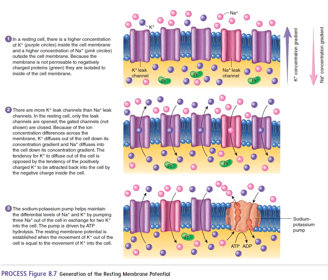
Resting membrane potential
The uneven distribution of charge across the cell membrane, with the inside being negatively charged compared to the outside.
Leak channels
Ion channels that are always open, allowing ions to "leak" across the membrane down their concentration gradient.
Gated channels
Ion channels that are closed until opened by specific signals
voltage-gated channels
opened by a change in membrane potential.
Generation of the Resting Membrane Potential
action potentials
action potential conduction
synapse
3 stages of neuron transmission
Action potential
A rapid change in the membrane potential of excitable cells, caused by the opening and closing of voltage-gated Na+ and K+ channels.
Action potential conduction
The propagation of action potentials along the cell membrane, either through continuous conduction in unmyelinated axons or saltatory conduction in myelinated axons.
continuous conduction
saltatory conduction
Pattern of action potential conduction along a neuron cell may occur in one of two ways:
continuous conduction
occurs in unmyelinated axons. An action potential in one part of a cell membrane stimulates local currents in adjacent parts of the cell membrane. The local currents in the adjacent membrane produce an action potential. The action potential is conducted along the entire axon cell membrane.
saltatory conduction
In myelinated axons, an action potential at one node of Ranvier causes a local current to flow through the surrounding extracellular fluid and through the cytoplasm of the axon to the next node, stimulating an action potential at that node of Ranvier. By this means, action potentials “jump” from one node of Ranvier to the next along the length of the axon.
Synapse
A junction where the axon of one neuron interacts with another neuron or with cells of an effector organ, such as a muscle or gland.
presynaptic terminal
postsynaptic membrane
synaptic cleft
3 major components of the structure if a synapse
Presynaptic terminal
The end of the axon that forms a synapse, where neurotransmitters are released.
Postsynaptic membrane
The membrane of the dendrite or effector cell that receives the neurotransmitters from the presynaptic terminal.
Synaptic cleft
The space separating the presynaptic and postsynaptic membranes.
Neurotransmitters
Chemical substances stored in synaptic vesicles in the presynaptic terminal, released into the synaptic cleft to bind to specific receptor molecules on the postsynaptic membrane.
Neurotransmitter
Chemical substances that transmit signals across synapses between neurons.
Hyperpolarized
A state in which the membrane potential of a cell becomes more negative than the resting membrane potential, inhibiting the occurrence of an action potential.
Acetylcholine (ACh)
A neurotransmitter substance involved in synaptic transmission, particularly in the neuromuscular junction.
Norepinephrine
A neurotransmitter substance that can be actively transported back into the presynaptic terminal or broken down by enzymes.
Acetylcholinesterase
An enzyme that breaks down acetylcholine, particularly in synapses where acetylcholine is the neurotransmitter.
Converging pathway
Diverging pathway
2 simplest neuronal pathways
Converging pathway
A neuronal pathway in which two or more neurons synapse with the same neuron, allowing information from multiple pathways to converge into a single pathway.
Diverging pathway
A neuronal pathway in which the axon from one neuron divides and synapses with more than one other neuron, allowing information from one pathway to diverge into multiple pathways.
Summation
The process of integrating multiple sub-threshold local potentials in neuronal pathways to bring the membrane potential to threshold and trigger an action potential.
Spatial Summation
2. temporal summation
2 types of summation
Spatial summation
occurs when the local potentials originate from different locations on the postsynaptic neuron— for example, from converging pathways.
occurs when local potentials overlap in time. This can occur from a single input that fires rapidly, which allows the resulting local potentials to overlap briefly.
Temporal summation
Central Nervous System (CNS)
The brain and spinal cord, which are responsible for processing and coordinating information.
brain
housed within the skull
spinal cord
in the vertebral column
Peripheral Nervous System (PNS)
All the nerves and ganglia outside the brain and spinal cord that collect and relay information to the CNS and regulate activity in muscles and glands.
12 pairs of cranial nerves
31 pairs of spinal nerves
nerves of the PNS can be divided into two groups
extends from the foramen magnum at the base of the skull to the second lumbar vertebra
spinal cord
Spinal cord
The part of the CNS that is housed within the vertebral column and is responsible for relaying information between the body and the brain.
Cauda Equina
The collective term for the inferior end of the spinal cord and the spinal nerves exiting there, resembling a horse's tail.
Dorsal Columns
Ventral Columns
Lateral Columns
white matter in each half of the spinal cord is organized into three columns:
Dorsal Columns
The organized white matter columns in the spinal cord located on the posterior side.
Ventral Columns
The organized white matter columns in the spinal cord located on the anterior side.