Advanced Imaging - Nuclear Medicine
1/23
There's no tags or description
Looks like no tags are added yet.
Name | Mastery | Learn | Test | Matching | Spaced | Call with Kai |
|---|
No analytics yet
Send a link to your students to track their progress
24 Terms
Disease will often start at cellular level, with small physiological changes. Imaging modalities, such as radiography, ultrasound, CT and MRI can only detect ..
Changes later as the disease alters the anatomy of organs or tissues
Nuclear medicine allows detection of metabolic changes occurring ..
At cellular level
Nuclear medicines is routinely used in ..
The staging and evaluation of many diseases in humans, especially cancer and come neurodegenerative disorders - often used in horses to assist in diagnosis of lameness
PetScan can be useful in identifying things like ..
Recurring cancers or metastasis of cancers
Nuclear medicine will enable ..
New cellular changes to be identified - treatment can start easier - can identify those changes but won’t till you what those cellular changes are - additional investigation to assess if it was cancer cells or if it was just an infection or something similar
Applications in Veterinary Medicine
Abdominal abscesses
Thyroid disorders - can actually be used to treat hyperthyroidism
Cancer investigations - looking for any signs of metastases
Portosystemic shunt
Lameness (particularly in equine)
Exercise Induct Pulmonary Haemorrhage (EIPH) in competition horses
Nuclear Imaging techniques (3)
1 - Scintigraphy
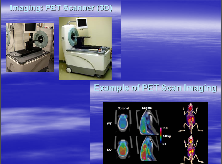
2 - Position Emission Tomography (PET) Scanning
3 - Single Positron Emission Computed Tomography (SPECT) Scanning
PET scans are often combined with either CT or MRI to show ..
Both anatomical and physiological changes side by side ‘fusion’ images
What does Nuclear Medicine Involve?
It is a non-invasive technique
The patient is dosed with a very small amount of ‘gamma ray emitting’ radioisotope (also known as a tracer)
The radioisotope may be injected, ingested or inhaled as appropriate for the study being performed
if looking at the lungs this would likely be inhaled, if it was the circulatory system this would be injected
Attach the tracer to something that has the affinity for the area of interest
The radioisotope is usually part of a larger molecule that has a specific affinity for the tissue or organ of interest. For instance ..
Some organic phosphonates (accumulate in bone) have an affinity for bone
Isotope bound to sulphur colloid will localise in the liver and spleen
Very few radioisotopes have direct affinity for a given tissue ..
Iodine is the notable exception and localises very strongly in the thyroid
Inhaled gases or aerosols localise in the ..
Airways and lungs and may or may not be absorbed into the bloodstream
In veterinary medicine, the most commonly used isotope is metastable technetium-99, although radioactive what can also be used in specific instances?
Iodine
Indium
Thallium
Once the radioisotope has been injected and travelled to the area of the interest. The radioisotope emits gamma radiation which can be imaged with a specialised machine called a Gamma camera. The radioisotope will collect in areas of higher chemical activity, this is helpful because ..
Certain tissues of the body, and certain diseases, have a higher level of chemical activity. These areas of disease will show up as ‘hot spots’
Difference between procedure - It is the TYPE of gamma camera used and how the image is generated that defines the different imaging modalities, i.e.
Scintigraphy used a 2D gamma camera
PET scanning used a 3D gamma camera (cross sectional images)
SPECT scanning also used a 3D camera (cross sectional images)
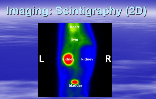
Imaging: Scintigraphy (2D)
Another image of Scintigraphy (2D)
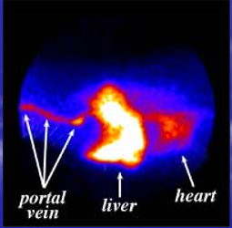
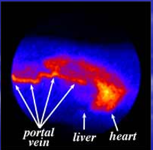
Scintigraphy Case Study - Portosystemic Shunt
Image - Composite image of a normal dog showing the uptake of radioactive dye into the portal vein and liver
image - Composite image showing the uptake of radioactive dye into the portal vein.Notice that the dye bypasses the liver and appears in the heart and lungs first
PET VS SPECT
Both use 3D gamma cameras
The main difference between SPECT and PET scans is the type of radiotracer used
PET Image
PET

SPECT Image
SPECT
Patient preparation - Companion animals
Starved overnight as sedation is usually required
Catheter placement in a peripheral vein away from the areas to be scanned
Disadvantages
Will show up a lesion only, not what TYPE of lesion - so not a definite diagnosis
False positives can occur
Dose to patient on average 3mSv (MPD = 20)
The major issue with using nuclear medicine imagine in veterinary medicines is not the affability of the gamma cameras, or the technical expertise required to operate them. Cameras are readily available on the used market, and training of technologists to operate them is not prohibitively complex. Rather it is ..
The regulations surrounding the acquisition and use of radiopharmaceuticals in animals. All use must be strictly documented and, unlike in human medicine, the veterinary patient generally must remain in the hospital after the study is performed to allow any elimination of radionuclides from the body to be essentially complete. This is done in order to limit exposure of owners to the radionuclides
Health and Safety
Use of radioactive isotopes as governed by the Radioactive Substances Act 1993
Licences must be obtained from the Environment Agency for such activity, one for keeping and using and another for accumulation and disposal
Local rules expected to be in place, must the same as with ionising radiation