Chapter 15 - The Spinal Cord: Meninges, Spinal Nerves, and Spinal Tracts
1/14
There's no tags or description
Looks like no tags are added yet.
Name | Mastery | Learn | Test | Matching | Spaced | Call with Kai |
|---|
No analytics yet
Send a link to your students to track their progress
15 Terms
The Spinal Cord
The spinal cord descends within the vertebral canal (bony canal formed by vertebrae)
Spinal cord and its covering meninges are separated from bony vertebrae by fat-filled epidural space
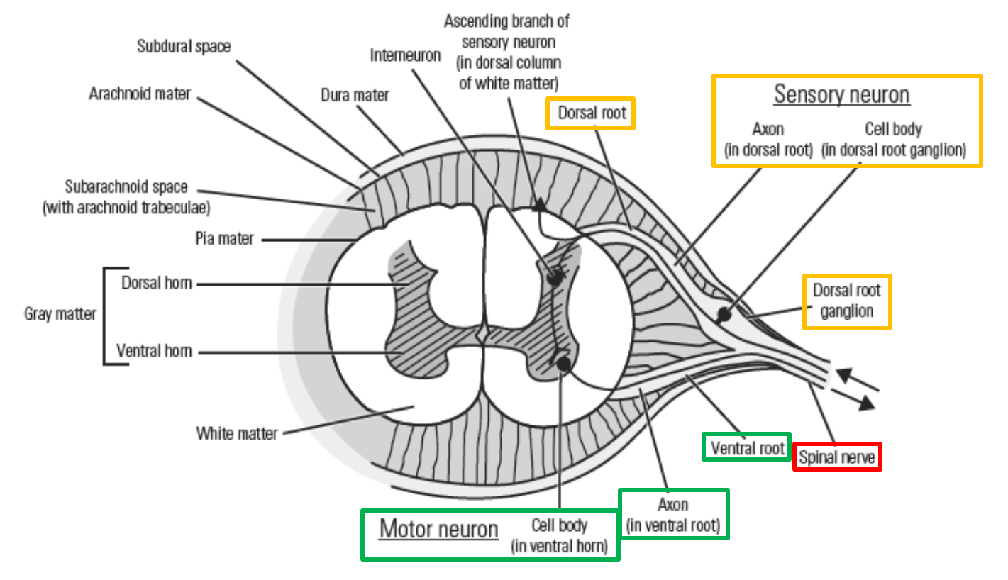
Spinal cord is surrounded by three layers of meninges:
Dura mater - outermost layer
Arachnoid mater
Pia mater - innermost layer
Dura mater
Fibrous, outermost meningeal layer
Separated from bony vertebrae by epidural space
Forms continuous tube around spinal cord
Anchored to margins of foramen magnum of skull - continuous with dura mater of cranial cavity
Extends laterally to cover each spinal nerve - anchored to bony margins of intervertebral foramen
Terminates inferiorly by attaching to coccyx
Arachnoid mater
Middle meningeal layer
Separated from dura mater by very thin subdural space
Subarachnoid space
Filled with cerebrospinal fluid - provides fluid cushion around spinal cord
Thin arachnoid trabeculae pass through space (between arachnoid mater and pia mater)
Pia mater
–Innermost meningeal layer
Separated from arachnoid mater by subarachnoid space
–Tightly adherent to outer surface of spinal cord
–Terminates as filum terminale
At inferior end of spinal cord, pia mater narrows to thin cord-like filum terminale that continues beyond end of spinal cord
Filum terminale runs inferiorly to attach to coccyx
Serves to anchor inferior end of spinal cord
Spinal Cord - Cross Section
Spinal cord is part of central nervous system
–Consists of gray matter and white matter
Gray matter - located centrally
→ Contains neuron cell bodies
White matter - located peripherally
→ Consists of neuronal axons
–Spinal cord enlargements
Gray matter
–Forms central, “H”-shaped region
–Divided into dorsal horn and ventral horn
–Dorsal horn
Located on dorsal (posterior) side of spinal cord
Receives axons from sensory neurons
–Ventral horn
Located on ventral (anterior) side of spinal cord
Contains cell bodies of motor neurons
White matter
–Peripherally located
–Consists of neuronal axons, running vertically (up or down spinal cord)
Tract - localized bundle of functionally related axons within white matter
Tracts contain descending motor axons (running downward, from brain to spinal cord) or ascending sensory axons (running upward, from body to brain)
Spinal cord enlargements
–Two regions of the spinal cord are enlarged (have a wider diameter) due to an increased number of neurons
More neurons required for innervation of the upper or lower limbs
–Cervical enlargement (C5-T1 spinal cord levels)
Gives rise to the brachial plexus for innervation of the upper limbs
–Lumbosacral enlargement (L1-S4 spinal cord levels)
Gives rise to the lumbar and sacral plexuses that innervate the lower limbs
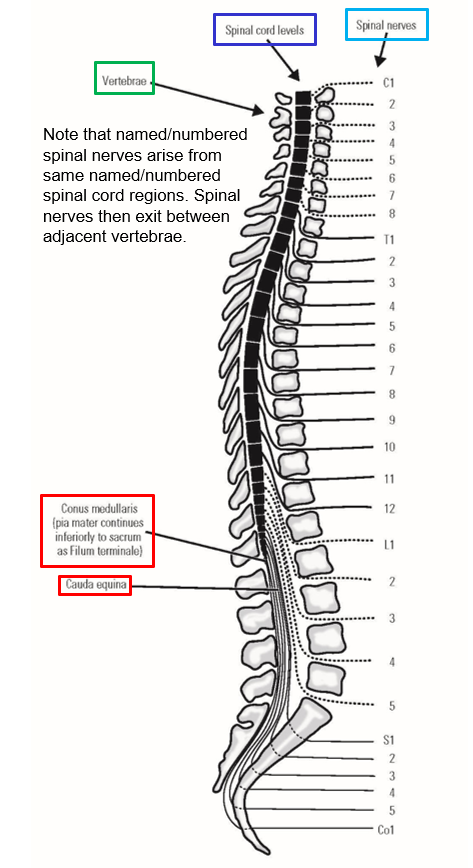
Spinal Nerves
Spinal nerves exit vertebral column between adjacent vertebrae via intervertebral foramen
–Nerves are named and numbered according to the vertebral level of their intervertebral foramen:
C1-C8 cervical spinal nerves
→ C1 spinal nerve exits between skull & C1 vertebra
→ C8 spinal nerve exits between C7 & T1 vertebrae
T1-T12 thoracic spinal nerves
L1-L5 lumbar spinal nerves
S1-S5 sacral spinal nerves
Co1 - single pair of coccygeal spinal nerves
–Spinal cord region that gives rise to each spinal nerve are also named and numbered in same manner
–Each spinal nerve is formed by the junction of a dorsal root and a ventral root
–Dorsal root
Structure that carries only axons of sensory (afferent) neurons into the spinal cord
Sensory neuron cell body located in dorsal root ganglion (associated with dorsal root, outside of the spinal cord)
Dorsal root located on dorsal (posterior) side of spinal cord
Sensory axons enter dorsal horn of spinal cord gray matter
–Ventral root
Structure that carries only axons of motor (efferent) neurons out of the spinal cord
Motor neuron cell body located in ventral horn of spinal cord gray matter
Ventral root exits on ventral (anterior) side of spinal cord
Mnemonic for spinal nerves: SAME DAVE
Sensory = Afferent
Motor = Efferent
Dorsal (root) = Afferent
Ventral (root) = Efferent
–Each spinal nerve is formed by the junction of a dorsal root and a ventral root
–Thus, each spinal nerve carries axons from both afferent neurons and efferent neurons
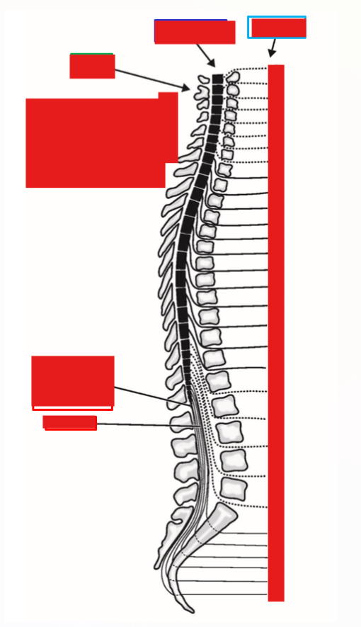
Spinal Cord - Longitudinal
Spinal cord extends inferiorly within vertebral column
Begins at foramen magnum of skull (continuous with medulla oblongata of brain stem)
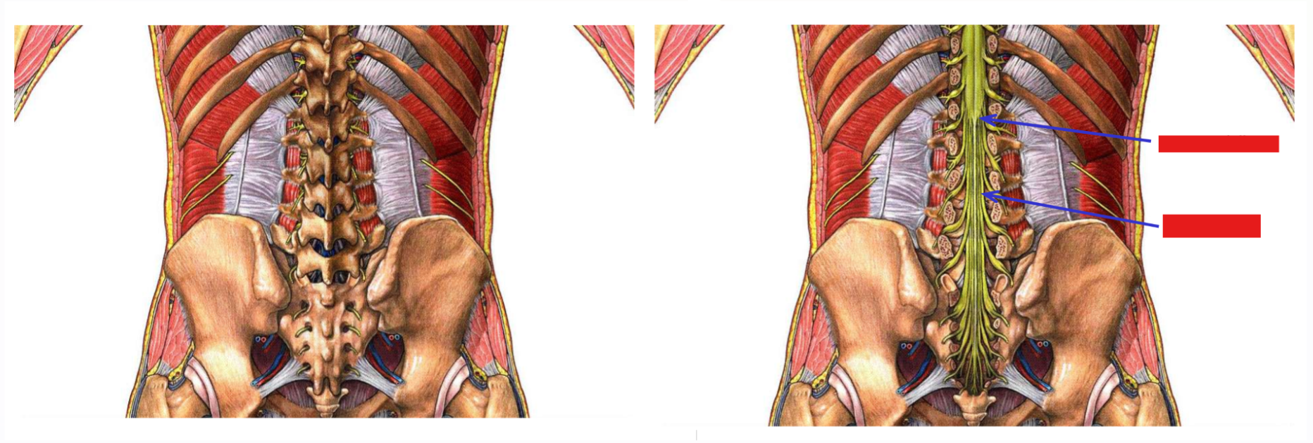
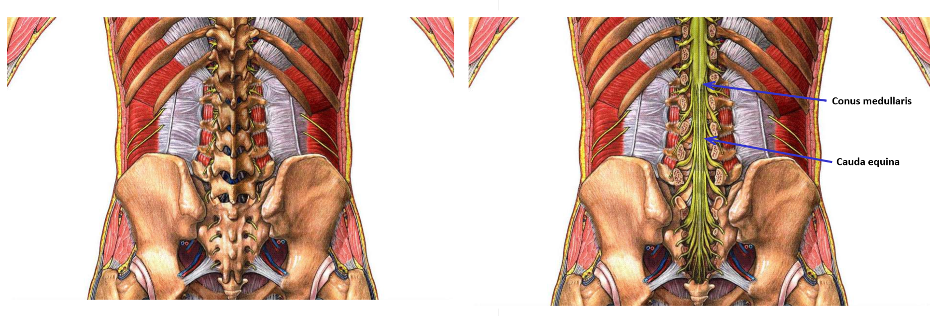
Conus medullaris - conical, terminal end of spinal cord
In adult, spinal cord terminates at L1-L2 vertebral level
Pia mater continues inferiorly as filum terminale
At birth, spinal cord and vertebral column are same length (spinal cord extends full length of vertebral column)
During childhood growth, spinal cord reaches adult size early
Vertebral column continues to grow in length
In adult, spinal cord does not extend full length of vertebral column
Conus medullaris located at L1-L2 vertebral level
Each spinal nerve still exits through its intervertebral foramen
In adult, only upper cervical nerves run laterally (from spinal cord to intervertebral foramen)
Other spinal nerves must descend before exiting vertebral column
Cauda equina
Large collection of descending spinal nerves located inferior to termination of spinal cord (below L2 vertebral level)