The heart and circulation in animals
1/12
There's no tags or description
Looks like no tags are added yet.
Name | Mastery | Learn | Test | Matching | Spaced |
|---|
No study sessions yet.
13 Terms
Explain the features that animal transport systems have
a suitable medium in which to carry materials
A pump eg. The heart for loving the blood
Valves to maintain one-directional bloood flow
Some also have
a respiratory pigment like in vertebrates and some invertebrates which increases volume of o2 that can be transported eg. Haemoglobin
A system of vessels with a branching network to distribute the transport medium to all parts of the body
Explain open circu systems
Open circulatory is when blood doesn’t move around the body in blood but it bathed the tissues directly while held in a cavity called the haemocoel.
An example is insects which have a long, dorsal, tube shaped heart that runs the length of their bodies.
It pumps blood out at low pressure to the haemocoel where materials are exchanged between blood and body cells. The blood then returns slowly to the heart and the circuit starts again
There is no respiratory pigment because oxygen diffuses directly from trachea and in a completely different system so the blood doesn’t contain oxygen only excretory products and nutrients
Explain the closed circulatory system
Closed circuiting- The blood moves in blood vessels. There are two types of close circulatory systems
Single circulation - eg. Fish and earthworm
in single circulation the blood moves through the heart once in its passage around the body
in a earthworm - blood moves forward in the dorsal vessel and back in the ventral vessel. 5 pairs of ‘pseudo hearts’ (thickened, muscular blood vessels) pump the blood from dorsal to ventral and keep it moving
In fish - the one ventricle of the heart pumps deoxygenated blood to the gills where its pressure falls. Oxygenated blood is carried to the tissues and from there deoxygenated blood returns to the one atria of the heart and circulation begins again. They have a low metabolic rate so okay
Double - In double circulation the blood passes through the heart twice in its circuit around the body - eg. Mammals
blood is pumped by a muscular heart at high pressure by giving it a rapid flow rate through blood vessels. Organs aren’t in direct contact with the blood but are bathed by tissues fluid which seeps out of the capillaries. Haemoglobin carries o2. Blood pressure is reduced in the lungs and it’s pressure would be low low to make the circuit round the hole body so it returns to the heart which raises pressure and generated again to supple materials to the rest of the body
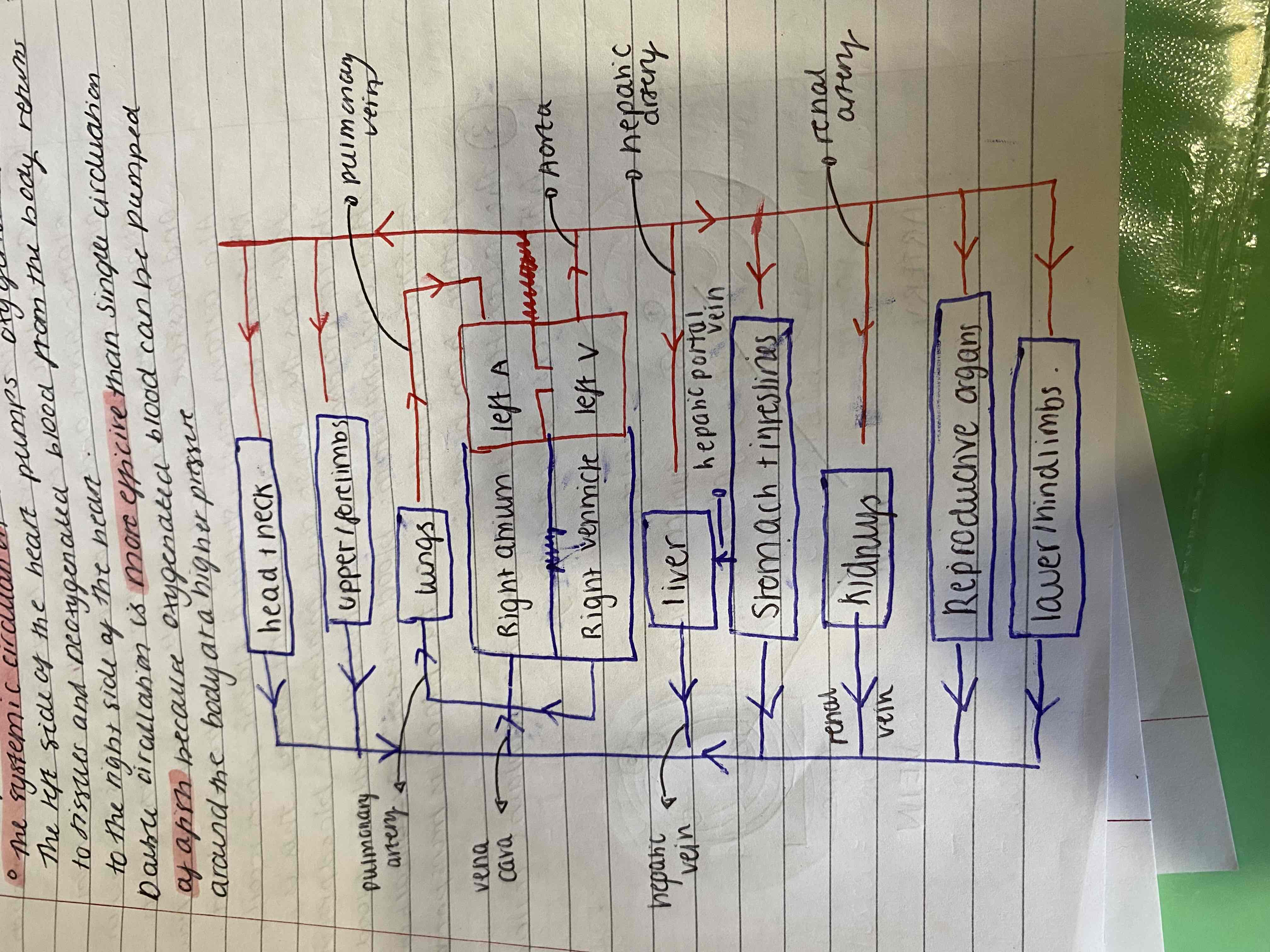
Double circulation is more effective than single circulation of a fish because oxygenated blood can be pumped around the body at a higher pressure
Describe the double circulation system
It can be described as two systems
Pulmonary circulation - serves the lungs. The right side of the heart pumps deoxygenated blood to the lungs and oxygenated blood returns to the left side of the heart
Systemic circulation- served the body respiring tissues. The left side of the heart pumps oxygenated blood to the body tissues and deoxygenated blood from the body returns to the right side of the heart
explain blood vessels - arteries
Structure - there is a three layered structure
The inner most later is the ENDOTHELIUM which is one cell thick and is a smooth lining to reduce friction and minimal resistance to blood flow
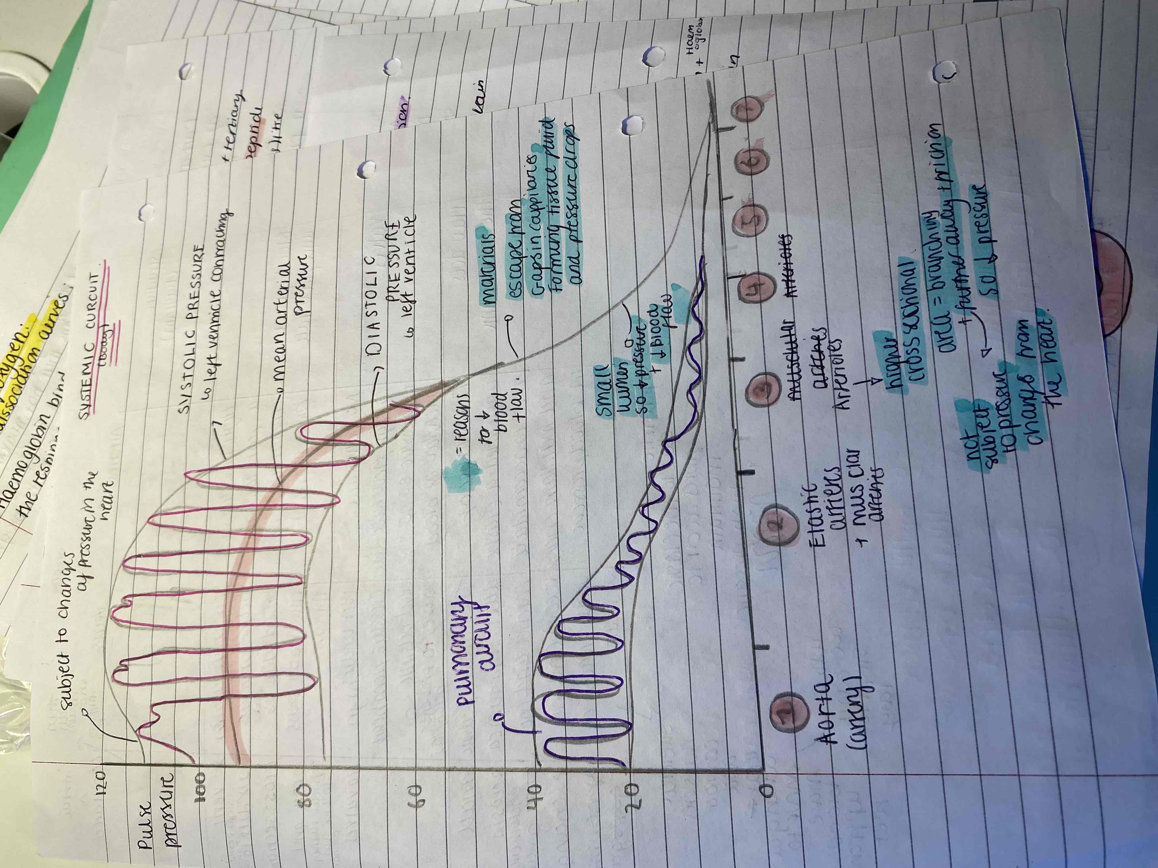
The middle layer is the TUNIC MEDIA which is the thickest and contains elastic fibres which allow stretching to accommodate changes in blood flow and pressure as the blood is pumped from heart. At a certain point the stretches elastic fibres recoil due to elastic potential energy this pushes blood through artery. This is felt as the pulse and maintains blood pressure. The tunica media also contains smooth muscle which contracts to regulate blood flow and pressure as the blood is transited further from heart. - PULSITILE
The outer layer is the TUNICA EXTERNA - contains collagen and fibres to resist overstretching
The size of the linen is far smaller to support the high pressure derived from the heartof the blood pumping around the whole body
no valves due to high blood pressure so no back flow
Function - they carry blood away from the heart and branch into smaller vessels called arterioles that further subdivide into capillaries
They have a high pressure and fast flow rate maintained by pressure
Explain the blood vessels - veins
Structure - they have the same structure as arteries however they have a larger diameter lumen and thinner less muscular walls of tunica media so the pressure is low to reduce resistance and increase blood flow which is slow due to not pusitile and not under influence of the heart (so the tunica media can’t recoil). So to get blood back up the sketetal muscles contract and relax when you move to move blood up to heart (why you can’t sit for too long)
for veins above the heart blood returns to the Heart by gravity as it moves through other veins by the pressure from surrounding muscles. Veins have semi-lunar valves to endure one-directional blood flow and no back flow of blood. - faulty valves can result in varicose veins and heart failure.
Functions - They return blood usually deoxygenated to the heart.
Explain blood vessels - capillaries
Structure - They form a vast network of cell cell thick endothelium capillaries. They have pores between cells make the capillaries permeable to water and solutes eg. glucose. they have a smaller lumen diameter and cross sectional area is high to increase time for material exchange so rate of blow flow is slow. There are so many capillaries in a capillary bed that reducing the rate doesn’t effect.
Function - Pernitrates all tissues and organs. Blood from the capillaries collects into venules which take blood into veins which return it to the heart. The pore mean that they can exchange materials into cells.
Explain the structures and functions of the heart and the path of one circuit
Structure - It is made of cardiac muscle which is a specialized tissue with myogenic contraction. There are two atria on top of two (pumping chambers) ventricles to completely separate o2 and not o2 blood to not contaminate the o2 blood. The heart also contains chordae tendinea to prevent the heart turning inside out during low pressure in diastole
Functions - it is a pump to circulate blood. One pump deals with oxygenated blood and one pump deals with deoxygenated blood. It is myogenic which means that heartbeat is not under nervous control and it is initiated within the muscle. However the heartrate can be modified by nervous and hormonal stimulation. It also never tires.
Circuit - vena cava (superior and inferior), semi-lunar valves, right atrium, Atria-ventricular tricuspid valve, right ventricle, semi-lunar valve, pulmonary artery, lungs, pulmonary vein, semi-lunar valve, left atrium, atria-ventricular valve bicuspid, left ventricle, semi -lunar valve, aorta, body
What is the cardiac cycle and briefly outline the stages
The cardiac cycle describes the sequence of events in one heart beat which normally lasts in an adults heart for 0.8 seconds.
It consist of alternating contractions (systole) and relaxations (diastole) that change volume in the heart to change pressure to change blow flow rate
The three stages are
Atrial systole - 0-1.8 secs
ventricular systole - 1.8-0.4 secs
diastole - 0.4-0.8 secs
explain each stage of the cardiac cycle
Atrial systole - atria walls contract so blood volume decrease so blood pressure in atria increases. This pushes blood into the relaxed ventricles - lower pressure- via AV valves
Ventricular systole - Th ventricle walls contract and this decreases volume so pressure increases which forces blood up through semi-lunar valves out of the heart and into pulmonary artery and aorta. AV valves are closed due to rise in ventricular pressure in front building up.
Diastole - the ventricles relax and volume in the ventricles increases so pressure falls. Semi lunar valves close to prevent backflow of blood from aorta and pulmonary artery. The atria also relax during diastole so blood from the vena cava can enter because of increased volume and decreased pressure.
Explain the stages of the flow of blood in the cardiac cycle on the left side of the heart and how that relates to the heart in general
The left side of the atria relaxes and revived o2 ated blood from pulmonary artery.
When full this closes the bicuspid valve
Relaxation of left ventricle draws blood from left atrium at high pressure to low pressure in left ventricle
Left atrium contracts pushing remaining blood into ventricle
With the left atrium relaxed and biscuspid valve shut left ventricle contracts and its strong muscular walls exert a large pressure.
This pressure pushes the blood up and out the heart through semi lunar valves into aorta and closes semi lunar
The two sides of the heart work together and the atria contact rhythmically with each other and the ventricles.
A complete contraction is called a heartbeat
When they contract it’s empty and full is relaxed
Left Ventricle Have more muscle as need to pump to rest of body and tissues and ventricles in general have more muscle as pump to other parts of body like lungs
Explain the purpose of valves and the valve graph with relation to pressure
They prevent back flow.
They close if the pressure I front of them is higher than behind and open when pressure is higher behind than in front
Semi lunar and AV bicuspid and tricuspid are the two types in heart
Eg. If atrium pressure was lower than pressure in ventricle the bicuspid valve would close

Explain the graphs for pressure changes in systemic circuit and pulmonary circuit in terms of blood vessels