RAD Safety Part 1/4
1/59
There's no tags or description
Looks like no tags are added yet.
Name | Mastery | Learn | Test | Matching | Spaced |
|---|
No study sessions yet.
60 Terms
What type of personnel radiation monitoring device has a gas-filled ionization chamber and provides a reading at any time via Internet connection?
A. Pocket dosimeter
B. Direct ion storage dosimeter
C. Optically stimulated luminescence dosimeter
D. Thermoluminescent dosimeter
(B) The newest type of personnel monitoring device is the direct ion storage dosimeter (DIS); it is a digital ionization dosimeter. The DIS eliminates the need to collect and send dosimeters for monthly or quarterly processing. The user wears the DIS, which looks like a small flash drive, in the same way as other monitors such as an OSL (optically stimulated luminescence) dosimeter. The DIS has a gas-filled ionization chamber within and uses Bluetooth technology to relate its raw data via mobile device or any computer with Internet access and a USB connection. Occupational exposure can be read, and reread, at any time without loss of information.
The pocket dosimeter, or pocket ionization chamber, resembles a penlight and has a thimble ionization chamber within. Ions are counted and radiation quantity is registered in milliroentgens (mR). The use of the pocket dosimeter is indicated when working with high exposures or large quantities of radiation for short periods of time, so that an immediate reading is available to the user. The pocket dosimeter is sensitive and accurate but has limited application in diagnostic radiography.
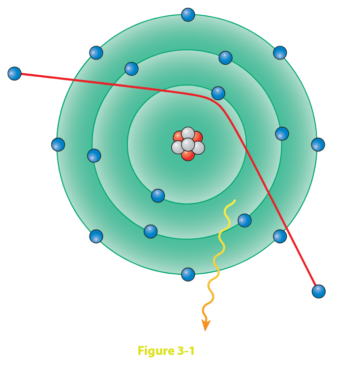
Figure 3-1 is representative of:
A. the production of Compton scatter
B. the photoelectric effect
C. the production of Bremsstrahlung x-ray photons
D. the production of characteristic x-rays
(C) Bremsstrahlung (braking or Brems) radiation is one of the two types of x-ray photons produced at the x-ray tube tungsten target. When high-speed electrons coming from the cathode filament pass near or through a tungsten atom, they can be attracted by the positively charged nucleus, slowed down/braked, and deflected from their course with a resulting loss of energy.
This energy is given up in the form of an x-ray photon.
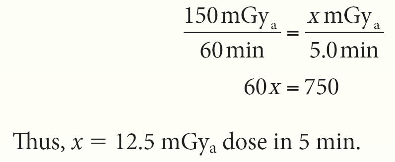
A time of 5 min is required for a particular fluoroscopic examination, whose exposure rate is 150 mGyₐ/h. What is the approximate radiation exposure for the radiologic staff present in the fluoroscopy room during the examination?
A. 75 mGyₐ
B. 37.5 mGyₐ
C. 12.5 mGyₐ
D. 3.75 mGyₐ
(C) If the exposure rate for the examination is 150 mGyₐ/h (60 min), then a 5-min examination would be proportionally less—as the following equation illustrates:
Stochastic/probabilistic effects of radiation are those that:
have a threshold
may be described as “all-or-nothing” effects
are late effects
A. 1 only
B. 1 and 2 only
C. 2 and 3 only
D. 1, 2, and 3
(C) Late effects of radiation can occur in cells that have survived a previous irradiation months or years earlier. These late effects, such as carcinogenesis and genetic effects, are “all-or-nothing” effects—either the organism develops cancer or it does not.
Most late effects do not have a threshold dose; that is, any dose, however small, theoretically can induce an effect. Increasing that dose will increase the likelihood of the occurrence but will not affect its severity; these effects are termed stochastic/probabilistic.
Nonstochastic/deterministic effects are those that will not occur below a particular threshold dose and that increase in severity as the dose increases.
The uppermost collimator shutter functions to eliminate:
A. off-focus radiation
B. leakage radiation
C. scatter radiation
D. primary radiation
(A) The collimator is the most practical and efficient beam-restricting device; the collimator box is attached to the tube head. Adjustable lead shutter collimators are used to define the size and shape of the x-ray field that emerge from the x-ray tube port window.
The fixed diaphragm located just outside the x-ray tube’s port window functions to significantly reduce the effect of off-focus radiation. Off-focus (extrafocal or stem) radiation is produced when electrons strike surfaces other than the focal track.
The next set/stage of lead shutters consists of two pairs of adjustable shutters—one pair for field length and another pair for field width. These shutters are used to regulate the length and width of the irradiated field.
Somatic effects of radiation refer to effects that are manifested:
A. in the descendants of the exposed individual
B. during the life of the exposed individual
C. in the exposed individual and his or her descendants
D. in the reproductive cells of the exposed individual
(B) Somatic effects of radiation refer to those effects experienced directly by the exposed individual, such as erythema, epilation, and cataracts. Genetic effects of radiation exposure are caused by irradiation of the reproductive cells of the exposed individual and are transmitted from one generation to the next.
Guidelines for the use of protective shielding state that gonadal shielding should be used:
if the patient has reasonable reproductive potential
when the gonads are within 5 cm of the collimated field
when tight collimation is not possible
A. 1 only
B. 1 and 2 only
C. 1 and 3 only
D. 2 and 3 only
(B) It is our professional responsibility to minimize exposure dose to both patients and ourselves, and one of the most important ways is with a closely collimated radiation field. Gonadal shielding should be used when the patient is of reproductive age or younger, when the gonads are in or within 5 cm of the collimated field, and when the clinical objectives will not be compromised.
The skin response to radiation exposure, which appears as reddening of the irradiated skin area, is known as:
A. dry desquamation
B. moist desquamation
C. erythema
D. epilation
(C) The first noticeable skin response to excessive irradiation would be erythema, a reddening of the skin very much like sunburn. Dry desquamation, a dry peeling of the skin, may follow. Moist desquamation is peeling with associated pus-like fluid.
Epilation, or hair loss, may be temporary or permanent depending on sensitivity and dose.
Increasing field size, while leaving technical factors unchanged, will:
A. decrease the DAP
B. decrease SR production
C. increase the DAP
D. increase the EFS size
(C) Dose area product (DAP) expresses the dose of radiation to a particular volume of tissue, thereby being a potentially better indicator of risk than dose values alone. DAP is expressed in terms of cGy-cm².
An increased field size will increase the DAP even if the technical factors (dose) remain unchanged. As field size decreases, the amount of exposed tissue decreases, and DAP is decreased.
DAP can be monitored using a DAP meter in both radiographic and fluoroscopic procedures. The DAP meter is radiolucent and is mounted just below the radiographic collimator, measuring x-radiation before it reaches the part.
Skin dose can be determined by dividing the skin area exposed by the DAP measurement. This value represents potential deterministic effect to that tissue. Increasing field size increases the production of scattered radiation. Focal spot size is unrelated to dose.
The single most important source of scattered radiation in both radiography and fluoroscopy is the:
A. x-ray table
B. x-ray tube
C. patient
D. IR
(C) The patient, as the first scatterer, is the most important scatterer. At 1 m from the patient, the intensity of the scattered beam is 0.1% of the intensity of the primary beam.
Compton scatter emerging from the patient is almost as energetic as the primary beam entering the patient.
Which acute radiation syndrome requires the largest exposure before the associated effects become apparent?
A. Hematopoietic
B. Gastrointestinal
C. Cerebrovascular
D. Skeletal
(C) Radiation effects that appear days or weeks following exposure (early effects) are in response to high radiation doses; this is called acute radiation syndrome. These effects should never occur in diagnostic radiology; they occur only in response to much greater, usually whole-body, doses.
Sufficient exposure of the hematologic system to ionizing radiation can result in nausea, vomiting, diarrhea, decreased blood cell count, and infection. Very large exposure of the GI system (6–10 Gy) causes severe damage to the (stem) cells lining the GI tract.
This can result in nausea, vomiting, diarrhea, blood changes, and hemorrhage. Exposure greater than 50 Gy is required to cause cerebrovascular syndrome, affecting the normally resilient CNS and cardiovascular systems.
How are kV and HVL related?
A. Directly
B. Inversely
C. Inverse squared
D. Direct squared
(A) HVL is defined as the thickness of any absorber that will reduce x-ray beam intensity to one-half of its original value. It is influenced by the type of rectification, total filtration, and kilovoltage.
As kilovoltage increases, half-value layer increases—thus making it a direct relationship. An x-ray tube HVL should remain almost constant. If HVL decreases, it is an indication of a decrease in the actual kilovoltage.
If the HVL increases, it indicates the deposition of vaporized tungsten on the inner surface of the glass envelope (as a result of tube aging) or an increase in the actual kilovoltage.
Occupational radiation monitoring is required when it is possible that the individual might receive more than:
A. 0.05 mSv
B. 0.1 mSv
C. 5 mSv
D. 50 mSv
(C) Different types of monitoring devices are available for the occupationally exposed, and anyone who might receive more than one-tenth the annual dose limit (of 50 mSv, i.e., 5 rem) must be monitored. Ionization is the fundamental principle of operation of both the film badge (film emulsion) and the pocket dosimeter (air chamber).
TLDs are radiation monitors that use lithium fluoride crystals. Once exposed to ionizing radiation and then heated, these crystals give off light in proportion to the amount of radiation received. OSL dosimeters are radiation monitors that use aluminum oxide crystals. These crystals, once exposed to ionizing radiation and then subjected to a laser, give off luminescence proportional to the amount of radiation received.
Sources of natural background radiation contributing to whole-body radiation dose include:
dental x-rays
terrestrial radionuclides
internal radionuclides
A. 1 only
B. 1 and 2 only
C. 2 and 3 only
D. 1, 2, and 3
(C) The entire population of the world is exposed to varying amounts of background (environmental) radiation. Sources of background radiation are either natural or man-made.
Exposure to natural background radiation is a result of cosmic radiation from space (external terrestrial) and naturally radioactive elements within the earth’s crust (internal terrestrial) and our own bodies (internal sources, from ingested materials).
Naturally, the closer we are to the cosmic radiations from space, the greater our personal exposure will be; living at higher elevations and air travel expose us to greater amounts of radiation. Living or working in a building made of materials derived from the ground exposes us to some background radiation from the naturally radioactive elements found in the earth’s crust.
The food we eat, the water we drink, and the air we breathe all contribute to the quantity of radiation we ingest and inhale. Man-made radiation, however, is the type of background radiation over which we have some control. Medical and dental x-rays, nuclear power plant environs, and nuclear medicine contribute to our exposure to man-made background radiation.
According to the BEIR VII report, medical and dental x-rays and nuclear medicine studies account for approximately 79% of the man-made radiation exposure in the United States. In addition, NCRP Reports No. 160 and 184 indicate that medical radiation exposure now contributes 50% of the public’s exposure to ionizing radiation. NRC (Nuclear Regulatory Commission) regulations and radiation exposure limits are published in Title 10 of the Code of Federal Regulations (CFR), Part 20.
Which of the following tissues is/are considered to be particularly radiosensitive?
Intestinal mucous membrane
Epidermis of extremities
Optic nerves
A. 1 only
B. 1 and 2 only
C. 2 and 3 only
D. 1, 2, and 3
(A) The most radiosensitive portion of the GI tract is the small bowel. Projecting from the lining of the small bowel are villi, from the epithelial crypts of Lieberkühn, which are responsible for the absorption of nutrients into the bloodstream.
Because the epithelial cells of the villi are continually being cast off, new cells must continually arise from the crypts of Lieberkühn. These new cells are highly mitotic undifferentiated stem cells and therefore very radiosensitive.
Thus, the small bowel is the most radiosensitive portion of the GI tract. In adults, the CNS is the most radioresistant system, and the epidermis is composed of radioresistant, mature postmitotic cells.
Diagnostic x-radiations are correctly described as:
A. low energy, low LET
B. low energy, high LET
C. high energy, low LET
D. high energy, high LET
(A) X-radiation used for diagnostic purposes is of relatively low energy. Kilovoltages of up to 150 kV are used, as compared with radiations having energies of up to several million volts.
Linear energy transfer (LET) refers to the rate at which energy is transferred from ionizing radiation to soft tissue. Particulate radiations, such as alpha particles, have mass and charge and, therefore, lose energy rapidly as they penetrate only a few centimeters of air.
X- and gamma radiations, having no mass or charge, are low-LET radiations.
What is used to account for differences in tissue characteristics when determining effective dose to biologic material?
Tissue weighting factors (Wₜ)
Radiation weighting factors (Wᵣ)
Absorbed dose
A. 1 only
B. 1 and 2 only
C. 2 and 3 only
D. 1, 2, and 3
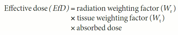
(A) The tissue weighting factor (Wₜ) represents the relative tissue radiosensitivity of irradiated material (e.g., muscle vs. intestinal epithelium vs. bone).
The radiation weighting factor (Wᵣ) is a number assigned to different types of ionizing radiations to better determine their effect on tissue (e.g., x-ray vs. alpha particles). The Wᵣ of different ionizing radiations depends on the LET of that particular radiation.
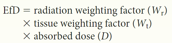
The following formula is used to determine effective dose (EfD):
The x-ray interaction with matter that is responsible for the majority of scattered radiation reaching the image receptor (IR) is:
A. the photoelectric effect
B. Compton scatter
C. classical scatter
D. Thompson scatter
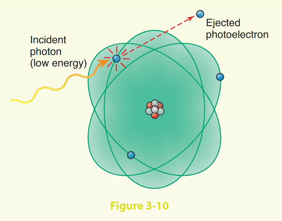
(B) In the photoelectric effect, a relatively low-energy photon uses all its energy to eject an inner-shell electron, leaving a vacancy. An electron from the shell above drops down to fill the vacancy and in so doing gives up a characteristic ray.
This type of interaction is most harmful to the patient because all the photon energy is transferred to the tissue. In Compton scatter, a high-energy incident photon ejects an outer-shell electron. In so doing, the incident photon is deflected with reduced energy, but it usually retains most of its energy and exits the body as an energetic scattered ray.
This scattered ray will either contribute to image fog or pose a radiation hazard to personnel depending on its direction of exit. In classic scatter, a low-energy photon interacts with an atom but causes no ionization; the incident photon disappears into the atom and then is released immediately as a photon of identical energy but with changed direction. Thompson scatter is another name for classic scatter.
The exposure rate to a body 5 m from a source of radiation is 50 mGyₐ/h. Which of the following distances would best decrease the exposure to 10 mGyₐ/h?
A. 2.2 m
B. 5 m
C. 11 m
D. 25 m
(C) The relationship between x-ray intensity and distance from the source is expressed by the inverse-square law of radiation. The formula is:
Thus, x = 11.1 m (36 feet) is necessary to decrease the exposure to 10 mGya/h. Note that in order for the exposure rate to decrease, the distance from the source of radiation must increase; as distance decreases, exposure rate increases.
Types of secondary radiation barriers include:
The control booth
Lead aprons
The x-ray tube housing
A. 2 only
B. 1 and 2 only
C. 2 and 3 only
D. 1, 2, and 3
(D) Secondary radiation includes leakage and scattered radiation. Secondary barriers are those that should never be struck by the primary beam. The control booth wall is a secondary barrier; therefore, the primary beam must never be directed toward it.
The x-ray tube housing must reduce leakage radiation to less than 1 mGya/h (100 mR/h) at a distance of 1 m from the housing. Lead aprons, lead gloves, portable x-ray barriers, and so on are also designed to protect the user from exposure to scattered radiation and will not protect the individual from the primary beam.
Late radiation–induced somatic effects include:
Thyroid cancers
Cataractogenesis
Reduced fertility
A. 1 only
B. 1 and 2 only
C. 2 and 3 only
D. 1, 2, and 3
(D) Late somatic effects are those that can occur years after initial exposure and are caused by low, chronic exposures. Occupationally exposed personnel are concerned with the late effects of radiation exposure.
Bone malignancies, thyroid cancers, leukemia, and skin cancers are examples of carcinogenic somatic effects of radiation. Another example of somatic effects of radiation is cataract formation in the lenses of eyes of individuals accidentally exposed to sufficient quantities of radiation.
The lives of many of the early radiation workers were several years shorter than the lives of the general population. Statistics revealed that radiologists, for example, had a shorter life span than physicians of other specialties. Life span shortening, then, was another somatic effect of radiation.
Certainly, these effects should never be experienced nowadays. The human reproductive organs are particularly radiosensitive. Fertility and heredity can be greatly affected by the germ cells produced within the testes (spermatogonia) and ovaries (oogonia). Excessive radiation exposure to the gonads can cause reduced fertility or permanent sterility and/or genetic mutations.
Each time an x-ray photon scatters, its intensity at 1 m from the scattering object is what fraction of its original intensity?
A. 1/10
B. 1/100
C. 1/500
D. 1/1000
(D) One of the radiation protection guidelines for the occupationally exposed is that x-ray photons should scatter twice before reaching the operator. Each time an x-ray photon scatters, its intensity at 1 m from the scattering object is one-thousandth of its original intensity.
Of course, the operator should be behind a protective shield while making the exposure, but multiple scatterings further reduce the danger of exposure from the scattered radiation.
The law of Bergonié and Tribondeau states that cells are more radiosensitive if they are:
Highly proliferative
Highly differentiated
Immature
A. 1 only
B. 1 and 2 only
C. 1 and 3 only
D. 1, 2, and 3
(C) Bergonié and Tribondeau were French scientists who, in 1906, theorized what has now become verified law. Cells are more radiosensitive if they are immature (undifferentiated or stem) cells, if they are highly mitotic (having a high rate of proliferation), and if the irradiated tissue is young.
Cells and tissues that are still undergoing development are more radiosensitive than fully developed tissues.
A thermoluminescent dosimetry system uses which of the following crystals?
A. Silver halide
B. Sodium thiosulfate
C. Lithium fluoride
D. Aluminum oxide
(C) TLDs are personnel radiation monitors that use lithium fluoride crystals. Once exposed to ionizing radiation and then heated, these crystals give off light proportional to the amount of radiation received. TLDs are very accurate personnel monitors.
Even more accurate are optically stimulated luminescence (OSL) dosimeters. OSL dosimeters use aluminum oxide as their sensitive crystal. Silver halide is in film emulsion and sodium thiosulfate is in fixer solution.
A controlled area is one that is occupied by:
Radiology personnel
Patients
Anyone
A. 1 only
B. 2 only
C. 1 and 2 only
D. 3 only
(C) A controlled area is occupied by radiology personnel and patients. The radiology personnel are trained in radiation safety and wear radiation monitors. The radiation barriers in a controlled area must keep the weekly dose to radiation workers to less than 1 mSv/week (100 mrem).
That limit is based on the annual occupational dose limit of 50 mSv/year. The occupancy factor in a controlled area is considered to be 1, indicating that the area may always be occupied and, therefore, requires maximum shielding.
An uncontrolled area is one that is occupied by anyone; the maximum exposure permitted in an uncontrolled area is 20 μSv (2 mrem)/week. That limit is based on the recommended annual dose limit to the general population of 1 mSv (100 mrem)/year. Shielding requirements vary according to several factors, one being occupancy factor.
All of the following have an effect on patient dose, except:
A. Kilovoltage
B. Milliampere seconds
C. Focal spot size
D. Inherent filtration
(C) The selected milliampere seconds are directly related to patient dose, that is, if milliampere seconds are doubled, patient dose is doubled. Similarly, if milliampere seconds are cut in half, patient dose is cut in half.
The selected kilovolts peak is inversely related to patient dose, that is, if the kilovolts peak is increased, patient dose can be decreased because more x-ray photons are transmitted through the patient rather than being absorbed.
Inherent filtration is provided by materials that are a permanent part of the tube housing, that is, the glass envelope of the x-ray tube and the oil coolant. Added filtration, usually thin sheets of aluminum, is present to make a total of 2.5-mm Al equivalent for equipment operated above 70 kVp. Filtration is used to decrease patient dose by removing the weak x-rays that have no value but contribute to the skin dose.
The effect of focal spot size is principally on radiographic sharpness; it has no effect on patient dose.
The degree of x-ray attenuation is directly related to what quality of the radiographed part?
A. Valence electrons
B. Binding energy
C. Atomic number
D. Electron shells
(C) The radiologic image is obtained as a result of the attenuation processes occurring in the body. Tissues having a high atomic number (e.g., bone) are very dense and will allow little or no passage of x-rays. Those tissues appear white or light on the image as a result of photoelectric interactions.
Other tissues are easier for x-ray photons to penetrate—those tissues appear darker because a greater number of photons reached the IR. Valence electrons are those in the outermost atomic shell. Electron binding energy is related to its proximity to the atomic nucleus; inner-shell electrons have a higher binding energy than outer electrons.
An increase in total filtration of the x-ray beam will increase:
A. Patient skin dose
B. Beam HVL
C. Image contrast
D. Milliroentgen (mR) output
(B) Aluminum filters are used to decrease patient skin dose by absorbing the low-energy photons (therefore, decreased mGya output) that do not contribute to the image but do contribute to patient skin dose.
HVL is defined as that thickness of any absorber that will decrease the intensity of a particular beam to one-half of its original value. As filtration of an x-ray beam is increased, the overall average energy of the resulting beam is greater (because the low-energy photons have been removed)—and, therefore, the HVL thickness required would be greater.
In radiation protection, the product of absorbed dose, tissue weighting factor, and radiation weighting factor is used to determine:
A. C/kg
B. mR
C. EₓD
D. QF
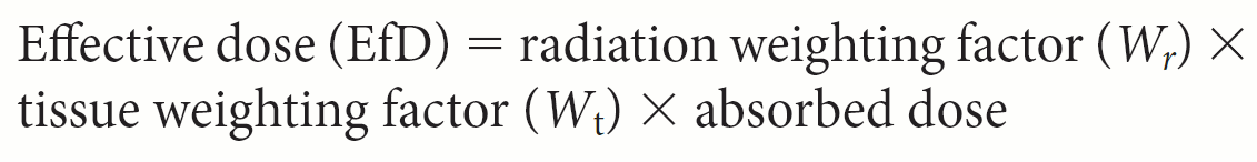
(C) The tissue weighting factor (Wₜ) represents the relative tissue radiosensitivity of irradiated material (e.g., muscle vs. intestinal epithelium vs. bone). The radiation weighting factor (Wᵣ) is a number assigned to different types of ionizing radiations to better determine their effect on tissue (e.g., x-ray vs. alpha particles).
The Wᵣ of different ionizing radiations depends on the LET of that particular radiation. The following formula is used to determine effective dose (EfD):
Effective dose (EfD) = radiation weighting factor (Wᵣ) × tissue weighting factor (Wₜ) × absorbed dose
Which of the following is recommended for the pregnant radiographer?
A. Change dosimeters weekly
B. Wear a second dosimeter under the lead apron
C. Wear two dosimeters and switch their positions appropriately
D. Leave radiation areas for the duration of the pregnancy
(B) Special arrangements are required for occupational monitoring of the pregnant radiographer. The pregnant radiographer will wear two dosimeters—one in its usual place at the collar and the other, a baby/fetal dosimeter, worn over the abdomen and under the lead apron during fluoroscopy.
The baby/fetal dosimeter must be identified as such and always must be worn in the same place. Care must be taken not to mix the positions of the two dosimeters. The dosimeters are read monthly, as usual.
The pregnant radiographer may not be made to leave the radiation area/department because of her pregnancy.
The annual dose limit for medical imaging personnel includes radiation from:
Occupational exposure
Background radiation
Medical x-rays
A. 1 only
B. 1 and 2 only
C. 2 and 3 only
D. 1, 2, and 3
(A) Occupationally, exposed individuals are required to use devices that will record and provide documentation of the radiation they receive over a given period of time, traditionally 1 month. The most commonly used personnel dosimeters are the OSL, the TLD, DOS dosimeter, and the film badge.
These devices must be worn only for documentation of occupational exposure. They must not be worn for any medical or dental x-rays one receives as a patient, and they are not used to measure naturally occurring background radiation.
Neurologic anomalies will most likely occur if an exposure dose of 40 mGy (40 rad) was delivered to a pregnant uterus during the:
A. 3rd week of pregnancy
B. 9th week of pregnancy
C. 15th week of pregnancy
D. 24th week of pregnancy
(B) Irradiation during pregnancy, especially in early pregnancy, must be avoided. The fetus is particularly radiosensitive during the first trimester, during much of which time pregnancy may not even be suspected.
High-risk examinations include pelvis, hip, femur, lumbar spine, cystograms and urograms, and upper and lower gastrointestinal (GI) series. During the first trimester, specifically the 2nd to 10th weeks of pregnancy (i.e., during major organogenesis), if the radiation dose is sufficient, fetal anomalies can be produced.
Skeletal and/or organ anomalies can appear if irradiation occurs in the early part of this time period, and neurologic anomalies can be formed in the latter part; mental retardation and childhood malignant diseases, such as cancers or leukemia, and retarded growth/development also can result from irradiation during the first trimester. Fetal irradiation during the second and third trimesters is not likely to produce anomalies but rather, with sufficient dose, some type of childhood malignant disease.
Fetal irradiation during the first 2 weeks of gestation can result in embryonic resorption or spontaneous abortion. It must be emphasized, however, that the likelihood of producing fetal anomalies at doses less than 200 mGy (20 rad) is exceedingly small and that most general diagnostic examinations are likely to deliver fetal doses of less than 10–20 mGy (1–2 rad).
If a quantity of ionizing radiation is delivered to a body over a long period of time, the effect:
A. Will be greater than if it were delivered all at one time
B. Will be less than if it were delivered all at one time
C. Has no relation to how it is delivered in time
D. Depends solely on the radiation quality
(B) The effects of a quantity of radiation delivered to a body depend on the amount of radiation received, the size of the irradiated area, and how the radiation is delivered in time.
If the radiation is delivered in portions over a period of time, it is said to be fractionated and has a less harmful effect than if it were delivered all at once because cells have an opportunity to repair, and some recovery occurs between doses.
Moving the image intensifier closer to the patient during fluoroscopy:
Decreases patient dose
Improves image quality
Decreases the SID
A. 1 only
B. 1 and 2 only
C. 1 and 3 only
D. 1, 2, and 3
(B) X-ray intensity is inversely related to SID; as SID increases, x-ray intensity decreases. The x-ray intensity at the image intensifier also depends on the SID.
Moving the image intensifier closer to the patient decreases the patient entrance dose and increases x-ray beam intensity at the input phosphor. The automatic brightness control (ABC) then decreases the milliamperes again decreasing the patient dose.
Moving the image intensifier closer to the patient also results in a shorter OID, therefore decreased magnification and improved image quality.
Which of the following types of radiation is/are considered electromagnetic?
X-ray
Visible light
Gamma
Ultraviolet light
Beta
A. 1, 3, and 5
B. 2, 3, 4, and 5
C. 1, 2, 3, and 4
D. 2, 3, and 5
E. 3, 4, and 5
(C) Gamma and x-radiations are electromagnetic, having wavelike fluctuations like other radiations of the electromagnetic spectrum (e.g., visible light, radio waves, ultraviolet, and infrared).
Alpha and beta radiations are particulate radiations; alpha is composed of two protons and two neutrons, and beta is identical to an electron.
How does filtration affect the primary beam?
It increases the average energy of the primary beam
It decreases the average energy of the primary beam
It makes the useful beam more penetrating
It increases the intensity of the primary beam
It decreases x-ray beam quality
It decreases patient dose
A. 1, 4, and 5
B. 1, 3, and 6
C. 2, 3, and 5
D. 3, 5, and 6
E. 4, 5, and 6
(B) X-rays produced at the tungsten target make up a heterogeneous primary beam. Filtration serves to eliminate the softer, less penetrating, low-energy photons, leaving an x-ray beam of higher average energy.
Because x-ray photons were removed from the beam, it has a decreased intensity/quantity. The overall x-ray beam is now more penetrating. Filtration is important in patient protection, because unfiltered, low-energy photons that are not energetic enough to reach the IR stay in the body and contribute to total patient dose.
What is the minimum lead requirement for lead aprons, according to the NCRP?
A. 1.0-mm Pb equivalent
B. 0.25-mm Pb equivalent
C. 0.50-mm Pb equivalent
D. 0.05-mm Pb equivalent
(C) Lead aprons are secondary radiation barriers and must contain at least 0.25-mm Pb equivalent (according to CFR 20), usually in the form of lead-impregnated vinyl.
Many radiology departments routinely use lead aprons containing 0.5 mm Pb (the NCRP recommends 0.5-mm Pb equivalent minimum). These aprons are heavier, but they attenuate a higher percentage of scattered radiation.
Calculation of effective dose (EₓD) requires:
Absorbed dose (D)
Type of radiation (Wᵣ)
Tissue exposed (Wₜ)
A. 1 only
B. 2 only
C. 1 and 2 only
D. 1 and 3 only
E. 1, 2, and 3
(E) The term effective dose (EfD) refers to the dose from radiation sources internal and/or external to the body and is expressed in units of Sievert or rem.
The factors used to determine effective dose (EfD) are as follows:
EfD = radiation weighting factor (Wᵣ)
× tissue weighting factor (Wₜ)
× absorbed dose (D)
The term equivalent dose (EqD) refers simply to the product of the absorbed dose (Gy/rad) and its radiation weighting factor (Wᵣ).
Immature cells are called:
Undifferentiated cells
Stem cells
Genetic cells
A. 1 only
B. 1 and 2 only
C. 1 and 3 only
D. 1, 2, and 3
(B) Cells are frequently identified by their stage of development. Immature cells may be called undifferentiated or stem cells.
Immature cells are much more radiosensitive than mature cells.
What is the term used to describe x-ray photon interaction with matter and the transference of part of the photon’s energy to matter?
A. Absorption
B. Scattering
C. Differential absorption
D. Divergence
(B) Scattering occurs when there is partial transfer of the proton’s energy to matter, as in the Compton effect.
Absorption occurs when an x-ray photon interacts with matter and disappears, such as in the photoelectric effect. Differential absorption describes the absorption characteristics of various tissues such as bone, muscle, and soft tissue.
The reduction in the intensity (quantity) of an x-ray beam as it passes through matter is termed attenuation. Divergence refers to a directional characteristic of the x-ray beam as it is emitted from the focal spot.
Advantages of anatomic compression during imaging include:
Decreased patient dose
Improved contrast resolution
Improved spatial resolution
A. 1 only
B. 1 and 2 only
C. 2 and 3 only
D. 1, 2, and 3
(D) Part compression, when possible, can improve spatial resolution by reducing part thickness and decreasing OID.
Contrast resolution is improved because less scattered radiation will be generated in a thinner part. Because the part is essentially thinner, technical factors can be reduced thereby decreasing patient dose.
To be in compliance with radiation safety standards, the fluoroscopy exposure switch must:
A. Sound during fluoro-on time
B. Be on a 6-foot-long cord
C. Terminate exposure after 5 min
D. Be the “dead-man” type
(D) For radiation safety, the fluoroscopy exposure switch must be of the “dead-man” type. When the foot is removed from the fluoro pedal, the “dead-man” switch will terminate the exposure immediately.
There must also be a fluoroscopy cumulative timer that will either sound or interrupt (it will not terminate/stop exposure) exposure after 5 min of fluoroscopy.
Any wall that the useful x-ray beam may be directed toward must be a:
A. Secondary barrier
B. Primary barrier
C. Leakage barrier
D. Scattered barrier
(B) Protective barriers are classified as either primary or secondary. Primary barriers protect from the useful, or primary, x-ray beam and consist of a certain thickness of lead.
They are located anywhere that the primary beam can possibly be directed, for example, the walls of the x-ray room. The walls of the x-ray room usually require a 1/16 inch (1.6 mm) thickness of lead and should be 7 feet high.
Secondary barriers protect from secondary (scattered and leakage) radiation. Secondary barriers are control booths, lead aprons, gloves, and the wall of the x-ray room above 7 feet. Secondary barriers require much less lead than primary barriers.
The annual dose limit for occupationally exposed individuals is valid for:
A. Alpha, beta, and x-radiations
B. X- and gamma radiations only
C. Beta, x-, and gamma radiations
D. All ionizing radiations
(C) The occupational dose limit is valid for beta, x-, and gamma radiations.
Because alpha radiation is so rapidly ionizing, traditional personnel monitors will not record alpha radiation. However, because alpha particles are capable of penetrating only a few centimeters of air, they are practically harmless as an external source.
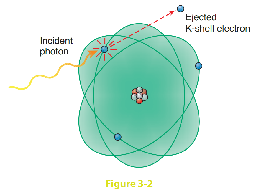
The interaction between x-ray photons and matter shown in Figure 3-2 is associated with:
An inner-shell electron
Photoelectric effect
Partial energy transfer from photon to electron
A. 1 only
B. 1 and 2 only
C. 1 and 3 only
D. 2 and 3 only
(B) Diagnostic x-ray photons interact with tissue in a number of ways, but most frequently they are involved in the production of Compton scatter or in a photoelectric interaction.
The photoelectric effect occurs when a relatively low-energy x-ray photon uses all its energy to eject an inner-shell electron. That electron is ejected (photoelectron) from the innermost (K) shell, leaving a “hole” in the K shell and producing a positive ion.
An L-shell electron then drops down to fill the K vacancy and in so doing emits a characteristic ray whose energy is equal to the difference between the binding energies of the K and L shells. The photoelectric effect occurs with high–atomic-number absorbers, such as bone and positive contrast media and is responsible for the production of contrast.
Therefore, its occurrence is helpful for the production of the radiographic image, but it contributes significantly to the dose received by the patient (because it involves complete absorption of the incident photon). Scattered radiation, which produces a radiation hazard to the radiographer (as in fluoroscopy), is a product of the Compton scatter interaction occurring with higher energy x-ray photons.
Patient dose increases as fluoroscopic:
A. FOV increases
B. FOV decreases
C. FSS increases
D. FSS decreases
(B) During fluoroscopic procedures, as field of view (FOV) decreases, magnification of the output screen image increases, and contrast and resolution improve.
The focal point on an image intensifier’s 6-inch field/mode is further away from the output phosphor than the focal point on the normal mode; therefore, the output image is magnified.
Because less minification takes place, the image is not as bright. Exposure factors are increased automatically to compensate for the loss in brightness with smaller FOVs. Focal spot size (FSS) is unrelated to patient dose.
Types of gonadal shielding include which of the following?
Flat contact
Shaped contact (contour)
Shadow
A. 1 only
B. 1 and 2 only
C. 2 and 3 only
D. 1, 2, and 3
(D) Gonadal shielding should be used whenever appropriate and possible during radiographic and fluoroscopic examinations.
Flat contact shields are useful for simple recumbent (AP and PA) studies, but when the examination necessitates obtaining oblique, lateral, or erect projections, they become less efficient.
Shaped contact (contour) shields are best because they enclose the male reproductive organs and remain in position in oblique, lateral, and erect positions. Shadow shields that attach to the tube head are particularly useful for surgical sterile fields.
What unit of measure is used to express ionizing radiation dose to biologic material?
A. Air kerma (Gyₐ)
B. GYₜ
C. Sv
D. RBE
(C) The Sievert (Sv) is the unit radiation dose to biologic material, and is used to express occupational exposure and effective dose.
The traditional unit rem is an acronym for radiation equivalent man; it includes the RBE specific to the tissue irradiated and can be used as a unit of measurement for the dose to biologic tissue.
The traditional unit rad is an acronym for radiation absorbed dose; it measures the energy deposited in any material. The SI unit used to express radiation dose is Gy<sub>t</sub>. Air kerma (Gya) is the SI unit of radiation exposure. Roentgen is a unit of exposure; it can be used to express the quantity of ionizations in air as a traditional unit of measure.
LET is best defined as:
A method of expressing radiation quality
A measure of the rate at which radiation energy is transferred to soft tissue
Transmission of polyenergetic radiation
A. 1 only
B. 1 and 2 only
C. 1 and 3 only
D. 1, 2, and 3
(B) When biologic material is irradiated, there are a number of modifying factors that determine what kind and how much response will occur in the material.
One of these factors is LET, which expresses the rate at which particulate or photon energy is transferred to the absorber. Because different kinds of radiation have different degrees of penetration in different materials, it is also a useful way of expressing the quality of the radiation.
For exposure to 10 mGy of each of the following ionizing radiations, which would result in the greatest dose to the individual?
A. External source of 1-MeV x-rays
B. External source of diagnostic x-rays
C. Internal source of alpha particles
D. Internal source of beta particles
(C) Electromagnetic radiations, such as x-rays and gamma rays, are considered low-LET radiations because they produce fewer ionizations than the highly ionizing particulate radiations, such as alpha particles.
Alpha particles are large and heavy (two protons and two neutrons), and although they possess a great deal of kinetic energy (approximately 5 MeV), their energy is lost rapidly through multiple ionizations (approximately 40,000 atoms/cm of air).
As an external source, alpha particles are almost harmless because they ionize the air very quickly and never reach the individual. As internal sources, however, they ionize tissues and are potentially the most harmful. It may be stated that the alpha particle has one of the highest LETs of all ionizing radiations.
The skin response to radiation exposure that appears as hair loss is known as:
A. Dry desquamation
B. Moist desquamation
C. Erythema
D. Epilation
(D) The various skin responses to irradiation include all four choices.
The first noticeable response would be erythema, a reddening of the skin very much like sunburn. Dry desquamation could follow; it is a dry peeling of the skin.
Moist desquamation is peeling with associated pus-like fluid. Epilation is hair loss; it can be temporary or permanent depending on sensitivity and dose.
Irradiation of macromolecules in vitro can result in:
Cleaved chromosome
Cross-linking
Mutation
A. 1 only
B. 1 and 2 only
C. 2 and 3 only
D. 1, 2, and 3
(D) Irradiation damage is a result of either the effects of irradiation on water (radiolysis) or its effects on macromolecules.
Effects on macromolecules include cleaved chromosomes, cross-linking, and mutations. Cleaved/broken chromosome is the result of a double-strand break on the same DNA “rung.” Cross-linking is incorrect joining of broken DNA fragments.
A mutation is the result of damage/alteration of nitrogenous base sequence as a result of irradiation. Because 80% of the body is made up of water, radiolysis of water is the predominant radiation interaction in the body.
The reduction in the intensity of an x-ray beam as it passes through material is termed:
A. Absorption
B. Scattering
C. Attenuation
D. Divergence
(C) The reduction in the intensity (quantity) of an x-ray beam as it passes through matter as a result of absorption and scatter is called attenuation.
Absorption occurs when an x-ray photon interacts with matter and disappears, such as in the photoelectric effect. Scattering occurs when there is partial transfer of energy to matter, such as in the Compton effect.
Which type of dose–response relationship represents radiation-induced leukemia and genetic effects?
A. Linear, threshold
B. Nonlinear, threshold
C. Linear, nonthreshold
D. Nonlinear, nonthreshold
(C) Radiation-induced malignancy, leukemia, and genetic effects are late effects (or stochastic/probabilistic effects) of radiation exposure.
These can occur years after survival of an acute radiation dose or after exposure to low levels of radiation over a long period of time. Radiation workers need to be especially aware of the late effects of radiation because their exposure to radiation is usually low level over a long period of time.
Occupational radiation protection guidelines, therefore, are based on late effects of radiation according to a linear, nonthreshold dose–response curve.
Which of the following groups of technical factors will deliver the least amount of exposure to the patient?
A. 400 mA, 0.25 s, 100 kVp
B. 600 mA, 0.33 s, 90 kVp
C. 800 mA, 0.5 s, 80 kVp
D. 800 mA, 1.0 s, 70 kVp
(A) The milliampere-second (mAs) setting regulates the quantity of radiation delivered to the patient, and the kilovolts peak (kVp) setting regulates the quality (i.e., penetration) of the radiation delivered to the patient.
Therefore, higher energy (more penetrating) radiation, accompanied by lower mAs, is the safest combination for the patient because more photons exit the patient rather than being absorbed.
Late effects of radiation, whose incidence is dose related and for which there is no threshold dose, are called:
A. Nonstochastic/deterministic
B. Stochastic/probabilistic
C. Chromosomal aberration
D. Hematologic depression
(B) Late or long-term effects of radiation can occur in tissues that have survived a previous irradiation months or years earlier.
These late effects, such as carcinogenesis and genetic effects, are stochastic/probabilistic, meaning they are “all-or-nothing”: either the organism develops cancer or it does not.
Most late effects have no threshold dose; any dose, however small, can induce an effect. Increasing the dose increases the likelihood of the effect but does not affect its severity.
Nonstochastic/deterministic effects (tissue responses) have a threshold and increase in severity as dose increases. Early effects occur after relatively high doses and should never happen in diagnostic radiology. Examples include hematologic effects (decreased WBCs, RBCs, platelets), gonadal effects (temporary infertility), and skin effects (epilation, erythema). Acute radiation lethality occurs after massive exposure and results in death in weeks or days.
Which of the following statements is/are true regarding the human gonadal cells?
The female oogonia reproduce only during fetal life
The male spermatogonia reproduce continuously
Both male and female stem cells reproduce only during fetal life
A. 1 only
B. 2 only
C. 1 and 2 only
D. 3 only
(C) The development of male and female reproductive stem cells has important radiation protection implications.
Male stem cells reproduce continuously. However, female stem cells develop only during fetal life; women are born with all the reproductive cells they will ever have.
It is exceedingly important to shield children whenever possible because they have their reproductive futures ahead of them.
Classify the following tissues in order of increasing radiosensitivity.
Liver cells
Intestinal crypt cells
Muscle cells
A. 1, 3, 2
B. 2, 3, 1
C. 2, 1, 3
D. 3, 1, 2
(D) According to Bergonié and Tribondeau, the most radiosensitive cells are undifferentiated, rapidly dividing cells, such as lymphocytes, intestinal crypt (of Lieberkühn) cells, and spermatogonia.
Liver cells are among the types of cells that are somewhat differentiated and capable of mitosis. These characteristics render them somewhat radiosensitive.
Muscle cells, as well as nerve cells and red blood cells, are highly differentiated and do not divide. Therefore, in order of increasing sensitivity (from least to greatest sensitivity), the cells are muscle, liver, and then intestinal crypt cells.
The largest amount of diagnostic x-ray absorption is most likely to occur in which of the following tissues?
A. Lung
B. Adipose
C. Muscle
D. Bone
(D) Our bodies contain a variety of tissues having a variety of tissue densities. These tissue densities afford differing degrees of resistance to the passage of x-ray photons.
Tissues having greater density absorb more of the x-ray beam (recall the photoelectric effect). Soft tissues are fairly easily penetrated to varying degrees, that is, lung and adipose are easier to penetrate than muscle.
Bone has much higher tissue mass density and, therefore, absorbs more of the x-ray beam.
According to NCRP regulations, leakage radiation from the x-ray tube must not exceed:
A. 0.1 mGyₐ/h (10 mR/h)
B. 1.0 mGyₐ/h (100 mR/h)
C. 0.1 mGyₐ/min (10 mR/min)
D. 1.0 mGyₐ/min (100 mR/min)
(B) X-ray photons produced in the x-ray tube can radiate in directions other than the one desired.
The tube housing, therefore, is constructed so that very little of this leakage radiation is permitted to escape.
The regulation states that leakage radiation must not exceed 1 mGy a/h (100 mR/h) at 1 m while the tube is operated at maximum potential.